Abstract
The inflammasome is a multi-protein complex which when activated regulates caspase-1 activation and IL-1β secretion. Inflammasome activation is mediated by NLR proteins that respond to stimuli. Among NLRs, NLRP3 senses the widest array of stimuli. NLRP3 inflammasome plays an important role in the development of many cancer types. However, Whether NLRP3 inflammasome plays an important role in the process of hepatocellular carcinoma (HCC) is still unknown. Here, the anticancer effect of luteoloside, a naturally occurring flavonoid isolated from the medicinal plant Gentiana macrophylla, against HCC cells and the underlying mechanisms were investigated. Luteoloside significantly inhibited the proliferation of HCC cells in vitro and in vivo. Live-cell imaging and transwell assays showed that the migration and invasive capacities of HCC cells, which were treated with luteoloside, were significantly inhibited compared with the control cells. The inhibitory effect of luteoloside on metastasis was also observed in vivo in male BALB/c-nu/nu mouse lung metastasis model. Further studies showed that luteoloside could significantly reduce the intracellular reactive oxygen species (ROS) accumulation. The decreased levels of ROS induced by luteoloside was accompanied by decrease in expression of NLRP3 inflammasome resulting in decrease in proteolytic cleavage of caspase-1. Inactivation of caspase-1 by luteoloside resulted in inhibition of IL-1β. Thus, luteoloside exerts its inhibitory effect on proliferation, invasion and metastasis of HCC cells through inhibition of NLRP3 inflammasome. Our results indicate that luteoloside can be a potential therapeutic agent not only as an adjuvant therapy for HCC, but also, in the control and prevention of metastatic HCC.
🔬 Techniques
💻 Software
✨ Fluorophores
🧪 Sample Preparation
🏭 Microscope Brands
🧪 Reagent Suppliers
🔎 Objectives
💻 Software Details
🏛️ Research Organizations (ROR)
Affiliated research institutions:
📋 Methods
Cell Lines and Reagents
The human HCC cell lines (Hep3B and SNU-449) were purchased from the American Type Culture Collection.
Human hepatoma cells
(Huh-7) was purchased from Japanese Collection of Research Bioresources (JCRB, Tokyo, Japan). The human hepatoma cell line SMMC-7721 was purchased from the Committee on Type Culture Collection of Chinese Academy of Sciences (Shanghai, China). The human HCC cell lines (MHCC-LM3 and MHCC97-H) were obtained from the Liver Cancer Institute of Zhongshan Hospital, Fudan University (Shanghai, China). Luteoloside ( Fig. 1A , Batch Number: 025-120622, Purity = 99.7%, purchased from Chengdu Herbpurify Co., Ltd., Chengdu, China), a naturally occurring flavonoid isolated from the medicinal plant Gentiana macrophylla , was dissolved at a concentration of 20 mM in 100% DMSO as a stock solution, stored at −20°C, and diluted with medium before each experiment. The final DMSO concentration did not exceed 0.1% throughout the study. 10.1371/journal.pone.0089961.g001 Figure 1 Luteoloside inhibits proliferation of HCC cells. (A) HPLC analysis of the purity of luteoloside used in the present study. Insert : The chemical structure of luteoloside. (B)–(C), comparative dose- and time-dependent effect of luteoloside on the proliferation potential of HCC cells. The percentage of cell viability in different treatment groups was determined using Cell Counting Kit-8 assay. *, P
📊 Figures
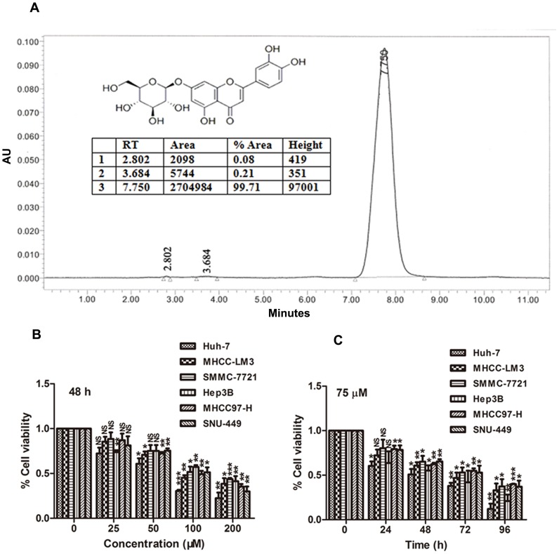
Figure 1
Luteoloside inhibits proliferation of HCC cells.
(A) HPLC analysis of the purity of luteoloside used in the present study. Insert : The chemical structure of luteoloside. (B)u2013(C), comparative dose- and time-dependent effect of luteoloside on the...
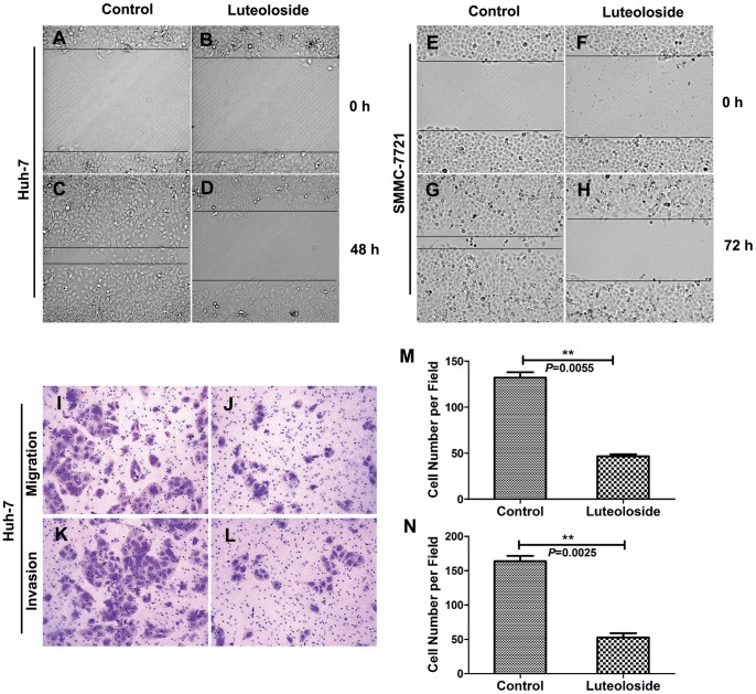
Figure 2
Luteoloside inhibits migration and invasion of HCC cells.
The migration of cells into the wound was monitored in multiple wells using a CellVoyager CV1000 confocal scanner system. The images were acquired every 0.5 hour for 48 hours (Huh-7 cells) or every ho...
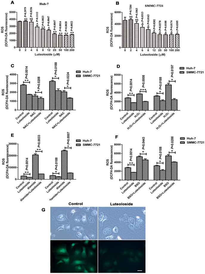
Figure 3
Luteoloside decreases intracellular ROS.
ROS levels were measured using the ROS assay with DCFH-DA fluorescence dye. (Au2013B) Cells were treated with luteoloside at the indicated concentration for 8 h, then reacted with 10 u00b5M DCFH-DA fo...
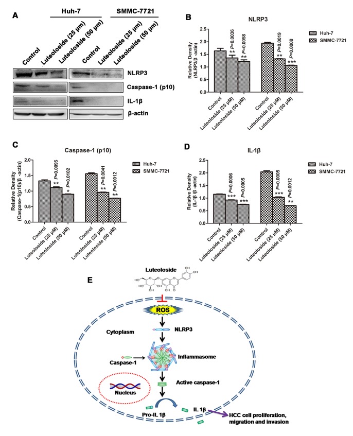
Figure 4
Luteoloside suppresses the NLRP3 inflammasome activation.
(A) Western blot analyses of NLRP3, Caspase-1 (p10) and IL-1u03b2 protein expression in Huh-7 and SMMC-7721 cells exposed two different concentrations of luteoloside for 48 h. (Bu2013D) Relative quant...
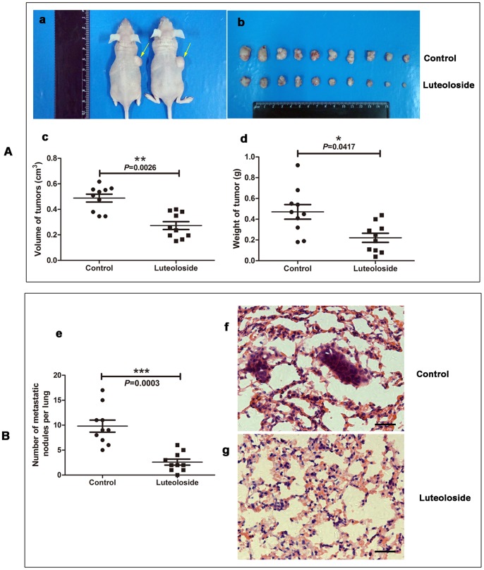
Figure 5
Luteoloside inhibits tumorigenic and spontaneous lung metastatic capabilities of SMMC-7721 cells.
(A) Subcutaneous injection of SMMC-7721 cells plus luteoloside treatment in nude mice inhibited tumor growth. (B) Tail vein injection of SMMC-7721 cells plus luteoloside treatment in nude mice inhibit...
Figure images are served from the NIH/NLM PubMed Central Open Access Subset or Europe PMC; copyright remains with the publishers and authors.
💬 Discussion
0 commentsNo comments yet. Be the first to start a discussion!
Leave a Comment