Abstract
With recent advances in high-throughput, automated microscopy, there has been an increased demand for effective computational strategies to analyze large-scale, image-based data. To this end, computer vision approaches have been applied to cell segmentation and feature extraction, whereas machine-learning approaches have been developed to aid in phenotypic classification and clustering of data acquired from biological images. Here, we provide an overview of the commonly used computer vision and machine-learning methods for generating and categorizing phenotypic profiles, highlighting the general biological utility of each approach.
🔬 Techniques
🧬 Organisms
💻 Software
✨ Fluorophores
🧪 Sample Preparation
🔬 Cell Lines
💻 Software Details
🏛️ Research Organizations (ROR)
Affiliated research institutions:
📊 Figures
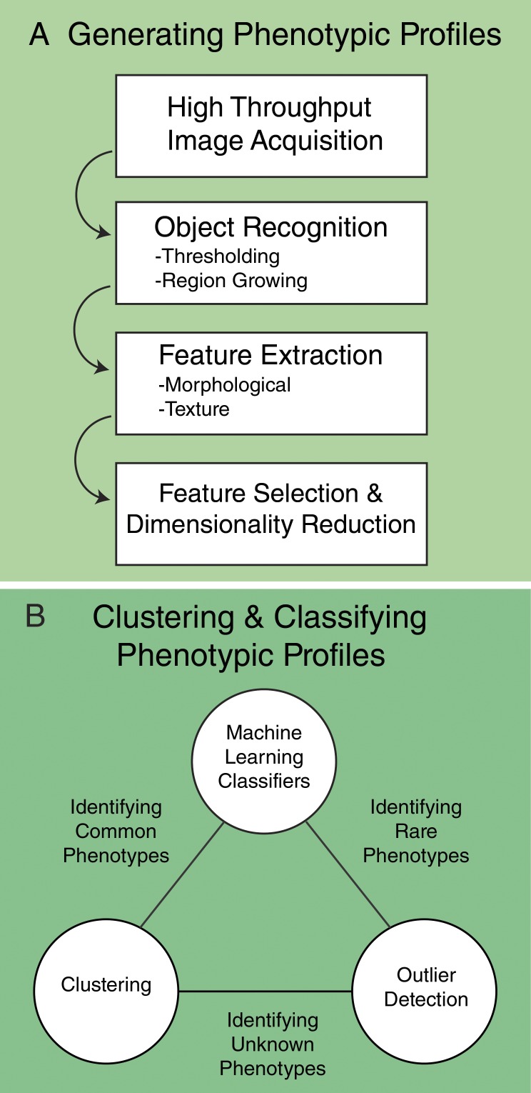
Figure 1.
General workflow for the generation and classification of phenotypic profiles. (A) Generation of phenotypic profiles involves high-throughput image acquisition, followed by segmentation, feature extra...
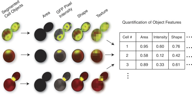
Figure 2.
Micrographs of individual budding yeast cells identified during segmentation, with illustrative examples of four types of features that could be identified during feature extraction. In these microgra...
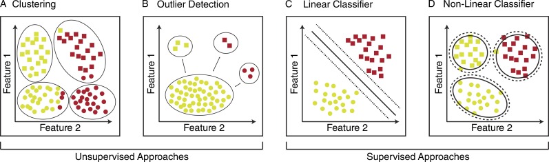
Figure 3.
Schematic representation of unsupervised and supervised methods to classify phenotypic profiles. (Au2013D) Each shape represents one object in the dataset. All features associated with each object are...
Figure images are served from the NIH/NLM PubMed Central Open Access Subset or Europe PMC; copyright remains with the publishers and authors.
💬 Discussion
0 commentsNo comments yet. Be the first to start a discussion!
Leave a Comment