Abstract
AbstractHigh-resolution imaging through turbid media is a fundamental challenge of optical sciences that has attracted a lot of attention in recent years for its wide range of potential applications. Here, we demonstrate that the resolution of imaging systems looking behind a highly scattering medium can be improved below the diffraction-limit. To achieve this, we demonstrate a novel microscopy technique enabled by the optical memory effect that uses a deconvolution image processing and thus it does not require iterative focusing, scanning or phase retrieval procedures. We show that this newly established ability of direct imaging through turbid media provides fundamental and practical advantages such as three-dimensional refocusing and unambiguous object reconstruction.
🔬 Techniques
💻 Software
🏭 Microscope Brands
💻 Software Details
🏛️ Research Organizations (ROR)
Affiliated research institutions:
📋 Methods
A coherent laser beam generated by a solid state continuous wave laser (532 nm) was transmitted through a rapidly rotating ground glass resulting in an incoherent illumination. As objects we used a USAF chart (Thorlabs) or fabricated custom patterns with standard photolithography masks. A scattering medium (diffusive tape or ground glass 120 Grit, Thorlabs) was placed at 45 to 160 mm from the object, and a single lens (f = 250 mm, 60 mm) was used to image the plane of the object onto the camera (Mightex MCE-B013). A typical integration time for a single shot needed in our experiments was 20 ms. The PSF of the system was measured by replacing the object with an iris of typical size 10–50 μm. Deconvolution post-processing was performed by applying the Richardson-Lucy MATLAB based code to the raw data, no other image processing methods were used in this paper. The number of applied deconvolution iterations varied between 100 and 400.
📊 Figures
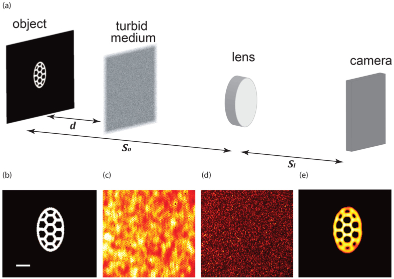
Figure 1
( a ) Optical setup. A mask is placed a distance d =u200916 cm behind a 120 grit ground glass diffuser. The plane of the object was imaged onto a CMOS camera using a 250u2009mm focal length lens. ( b ...
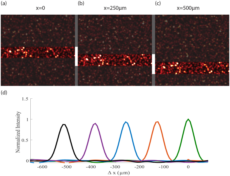
Figure 2
( au2013c ) PSF of the imaging system across the memory-effect field of view, measured by laterally shifting an iris in the object plane. The central area is highlighted to facilitate visualization of...
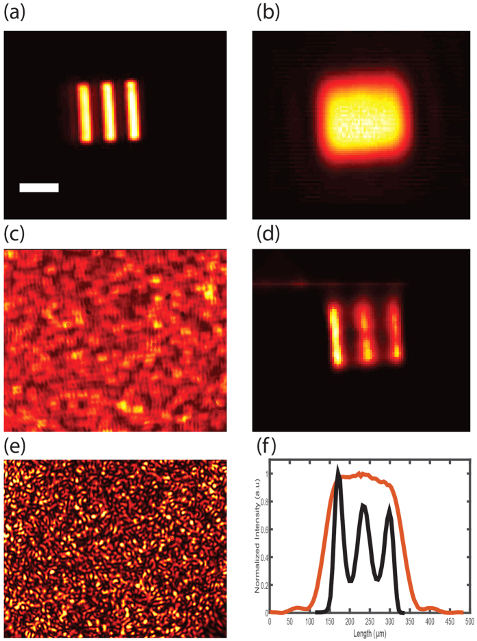
Figure 3
Super-resolution imaging through turbid medium.
( a ) Imaged object. Scale bar, 100u2009u03bcm. ( b ) Blurred image of the object with no turbid medium and a standard imaging system of low numerical aperture. ( c ) Image of the object hidden behind...
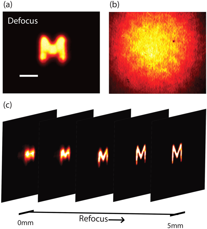
Figure 4
( a ) Defocused image of an object as seen through an imaging system (with no turbid medium) focused on a plane 5u2009mm away from the location of the object. Scale bar, 200u2009u03bcm. ( b ) The imag...
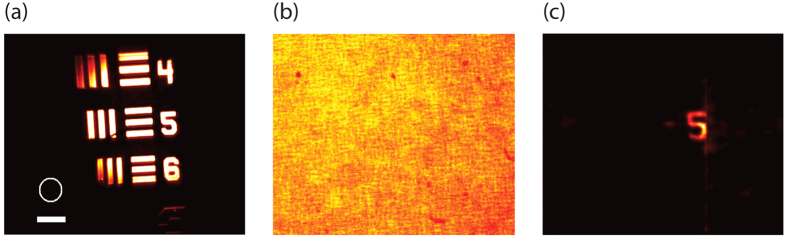
Figure 5
Large FOV illumination: ( a ) The object illuminated in the experiment (scale bar, 200u2009u03bcm), the white circle indicates the FOV permitted by the memory effect (this experiment was performed usi...
Figure images are served from the NIH/NLM PubMed Central Open Access Subset or Europe PMC; copyright remains with the publishers and authors.
💬 Discussion
0 commentsNo comments yet. Be the first to start a discussion!
Leave a Comment