Abstract
During protein synthesis, mRNA and tRNA undergo coupled translocation through the ribosome in a process that is catalyzed by elongation factor G (EF-G). On the basis of cryo-EM reconstructions, counterclockwise and clockwise rotational movements between the large and small ribosomal subunits have been implicated in a proposed ratcheting mechanism to drive the unidirectional movement of translocation. We used a combination of two fluorescence-based approaches to study the timing of these events, intersubunit fluorescence resonance energy transfer measurements to observe relative rotational movement of the subunits, and a fluorescence quenching assay to monitor translocation of mRNA. Binding of EF-G-GTP first induces rapid counterclockwise intersubunit rotation, followed by a slower, clockwise reversal of the rotational movement. We compared the rates of these movements and found that mRNA translocation occurs during the second, clockwise rotation event, corresponding to the transition from the hybrid state to the classical state.
🔬 Techniques
🧬 Organisms
✨ Fluorophores
🧪 Reagent Suppliers
💾 Data Repositories
🏛️ Research Organizations (ROR)
Affiliated research institutions:
📋 Methods
Materials and sample preparation tRNA fMet was purchased from MP Biomedicals; GTP, GDPNP, fusidic acid, spectinomycin, hygromycin B, tRNA Phe and tRNA Met were purchased from Sigma. GDP was further purified from contaminating GMP and GTP using ion-exchange chromatography as previously described 19 . Unlabeled mRNA (5′ GGC AAG GAG GUA AAA AUG UUU AAA CGU AAA UCU ACU 3′) and 3′ amino-modified mRNA (5′ GGC AAG GAG GUA AAA AUG UUU AAA 3′) were synthesized by Integrated DNA Technologies. 3′ amino-modified mRNA was conjugated with pyrene and subsequently purified according to published procedures 24 . fMet-tRNA fMet , N-Ac-Phe-tRNA Phe , and EF-G with a 6-His tag were prepared and purified as previously described 6 , 28 , 48 .
Ribosomes from E. coli strains
MRE600 (wild-type) and the fluorescently labeled 70S ribosome constructs S6-D/L9-A and S11-D/L9-A were prepared as described 19 , 49 – 51 . Stopped-flow kinetic experiments Ribosomal complexes were constructed and all stopped-flow experiments performed in buffer containing 20 mM Hepes·KOH (pH 7.5), 6 mM MgCl 2 , 150 mM NH 4 Cl, 6 mM β mercaptoethanol, 0.01% (w/v) Nikkol, 2 mM spermidine and 0.1 mM spermine. Pre-translocation complexes for FRET experiments were constructed by incubation of fluorescently-labeled 70S ribosomes (1 μM) with unlabeled mRNA (2 μM) and 2 μM deacylated tRNA Met (or deacylated tRNA fMet ) for 20 minutes at 37°C followed by incubation with N-Ac-Phe-tRNA Phe (1.5 μM) for 30 minutes at 37°C. For measurement of mRNA translocation kinetics, pre-translocation complexes were assembled similarly, except that unlabeled mRNA was replaced with pyrene-labeled mRNA (0.85 μM). In all experiments, pre-translocation ribosomes were mixed with EF-G and GTP (or GDPNP, GDP + fusidic acid, GTP + fusidic acid, depending on the experiment) using an Applied Photophysics stopped-flow fluorimeter. Final concentrations after mixing were: 35 nM ribosomes, 375 nM EF-G, 1 mM GTP, 0.2 mM GDP, 0.5 mM GDPNP, 0.1 mM fusidic acid. For the experiments in Fig. 4 , pre-translocation complexes (70 nM) were pre-incubated with either spectinomycin (2 mM ) or hygromycin B (0.4 mM) for 5 minutes at 37°C before mixing with EF-G and GTP. For FRET experiments, the donor dye (Alexa-488) was excited at 490 nm and the acceptor (Alexa-568) emission was detected using a 590 nM long-pass filter (Applied Photophysics). For the mRNA translocation assay, the pyrene dye was excited at 343 nm and fluorescence emission detected using a 375 nm long-pass filter. Monochromator slits were adjusted to 9.3 nm. All stopped-flow experiments were done at 23°C. Time traces were analyzed using Pro-Data-Viewer software (Applied Photophysics).
Show full methods section
Materials and sample preparation tRNA fMet was purchased from MP Biomedicals; GTP, GDPNP, fusidic acid, spectinomycin, hygromycin B, tRNA Phe and tRNA Met were purchased from Sigma. GDP was further purified from contaminating GMP and GTP using ion-exchange chromatography as previously described 19 . Unlabeled mRNA (5′ GGC AAG GAG GUA AAA AUG UUU AAA CGU AAA UCU ACU 3′) and 3′ amino-modified mRNA (5′ GGC AAG GAG GUA AAA AUG UUU AAA 3′) were synthesized by Integrated DNA Technologies. 3′ amino-modified mRNA was conjugated with pyrene and subsequently purified according to published procedures 24 . fMet-tRNA fMet , N-Ac-Phe-tRNA Phe , and EF-G with a 6-His tag were prepared and purified as previously described 6 , 28 , 48 .
Ribosomes from E. coli strains
MRE600 (wild-type) and the fluorescently labeled 70S ribosome constructs S6-D/L9-A and S11-D/L9-A were prepared as described 19 , 49 – 51 . Stopped-flow kinetic experiments Ribosomal complexes were constructed and all stopped-flow experiments performed in buffer containing 20 mM Hepes·KOH (pH 7.5), 6 mM MgCl 2 , 150 mM NH 4 Cl, 6 mM β mercaptoethanol, 0.01% (w/v) Nikkol, 2 mM spermidine and 0.1 mM spermine. Pre-translocation complexes for FRET experiments were constructed by incubation of fluorescently-labeled 70S ribosomes (1 μM) with unlabeled mRNA (2 μM) and 2 μM deacylated tRNA Met (or deacylated tRNA fMet ) for 20 minutes at 37°C followed by incubation with N-Ac-Phe-tRNA Phe (1.5 μM) for 30 minutes at 37°C. For measurement of mRNA translocation kinetics, pre-translocation complexes were assembled similarly, except that unlabeled mRNA was replaced with pyrene-labeled mRNA (0.85 μM). In all experiments, pre-translocation ribosomes were mixed with EF-G and GTP (or GDPNP, GDP + fusidic acid, GTP + fusidic acid, depending on the experiment) using an Applied Photophysics stopped-flow fluorimeter. Final concentrations after mixing were: 35 nM ribosomes, 375 nM EF-G, 1 mM GTP, 0.2 mM GDP, 0.5 mM GDPNP, 0.1 mM fusidic acid. For the experiments in Fig. 4 , pre-translocation complexes (70 nM) were pre-incubated with either spectinomycin (2 mM ) or hygromycin B (0.4 mM) for 5 minutes at 37°C before mixing with EF-G and GTP. For FRET experiments, the donor dye (Alexa-488) was excited at 490 nm and the acceptor (Alexa-568) emission was detected using a 590 nM long-pass filter (Applied Photophysics). For the mRNA translocation assay, the pyrene dye was excited at 343 nm and fluorescence emission detected using a 375 nm long-pass filter. Monochromator slits were adjusted to 9.3 nm. All stopped-flow experiments were done at 23°C. Time traces were analyzed using Pro-Data-Viewer software (Applied Photophysics).
Materials and sample preparation tRNA fMet was purchased from MP Biomedicals; GTP, GDPNP, fusidic acid, spectinomycin, hygromycin B, tRNA Phe and tRNA Met were purchased from Sigma. GDP was further purified from contaminating GMP and GTP using ion-exchange chromatography as previously described 19 . Unlabeled mRNA (5′ GGC AAG GAG GUA AAA AUG UUU AAA CGU AAA UCU ACU 3′) and 3′ amino-modified mRNA (5′ GGC AAG GAG GUA AAA AUG UUU AAA 3′) were synthesized by Integrated DNA Technologies. 3′ amino-modified mRNA was conjugated with pyrene and subsequently purified according to published procedures 24 . fMet-tRNA fMet , N-Ac-Phe-tRNA Phe , and EF-G with a 6-His tag were prepared and purified as previously described 6 , 28 , 48 .
Ribosomes from E. coli strains
MRE600 (wild-type) and the fluorescently labeled 70S ribosome constructs S6-D/L9-A and S11-D/L9-A were prepared as described 19 , 49 – 51 .
Supplementary Material 1
📊 Figures
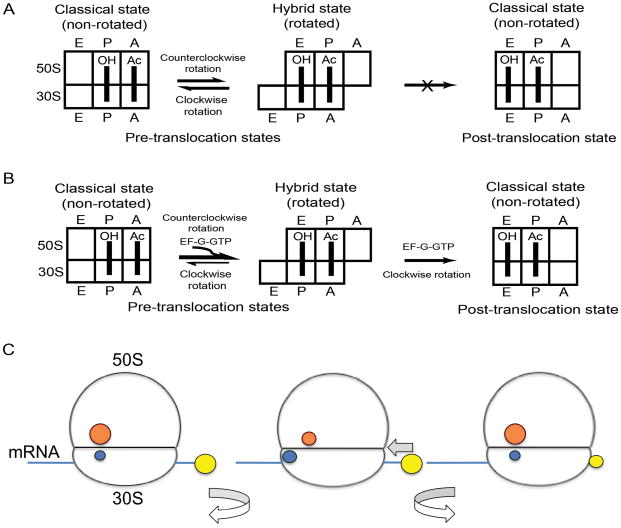
Figure 1
Hybrid-state model of translocation and experimental design. (A) Schematic depiction of intersubunit rotation and tRNA movement in pre-translocation ribosomes. Movement of mRNA is not depicted here. S...
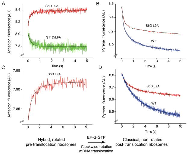
Figure 2
Kinetics of intersubunit rotation and mRNA translocation. (A,C) Kinetics of intersubunit rotation followed by FRET (acceptor fluorescence) (B,D) Kinetics of mRNA translocation measured by quenching of...
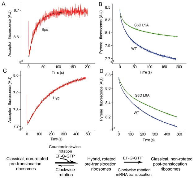
Figure 3
Resolution of the first and second steps of intersubunit rotation in the presence of antibiotics. (A,C) Kinetics of intersubunit rotation and (B,D) mRNA translocation in the presence of (A,B) spectino...

Figure 4
Stabilization of the rotated, hybrid-state conformation by binding EF-Gu00b7GDP in the presence of fusidic acid. Pre-translocation ribosomes containing initiator tRNA fMet in the P site and N-Ac-Phe-t...
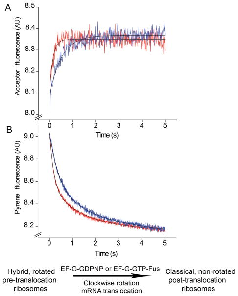
Figure 5
Effect of inhibition of EF-G release on the second (clockwise) step of intersubunit rotation and mRNA translocation. Pre-translocation ribosomes containing elongator tRNA Met in the P site and N-Ac-Ph...
Figure images are served from the NIH/NLM PubMed Central Open Access Subset or Europe PMC; copyright remains with the publishers and authors.
💬 Discussion
0 commentsNo comments yet. Be the first to start a discussion!
Leave a Comment