Abstract
Background: Ischemia reperfusion injury (I/RI) is a common complication of cardiovascular diseases. Resolution of detrimental I/RI-generated prothrombotic and proinflammatory responses is essential to restore homeostasis. Platelets play a crucial part in the integration of thrombosis and inflammation. Their role as participants in the resolution of thromboinflammation is underappreciated; therefore we used pharmacological and genetic approaches, coupled with murine and clinical samples, to uncover key concepts underlying this role. Methods: Middle cerebral artery occlusion with reperfusion was performed in wild-type or annexin A1 (AnxA1) knockout (AnxA1 −/− ) mice. Fluorescence intravital microscopy was used to visualize cellular trafficking and to monitor light/dye–induced thrombosis. The mice were treated with vehicle, AnxA1 (3.3 mg/kg), WRW4 (1.8 mg/kg), or all 3, and the effect of AnxA1 was determined in vivo and in vitro. Results: Intravital microscopy revealed heightened platelet adherence and aggregate formation post I/RI, which were further exacerbated in AnxA1 −/− mice. AnxA1 administration regulated platelet function directly (eg, via reducing thromboxane B 2 and modulating phosphatidylserine expression) to promote cerebral protection post-I/RI and act as an effective preventative strategy for stroke by reducing platelet activation, aggregate formation, and cerebral thrombosis, a prerequisite for ischemic stroke. To translate these findings into a clinical setting, we show that AnxA1 plasma levels are reduced in human and murine stroke and that AnxA1 is able to act on human platelets, suppressing classic thrombin-induced inside-out signaling events (eg, Akt activation, intracellular calcium release, and Ras-associated protein 1 [Rap1] expression) to decrease α IIb β 3 activation without altering its surface expression. AnxA1 also selectively modifies cell surface determinants (eg, phosphatidylserine) to promote platelet phagocytosis by neutrophils, thereby driving active resolution. (n=5–13 mice/group or 7–10 humans/group.) Conclusions: AnxA1 affords protection by altering the platelet phenotype in cerebral I/RI from propathogenic to regulatory and reducing the propensity for platelets to aggregate and cause thrombosis by affecting integrin (α IIb β 3 ) activation, a previously unknown phenomenon. Thus, our data reveal a novel multifaceted role for AnxA1 to act both as a therapeutic and a prophylactic drug via its ability to promote endogenous proresolving, antithromboinflammatory circuits in cerebral I/RI. Collectively, these results further advance our knowledge and understanding in the field of platelet and resolution biology.
🔬 Techniques
🔭 Microscopes
💻 Software
✨ Fluorophores
🧪 Sample Preparation
🔬 Cell Lines
🏭 Microscope Brands
🧪 Reagent Suppliers
💻 Software Details
💾 Data Repositories
🏛️ Research Organizations (ROR)
Affiliated research institutions:
📋 Methods
Middle cerebral artery occlusion with reperfusion was performed in wild-type or annexin A1 (AnxA1) knockout (AnxA1 −/− ) mice. Fluorescence intravital microscopy was used to visualize cellular trafficking and to monitor light/dye–induced thrombosis. The mice were treated with vehicle, AnxA1 (3.3 mg/kg), WRW4 (1.8 mg/kg), or all 3, and the effect of AnxA1 was determined in vivo and in vitro.
Methods
A detailed Methods section is provided in the online-only Data Supplement . The data that support the findings of this study are available from the corresponding author on reasonable request.
Animals
Animal experiments complied with ARRIVE (Animal Research: Reporting In Vivo Experiments) guidelines and followed the European Union Directive (2010/63/EU) or LSUHSC-S IACUC. Wild-type (WT) C57BL/6 mice or AnxA1 −/− mice 21 were used.
Human Samples
The study was approved by the institutional review board of the LSUHSC-S (STUDY00000572 and STUDY00000261) and conducted in accordance with the Declaration of Helsinki. Written informed consent was obtained from the participants.
Receptor Agonists and Drug Treatment
Vehicle (saline), whole protein AnxA1 (3.3 mg/kg, 21 Cambridge Research Biochemicals, Cleveland, UK), and WRW4 (1.8 mg/kg, 22 Tocris, Bristol, UK) were administered intravenously at the start of cerebral reperfusion. AnxA1 Quantification in Plasma Human or murine AnxA1 ELISA kits (MyBioSource) were used to quantify the plasma levels of AnxA1 (see online-only Data Supplement ). Transient Focal Middle Cerebral Artery Occlusion With Reperfusion Transient focal middle cerebral artery occlusion with reperfusion (tMCAo/R) was performed for 60 min followed by 4 or 24 h of reperfusion according to standard operating procedure in our laboratory. 9 Sham animals were subject to the same operative procedure without occlusion.
Show full methods section
Middle cerebral artery occlusion with reperfusion was performed in wild-type or annexin A1 (AnxA1) knockout (AnxA1 −/− ) mice. Fluorescence intravital microscopy was used to visualize cellular trafficking and to monitor light/dye–induced thrombosis. The mice were treated with vehicle, AnxA1 (3.3 mg/kg), WRW4 (1.8 mg/kg), or all 3, and the effect of AnxA1 was determined in vivo and in vitro.
Methods
A detailed Methods section is provided in the online-only Data Supplement . The data that support the findings of this study are available from the corresponding author on reasonable request.
Animals
Animal experiments complied with ARRIVE (Animal Research: Reporting In Vivo Experiments) guidelines and followed the European Union Directive (2010/63/EU) or LSUHSC-S IACUC. Wild-type (WT) C57BL/6 mice or AnxA1 −/− mice 21 were used.
Human Samples
The study was approved by the institutional review board of the LSUHSC-S (STUDY00000572 and STUDY00000261) and conducted in accordance with the Declaration of Helsinki. Written informed consent was obtained from the participants.
Receptor Agonists and Drug Treatment
Vehicle (saline), whole protein AnxA1 (3.3 mg/kg, 21 Cambridge Research Biochemicals, Cleveland, UK), and WRW4 (1.8 mg/kg, 22 Tocris, Bristol, UK) were administered intravenously at the start of cerebral reperfusion. AnxA1 Quantification in Plasma Human or murine AnxA1 ELISA kits (MyBioSource) were used to quantify the plasma levels of AnxA1 (see online-only Data Supplement ). Transient Focal Middle Cerebral Artery Occlusion With Reperfusion Transient focal middle cerebral artery occlusion with reperfusion (tMCAo/R) was performed for 60 min followed by 4 or 24 h of reperfusion according to standard operating procedure in our laboratory. 9 Sham animals were subject to the same operative procedure without occlusion.
Platelet and Leukocyte Labeling
Platelets were isolated from donor mice, ex vivo fluorescently labeled with carboxyfluorescein succinimidyl ester (90 µmol/L, 10 min, Sigma–Aldrich, St Louis, MO), and injected into recipient mice, followed by 0.02% rhodamine 6G (Sigma–Aldrich) to label circulating leukocytes in vivo. 9 Neutrophil Depletion Neutropenia was induced using mouse antineutrophil serum (1A8; BioXCell, West Lebanon, NH; 150 μg/mouse). 23 Neutrophil Isolation and Adoptive Transfer Neutrophils were isolated, labeled with CellTracker 24 (ThermoFisher, Waltham, MA), and treated with WRW4 (10 μmol/L, 10 min, Tocris) before injection into recipient neutropenic tMCAo/R mice. The mice were then treated with AnxA1 (see online-only Data Supplement ). 20 Cerebral Intravital Fluorescence Microscopy Intravital microscopy was performed according to standard operating procedure in our laboratory using a Zeiss Axioskop microscope (Zeiss, New York, NY; see online-only Data Supplement ). 9 Confocal Microscopy To visualize endothelial platelet–neutrophil aggregate (PNA) formation in vivo, the mice were injected with specific antibodies to label: neutrophils (eFluor 488 [green]–labeled anti-mouse Ly-6G, 2 μg/mouse; eBioscience, San Diego, CA) and platelets (Dylight 649 [red]-labeled antimouse CD42, 1 μg/mouse; Emfret Analytics, Eibelstadt, Germany; see online-only Data Supplement ). 9 Systemic PNA Assessment Systemic PNAs were assessed by flow cytometry in blood from tMCAo/R mice treated with either vehicle or AnxA1. Leukocytes were labeled with rat antimouse CD45.2–FITC, Gr-1–PE, and F4/80–eFluor450 and isotype controls (eBioscience), and platelets were labeled with CD41-APC (see online-only Data Supplement ). 21 Assessment of Activated Platelets by Flow Cytometry Two-color staining of activated murine and human α IIb β 3 , cell surface CD41a, P-selectin, and AnxAV (to measure PS) was performed using flow cytometry (see online-only Data Supplement ). 26 Platelet Aggregation Assay Platelet-rich plasma was freshly collected from tMCAo/R mice treated with either vehicle or AnxA1 and used to monitor platelet aggregation velocity after agonist exposure using a laser-particle analyzer (Lumex Ltd., St. Petersburg, Russia; see online-only Data Supplement ). 27 Thrombosis Thrombosis in cerebral vessels was induced using the light/dye thrombosis model (see online-only Data Supplement ). 25 Thirty minutes before onset of thrombosis, the mice were treated with vehicle or AnxA1 (1 µg/mouse). Ras-Associated Protein 1 Activity Ras-associated protein 1 (Rap1) activity assay was performed according to instructions from the supplier (see online-only Data Supplement ).
Western Blotting
Freshly prepared platelets (1×10 7 cells) were incubated with AnxA1 (100 ng/1×10 6 ) platelets for 15 min followed by thrombin stimulation (0.1 U) for 3 min. Platelet pellets were prepared for western blotting for Akt and FPR2 (see online-only Data Supplement ). Measurement of Intracellular Ca 2+ Levels in Fluo-3-Acetoxymethyl Ester Washed platelets (1×10 6 ) were stained with fluo-3-acetoxymethyl ester (5 μmol/L) for 30 min at 37°C as previously described, 28 and some samples were incubated with vehicle or AnxA1 (100 ng) before thrombin stimulation. Fluorescent intensity of fluo-3-acetoxymethyl ester–loaded platelets was immediately recorded using BD LSR II as previously described (see online-only Data Supplement ). 28 Phagocytosis Assay Neutrophil phagocytosis of platelets was performed using the IncuCyte ZOOM (Essen BioScience, Inc., Ann Arbor, MI; see online-only Data Supplement ). Cytokines in Plasma and Brain Tissue After 24 h of reperfusion, plasma and brain hemisphere homogenates were obtained. The levels of pro- and anti-inflammatory cytokines and thromboxane B 2 were measured using standard ELISAs (see online-only Data Supplement ). Infarct Volume After 24 h of reperfusion, the brains were removed and stained with 2% 2,3,5-triphenyltetrazolium chloride (Sigma–Aldrich). Sections were photographed, and digitized images of each brain section (and the infarcted area) were quantified using National Institutes of Health 1.57 Image software. 9 Neurological Score A 5-point neurological deficit score was used: 0 indicates no deficit; 1, failure to extend right paw; 2, circling to the right; 3, falling to the right; and 4, unable to walk spontaneously. 9 The mice were evaluated at 24 h of reperfusion. Blood–Brain Barrier Permeability Blood–brain barrier (BBB) permeability was assessed using Evans blue extravasation. 9 BBB permeability was normalized by dividing tissue Evans blue concentration (micrograms per gram of brain weight) by plasma concentration (micrograms per milliliter).
Thrombin–Antithrombin Complex Measurement
Plasma was collected from WT and AnxA1 −/− mice, and thrombin–antithrombin was measured using thrombin–antithrombin complex ELISAs (Assay Pro, Saint Charles, MO). D-Dimer Plasma was collected from WT and AnxA1 −/− mice, and D-dimer was measured using an Asserachrom D-dimer kit (no. 00947; Diagnostica Stago, Parsippany, NJ).
Statistical Analysis
All data were tested to follow a normal distribution using a Kolmogorov–Smirnov test of normality with a Dallal–Wilkinson–Lillie for the corrected p value. The data that passed the normality assumption was analyzed using Student t test (2 groups) or ANOVA with Bonferroni post-tests (more than 2 groups). The data that failed the normality assumption were analyzed using the nonparametric Mann–Whitney U test (2 groups) or Kruskal–Wallis with Dunn’s test (more than 2 groups). Analysis was performed using Graph Pad Prism5 software (San Diego, CA). The data are shown as mean values ± SEM or median with interquartile range (neurological score only). The differences were considered statistically significant at a value of P
📊 Figures
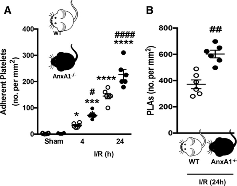
Figure 1.
Platelet and plateletu2013leukocyte interactions in the cerebral microcirculation are heightened in annexin A1 knockout (AnxA1 u2212/u2212 ) mice after ischemia reperfusion injury (I/R). Wild-type (WT...
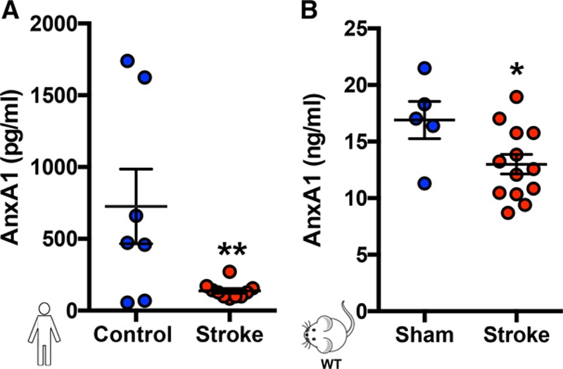
Figure 2.
Plasma levels of AnxA1 are reduced in human and murine stroke. Plasma was collected from control volunteers and stroke patients ( A ) and mice with and without stroke ( B , transient middle cerebral a...
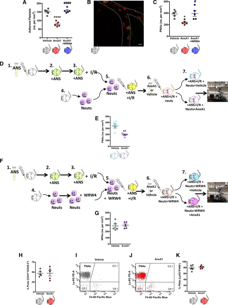
Figure 3.
Administration of exogenous annexin A1 (AnxA1) moderates platelet interactions in the brain microcirculation after ischemia reperfusion injury (I/R). Wild-type mice were subjected to transient middle ...
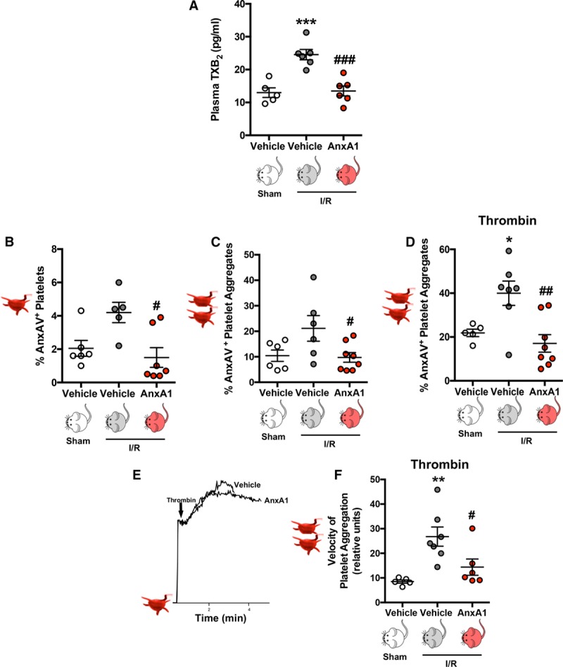
Figure 4.
Annexin A1 (AnxA1) reduces platelet activation/aggregation postischemia reperfusion injury (I/R). Wild-type mice were subjected to sham or transient middle cerebral artery occlusion for 60 min, follow...
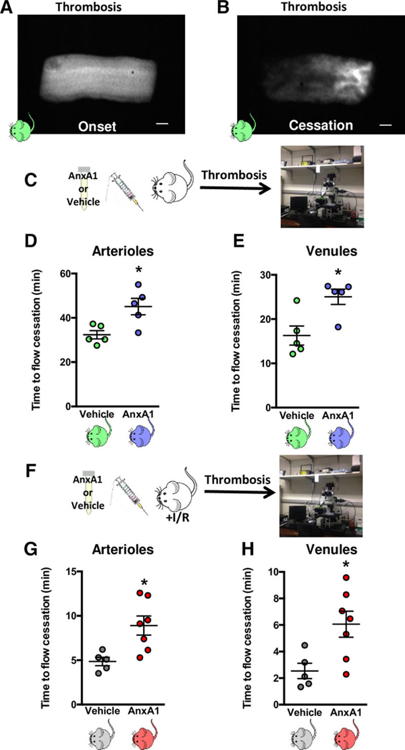
Figure 5.
Annexin A1 (AnxA1) protects against initial cerebral thrombosis and development of subsequent thrombotic events. A and B , Still images of cerebral pial vessels showing onset ( A ) and cessation ( B )...
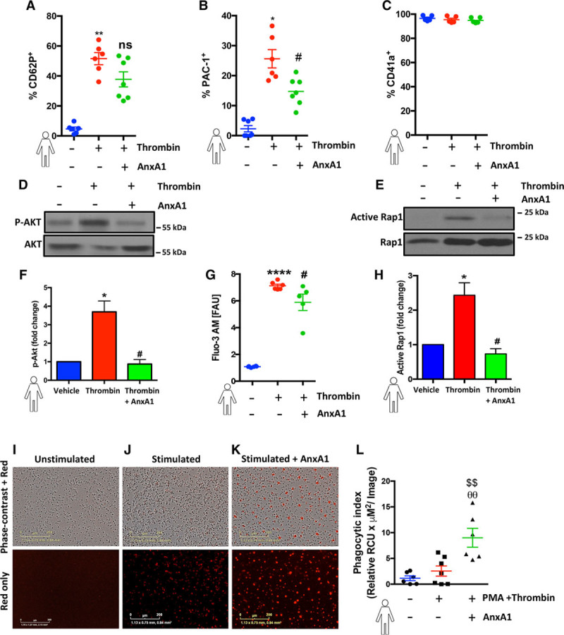
Figure 6.
Annexin A1 (AnxA1) decreases human platelet activation and promotes phagocytosis. A through C , Human platelets (1u00d710 6 ) were isolated, washed, preincubated with 100 ng of AnxA1 (30 min at 37u00b...
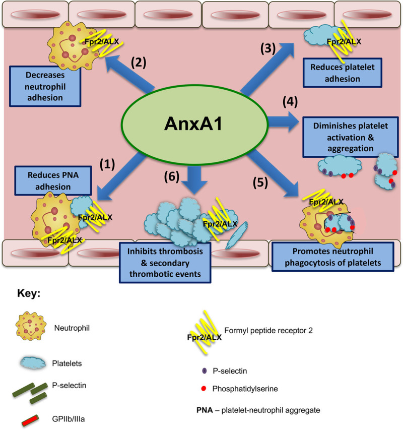
Figure 7.
Annexin A1 (AnxA1) mitigates thrombo inflammatory responses and promotes resolution during ischemia reperfusion injury .Schematic overview shows the effects of AnxA1 on both thrombotic and inflammator...
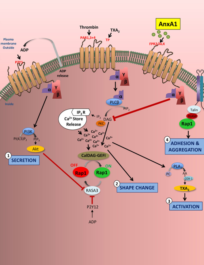
Figure 8.
Annexin A1 (AnxA1) reduces platelet activation and integrin activity. In thromboinflammation, platelets are stimulated via G-proteinu2013coupled receptors (eg, protease-activated receptors 1, 3, and 4...
Figure images are served from the NIH/NLM PubMed Central Open Access Subset or Europe PMC; copyright remains with the publishers and authors.
💬 Discussion
0 commentsNo comments yet. Be the first to start a discussion!
Leave a Comment