Abstract
The worldwide incidence of skin cancer has risen rapidly in the last decades, becoming one in three cancers nowadays. Currently, a person has a 4% chance of developing melanoma, the most aggressive form of skin cancer, which causes the greatest number of deaths. In the context of increasing incidence and mortality, skin cancer bears a heavy health and economic burden. Nevertheless, the 5-year survival rate for people with skin cancer significantly improves if the disease is detected and treated early. Accordingly, large research efforts have been devoted to achieve early detection and better understanding of the disease, with the aim of reversing the progressive trend of rising incidence and mortality, especially regarding melanoma. This paper reviews a variety of the optical modalities that have been used in the last years in order to improve non-invasive diagnosis of skin cancer, including confocal microscopy, multispectral imaging, three-dimensional topography, optical coherence tomography, polarimetry, self-mixing interferometry, and machine learning algorithms. The basics of each of these technologies together with the most relevant achievements obtained are described, as well as some of the obstacles still to be resolved and milestones to be met.
🔬 Techniques
🏭 Microscope Brands
📷 Detectors
🏛️ Research Organizations (ROR)
Affiliated research institutions:
📊 Figures
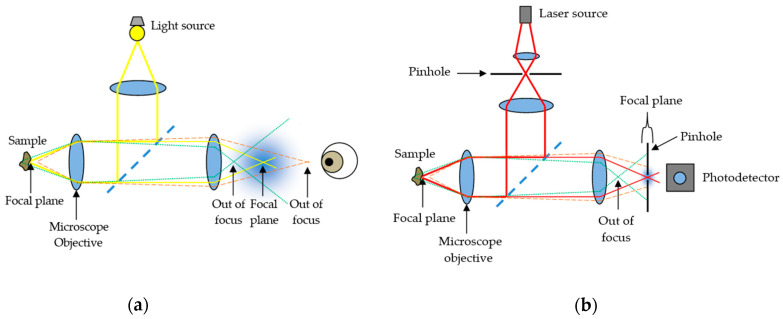
Figure 1
Comparison between ( a ) conventional optical microscopy and ( b ) confocal laser scanning microscopy.
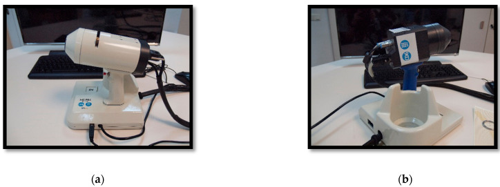
Figure 2
From authors Rey-Barroso et al. [ 43 ], multispectral (MS) imaging devices ( a ) General view of the previously developed handheld visible to the near-infrared (VIS-NIR) MS device. ( b ) General view ...
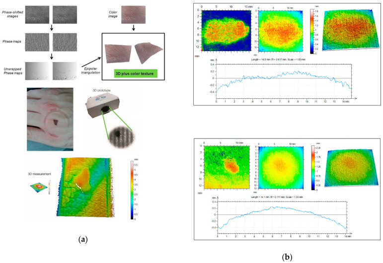
Figure 3
From Rey-Barroso et al. [ 65 ], ( a ) 3D fringe projector. The fringe images are reconstructed and unwrapped to obtain 3D images with superimposed color texture. A pigmented lesion and its heights map...
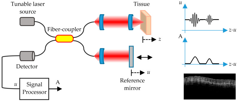
Figure 4
Schematic representation of an SS-OCT imaging system. u is the reference signal and A is the interference signal that produces the image of the different layers of the skin.
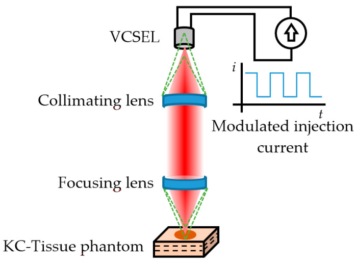
Figure 5
Confocal Laser Feedback Tomography as proposed by Mowla et al. [ 90 ]. The green dashed lines represent the out-of-focus reflections that are eliminated by the aperture of the 850 nm VCSEL acting as s...
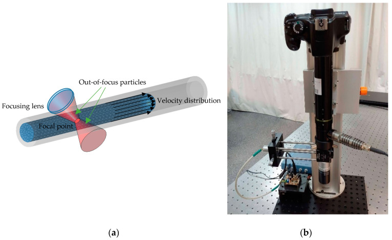
Figure 6
( a ) Diagram of the interaction of a focused laser beam on particles that flow at different velocities within a capillary. ( b ) Confocal SMI-based Doppler flowmeter (SMDF) developed by Yu00e1u00f1ez...
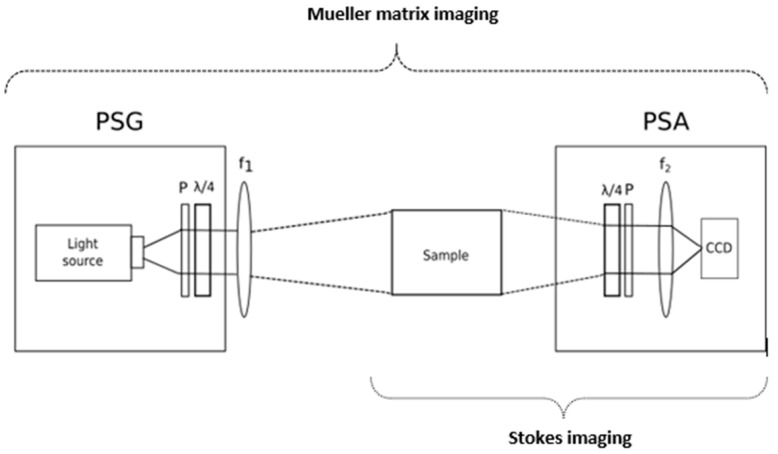
Figure 7
Stokes and Mueller matrix imaging (MMI) setup. The Stokes imaging setup included the sample and the polarization state analyzer (PSA). The PSA is a quarter wave plate (QWP) followed by a polarizer (P)...
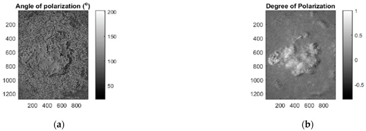
Figure 8
( a ) Map of the measured AOLP (angle of linear polarization) from a melanoma lesion. ( b ) Map of DOP (degree of polarization) from the melanoma lesion. Notice the lesion region with a higher DOP tha...
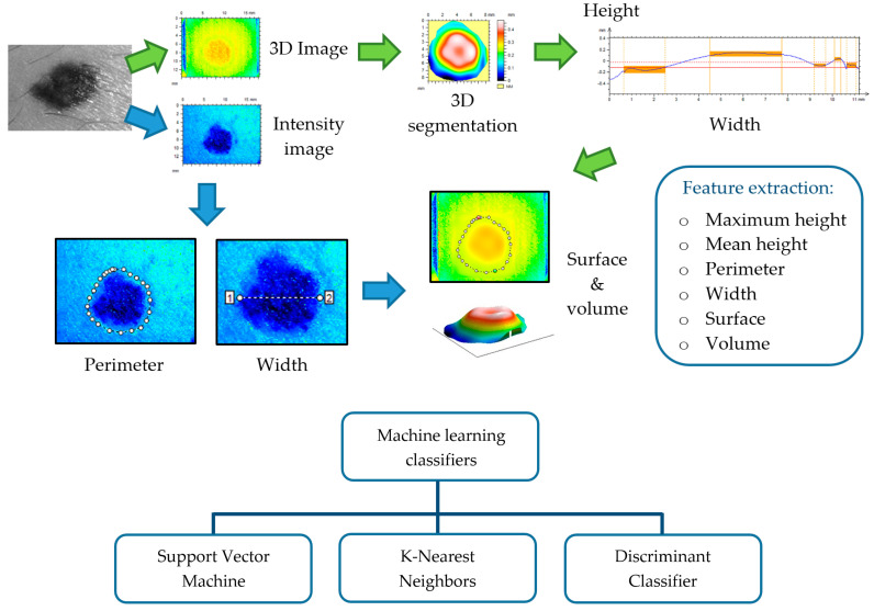
Figure 9
This illustrates the process of feature extraction, preceded by lesion segmentation from perilesional healthy skin, and feeding of machine learning classifiers with such features as descriptors.
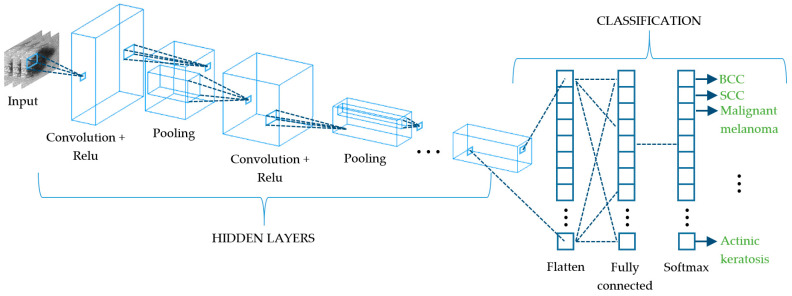
Figure 10
Schematic representation of a convolutional neural network (CNN) that extracts the different spatial characteristics of a skin cancer lesion at its subsequent layers.
Figure images are served from the NIH/NLM PubMed Central Open Access Subset or Europe PMC; copyright remains with the publishers and authors.
💬 Discussion
0 commentsNo comments yet. Be the first to start a discussion!
Leave a Comment