Abstract
Our understanding of how cells communicate has undergone a paradigm shift since the recent recognition of the role of exosomes in intercellular signaling. In this study, we investigated whether oxygen tension alters the exosome release and miRNA profile from extravillous trophoblast (EVT) cells, modifying their bioactivity on endothelial cells (EC). Furthermore, we have established the exosomal miRNA profile at early gestation in women who develop pre-eclampsia (PE) and spontaneous preterm birth (SPTB). HTR-8/SVneo cells were used as an EVT model. The effect of oxygen tension (i.e. 8% and 1% oxygen) on exosome release was quantified using nanocrystals (Qdot®) coupled to CD63 by fluorescence NTA. A real-time, live-cell imaging system (Incucyte™) was used to establish the effect of exosomes on EC. Plasma samples were obtained at early gestation (<18 weeks) and classified according to pregnancy outcomes. An Illumina TrueSeq Small RNA kit was used to construct a small RNA library from exosomal RNA obtained from EVT and plasma samples. The number of exosomes was significantly higher in EVT cultured under 1% compared to 8% oxygen. In total, 741 miRNA were identified in exosomes from EVT. Bioinformatic analysis revealed that these miRNA were associated with cell migration and cytokine production. Interestingly, exosomes isolated from EVT cultured at 8% oxygen increased EC migration, whilst exosomes cultured at 1% oxygen decreased EC migration. These changes were inversely proportional to TNF-α released from EC. Finally, we have identified a set of unique miRNAs in exosomes from EVT cultured at 1% oxygen and exosomes isolated from the circulation of mothers at early gestation, who later developed PE and SPTB. We suggest that aberrant exosomal signalling by placental cells is a common aetiological factor in pregnancy complications characterised by incomplete SpA remodeling and is therefore a clinically relevant biomarker of pregnancy complications.
🔬 Techniques
✨ Fluorophores
🧪 Sample Preparation
🔬 Cell Lines
🏭 Microscope Brands
🧪 Reagent Suppliers
💻 Software Details
💾 Data Repositories
🏛️ Research Organizations (ROR)
Affiliated research institutions:
📋 Methods
Cell culture
All experimental procedures were conducted within an ISO17025 accredited (National Association of Testing Authorities, Australia) research facility. All data was recorded within a 21 Code of Federal Regulation (CFR) part 11 compliant electronic laboratory notebook (Lab Archives, Carlsbad, CA 92008, USA). The HTR-8/SVneo cell line was kindly donated by Dr. Charles H. Graham (Queen's University, Ontario, Canada). HTR-8/SVneo was established by the transfection of trophoblast cells isolated from first trimester villous explants and containing a gene encoding the Simian virus 40 large T antigen to immortalize them [ 13 ]. HTR-8/SVneo is commonly used as an EVT model and HTR-8/SVneo authentication of human trophoblast cell line was performed with a STR DNA Profiling Analysis ( S1 Fig ). HTR-8/SVneo cells were maintained in phenol red-free RPMI 1640 medium supplemented with 10% heat-inactivated fetal bovine serum (FBS), 1% non-essential amino acids, 1 mM sodium pyruvate, 100 U/mL penicillin, and 100 mg/mL streptomycin. Cultures were maintained at 37°C and humidified under an atmosphere of 5% CO 2 -balanced N 2 and either 8% or 1% O 2 in an automated PROOX 110-scaled hypoxia chamber (BioSpherics ™ , Lacona, NY, USA). Cells were cultured in RPMI 1640 medium supplemented with 10% FBS-exosomes depleted for 48h before exosome isolation. Cells were sub-cultured with dissociation media, TrypLETM Express (Life technologies, USA). Cellular viability was determined by Trypan Blue exclusion solution and Countess ® Automated cell counter (Life Technologies, USA).
Show full methods section
Cell culture
All experimental procedures were conducted within an ISO17025 accredited (National Association of Testing Authorities, Australia) research facility. All data was recorded within a 21 Code of Federal Regulation (CFR) part 11 compliant electronic laboratory notebook (Lab Archives, Carlsbad, CA 92008, USA). The HTR-8/SVneo cell line was kindly donated by Dr. Charles H. Graham (Queen's University, Ontario, Canada). HTR-8/SVneo was established by the transfection of trophoblast cells isolated from first trimester villous explants and containing a gene encoding the Simian virus 40 large T antigen to immortalize them [ 13 ]. HTR-8/SVneo is commonly used as an EVT model and HTR-8/SVneo authentication of human trophoblast cell line was performed with a STR DNA Profiling Analysis ( S1 Fig ). HTR-8/SVneo cells were maintained in phenol red-free RPMI 1640 medium supplemented with 10% heat-inactivated fetal bovine serum (FBS), 1% non-essential amino acids, 1 mM sodium pyruvate, 100 U/mL penicillin, and 100 mg/mL streptomycin. Cultures were maintained at 37°C and humidified under an atmosphere of 5% CO 2 -balanced N 2 and either 8% or 1% O 2 in an automated PROOX 110-scaled hypoxia chamber (BioSpherics ™ , Lacona, NY, USA). Cells were cultured in RPMI 1640 medium supplemented with 10% FBS-exosomes depleted for 48h before exosome isolation. Cells were sub-cultured with dissociation media, TrypLETM Express (Life technologies, USA). Cellular viability was determined by Trypan Blue exclusion solution and Countess ® Automated cell counter (Life Technologies, USA).
Patients
A case control study design was used to establish pregnancy-associated changes in exosomal miRNA present in maternal plasma obtained from normal, preeclampsia and preterm birth pregnancies. Human plasma samples were obtained in accordance with the Declaration of Helsinki. This study was approved by the ethics committee of The University of Queensland (HREC/09/QRBW/14) and Ochsner Medical Foundation (USA). Women were recruited with informed written consent at the Ochsner Baptist Medical Center (New Orleans, USA). Samples were obtained before 20 weeks of gestation and classified according to the pregnancy outcomes in normal, PE or SPTB. PE was defined as new-onset hypertension [(BP) blood pressure ≥140/90mmHg on two separate occasions at least 6h apart or BP ≥160/110mmHg)] and proteinuria (>300mg/24h) after 20 weeks of gestation in previously normotensive women, according to the International Society for the Study of Hypertension in Pregnancy [ 14 ]. SPTB was defined as a birth before gestational week 37 + 0. The clinical characteristics of the patients included in this study are presented in Table 1 . 10.1371/journal.pone.0174514.t001 Table 1 Clinical characteristics of patients. Variables Normal (n = 6) PE (n = 6) SPTB (n = 6) Maternal age (years) 31 ± 2.9 (29–35) 32 ± 4.3 (28–33) 30 ± 5.4 (26–33) Height (cm) 165 ± 0.6 (160–170) 159 ± 0.7 (155–170) 160 ± 0.7 (150–165) Weight (Kg) 61 ± 5.3 (55–68) 63 ± 5.3 (55–70) 61 ± 4.6 (55–69) BMI (Kg/m 2 ) 23 ± 1.5 (20–26) 25 ± 1.3 (20–29) 24 ± 1.8 (20–27) Systolic/diastolic blood pressure (mm Hg) 109/65 ± 6.22/9.12 (90-120/50-81) 155/69 ± 10/8 * (>140/90) 111/67 ± 10/7 (90-120/50-80) Gestational age at sample collection (weeks) 17 ± 3.5 (15–18) 16 ± 2.6 (15–18) 16 ± 2.8 (15–18) Gestational age at delivery (weeks) 40 ± 1.2 (37–42) 38 ± 0.9 (37–39) 35 ± 1.1 (34–36) Data are presented as mean ± SD (range). PE = preeclampsia; SPTB = spontaneous preterm birth. *p
📊 Figures
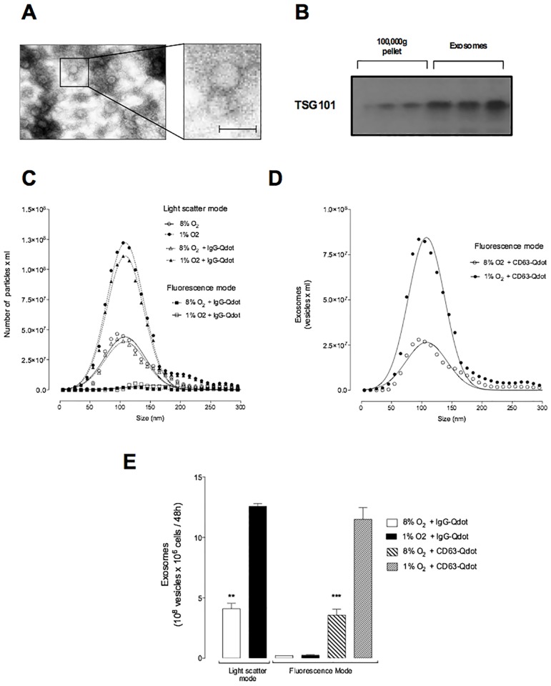
Fig 1
Effect of low oxygen tension on the release of exosomes.
The effect of oxygen tension (8% and 1% O 2 ) on the release of exosomes from EVT cells was quantified using NanoSight in light scatter and fluorescence mode. (A) Electron micrograph of exosomes isola...
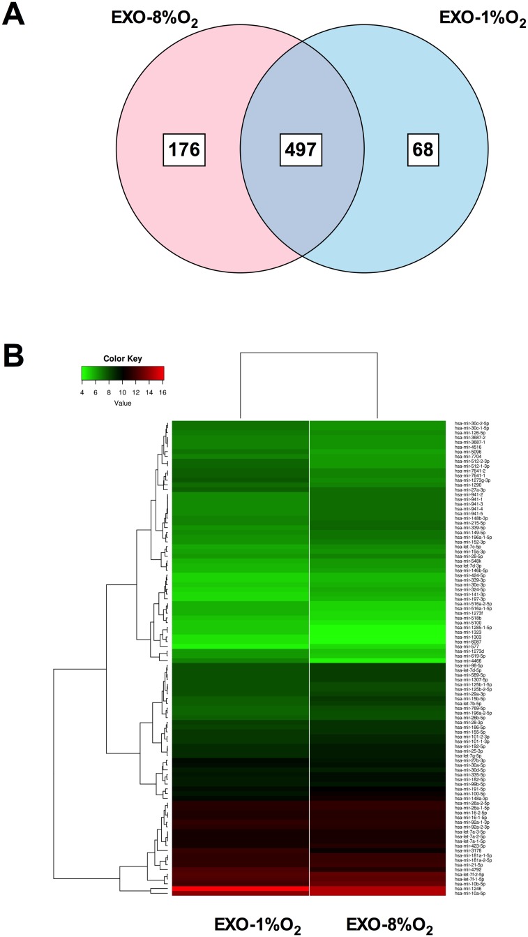
Fig 2
miRNA profile in EVT-derived exosomes under low oxygen tension.
(A) The Venn diagram depicts the distribution of common and unique miRNA identified in exosomes released from EVT exposed to 1% and 8% oxygen. (B) Top 100 abundant miRNAs classified by normalised coun...
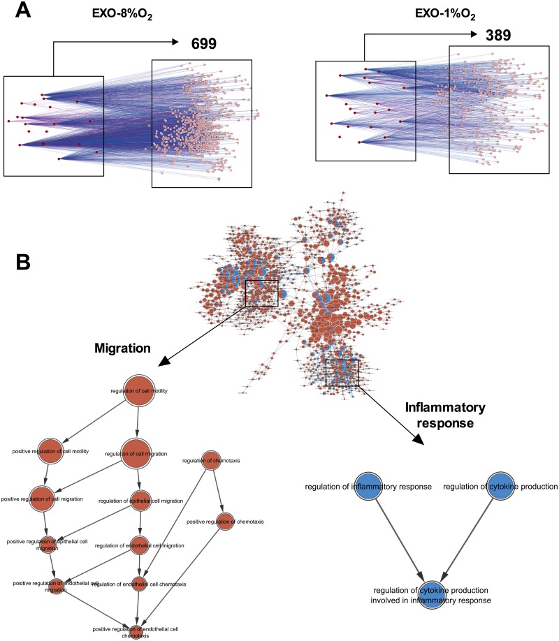
Fig 3
Target gene prediction of miRNAs identified in EVT-derived exosomes.
(A) Gene target identification using CyTargetLinker was performed on the top 20 miRNAs in exosomes from EVT cultured at 8% or 1% oxygen. The genes were identified to be regulated by at least two of ou...
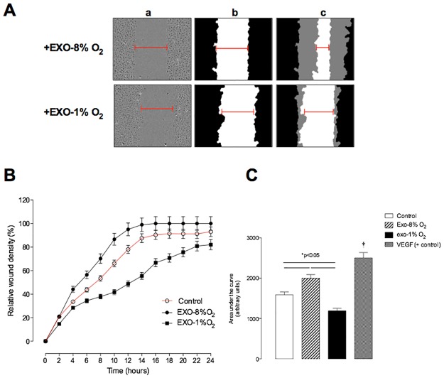
Fig 4
Effect of exosomes on endothelial cell migration.
Endothelial cells (EC) were grown to confluence in complete media, wounds were made using 96 well WoundMaker and culture in absence or presence of exosomes (100 u03bcg protein/ml equivalent to 5 x 10 ...
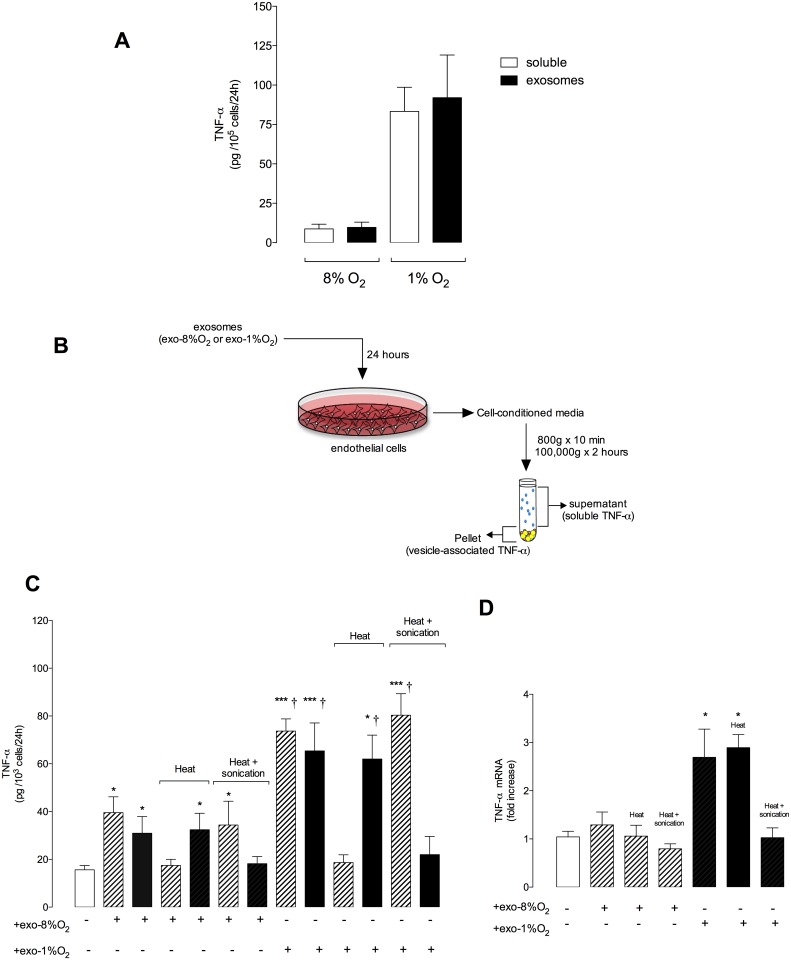
Fig 5
Levels of TNFu03b1 in exosome and cell-conditioned media.
The concentration of TNFu03b1 was quantified in exososomes and EC-consitioned media using an ELISA kit. (A) Exosomes were isolated from EVT cells cultured at 8% and 1% oxygen and the concentration of ...
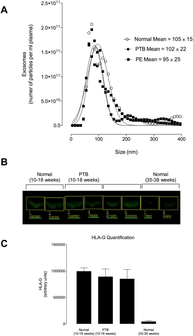
Fig 6
Exosomes present in maternal circulation before 18 weeks are positive for HLA-G.
Exosomes were isolated from maternal plasma before 18 weeks of gestation by differential and buoyant density centrifugation. (A) Representative exosomes size distribution isolated from maternal plasma...
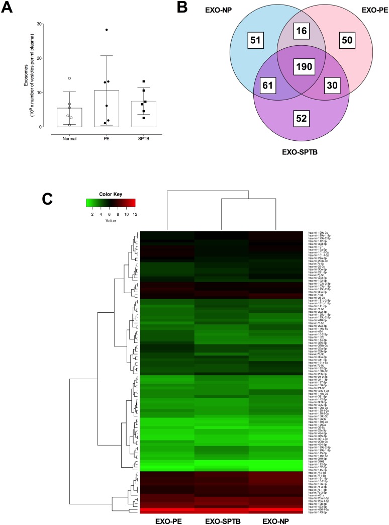
Fig 7
miRNA profile in exosome present in maternal circulation at early gestation.
(A) number of exosomes in normal, preeclampsia (PE) and spontaneous preterm birth (SPTB) at early gestation. (B) The Venn diagram shows the distribution of common and unique miRNA identified in exosom...
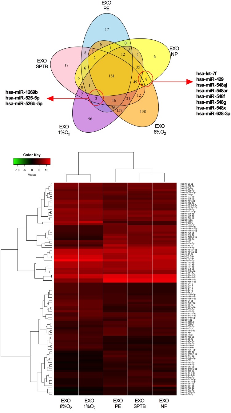
Fig 8
Comparison analysis of miRNA profile between exosomes from EVT and present in maternal circulation at early gestation.
(A) Venn diagrams showing unique and common miRNA detected in different exosomes preparations. (B) Highly abundant miRNAs identified in exosomes isolated from EVT cultured under 8% (EXO-8%O 2 ) or 1% ...
Figure images are served from the NIH/NLM PubMed Central Open Access Subset or Europe PMC; copyright remains with the publishers and authors.
💬 Discussion
0 commentsNo comments yet. Be the first to start a discussion!
Leave a Comment