Abstract
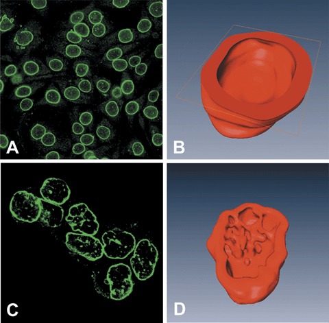
AbstractIn routine practice, nuclear pleomorphism of tumours is assessed by haematoxylin staining of the membrane‐bound heterochromatin. However, decoration of the nuclear envelope (NE) through the immunofluorescence staining of NE proteins such as lamin B and emerin can provide a more objective appreciation of the nuclear shape. In breast cancer, nuclear pleomorphism is one of the least reproducible parameters to score histological grade, thus we sought to use NE proteins to improve the reproducibility of nuclear grading. First, immuno‐fluorescence staining of NE as well as confocal microscopy and three‐dimensional reconstruction of nuclei in cultured cells showed a smooth and uniform NE of normal breast epithelium in contrast to an irregular foldings of the membrane and the presence of deep invaginations leading to the formation of an intranuclear scaffold of NE‐bound tubules in breast cancer cells. Following the above methods and criteria, we recorded the degree of NE pleomorphism (NEP) in a series of 273 invasive breast cancers tested by immunofluorescence. A uniform nuclear shape with few irregularities (low NEP) was observed in 135 cases or, alternatively, marked folds of the NE and an intranuclear tubular scaffold (high NEP cases) were observed in 138 cases. The latter features were significantly correlated (P‐value <0.002) with lymph node metastases in 54 histological grade 1 and in 173 cancers with low mitotic count. Decoration of the NE might thus be regarded as a novel diagnostic parameter to define the grade of malignancy, which parallels and enhances that provided by routine histological procedures.
🔬 Techniques
✨ Fluorophores
🧪 Sample Preparation
🔬 Cell Lines
🏭 Microscope Brands
🧪 Reagent Suppliers
💻 Software Details
🏛️ Research Organizations (ROR)
Affiliated research institutions:
📋 Methods
Cell lines
Breast cancer cell lines MCF7, BT474 and SKBR3 were obtained from the American Type Culture Collection (Manassas, VA, USA). Cells were grown in IDMEM (Iscove Dulbecco's Modified Eagle's medium) (Sigma Aldrich, St. Louis, MO, USA) supplemented with 10% foetal calf serum (Seromed, Berlin, Germany), 2 mmol/l glutamine, 100 U/ml penicillin and 100 μg/ml streptomycin (all from Sigma), and incubated at 37°C, 5% CO 2 . Primary cultures of breast epithelium were obtained from reduction mammoplasties [ 21 ]. Tissues were digested with collagenase IA 200 U/ml and hyaluronidase 100 U/ml (all from Sigma). Purification was then completed following the Dynabeads Magnetic Separation method (Dybal Biothec, Oslo, Norway). Cells were grown in DMEM/Ham's F12 (Dulbecco's Modified Eagle's medium, Sigma) supplemented with 10% foetal calf serum (Seromed), 0.01 mg/ml insulin, 500 ng/ml hydrocortisone and 20 ng/ml Epidermal Growth Factor (all from Sigma). For immunofluorescence (IF) staining, cells were plated on multi-chamber slides (Becton Dickinson, San Jose, CA, USA) and cultured in Iscove – Dulbecco's Modified Eagle's medium (IDMEM) until a 70% cellular confluence was reached. Cells were then fixed in methanol for 5 min. at –20°C and permeabilized in acetone for 5 sec. at –20°C, then brought to phosphate-buffered saline (PBS).
Histological specimens
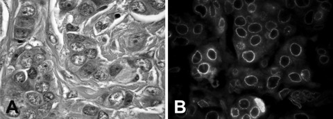
A preliminary investigation was conducted in five cases of histologically normal breast tissue and five cases of invasive breast cancer. Additionally, a series of consecutive 304 cases of unifocal invasive breast cancer, processed following the tissue micro array procedure as outlined by Sapino et al. [ 22 ] were included in the study. The NG of such cases had originally been established according to Elston and Ellis [ 1 ]. Briefly, for each case, the selection of at least four fields was carried out on the original haematoxylin and eosin stained slides during assessment of the histological grade, in order to guarantee that the most significant intratumoral variations in terms of tubular formation, nuclear atypia and mitosis number were identified. The number of selected fields varied from 4 to 6 depending on the heterogeneity both of the histological pattern and of the grade of differentiation of the invasive breast cancer component. Four micrometre thick sections of the paraffin block were collected onto poly-L-lysine coated slides. Sections were dehydrated in graded alcohols and processed for IF. Marked and diffuse irregularities of the nuclear membrane leading to the building up of a scaffold of intranuclear foldings were classified as high NEP, while mild-to-moderate NE irregularities with rare or absent intranuclear invaginations were classified as low NEP ( Table 1 ). Classification of cases into either high or low NEP categories was independently determined by the two senior authors blinded to the original histological grade. Cases were evaluated as tissue arrays and in each case 4 core, representative of different tumour foci, were selected. As a rule (in more than 80% of the cases) the nuclear shape was similar and homogeneous (either low or high NEP) in all cores of each case. In case of variability within different cores of the same case the high NEP category was assigned, whereas interobserver discrepancies, which occurred in 21 cases, were solved by consensus. We can roughly reckon that each core (diameter 1 mm) was presenting approximately 1000 cancer cells. More than 50% of cancer nuclei had to present a high grade of NE pleomorphism in order the case to be considered as high NEP. 1 Nuclear envelope pleomorphism (NEP) categories as defined by immunofluorescence and nuclear grade (NG) score values as defined by haematoxylin and eosin. NEP Low Mild or moderate NE irregularities, with rare or absent strands or intranuclear invaginations (see Figs 2A and 4B ) High Marked irregularities of the nuclear membrane and frequent strands or intranuclear invaginations (see Figs 2C and 3A ) NG * Score 1 Nuclei small with little increase in size in comparison with normal breast epithelial cells, regular outlines, uniform nuclear chromatin, little variation in size Score 2 Cells larger than normal with open vesicular nuclei, visible nucleoli and moderate variability in both size and shape Score 3 Vesicular nuclei, often with prominent nucleoli, exhibiting marked variation in size and shape, occasionally with very large and bizarre forms * Reference [ 7 ]. 4 Haematoxylin and eosin staining of G3 invasive carcinoma: the nuclei meet the criteria for score 3, being vesicular, with prominent nucleoli and exhibiting marked variation in size ( A ). NE staining with anti-emerin antibody in IF shows instead a smooth profile ( B ), corresponding to low NEP.
Show full methods section
Cell lines
Breast cancer cell lines MCF7, BT474 and SKBR3 were obtained from the American Type Culture Collection (Manassas, VA, USA). Cells were grown in IDMEM (Iscove Dulbecco's Modified Eagle's medium) (Sigma Aldrich, St. Louis, MO, USA) supplemented with 10% foetal calf serum (Seromed, Berlin, Germany), 2 mmol/l glutamine, 100 U/ml penicillin and 100 μg/ml streptomycin (all from Sigma), and incubated at 37°C, 5% CO 2 . Primary cultures of breast epithelium were obtained from reduction mammoplasties [ 21 ]. Tissues were digested with collagenase IA 200 U/ml and hyaluronidase 100 U/ml (all from Sigma). Purification was then completed following the Dynabeads Magnetic Separation method (Dybal Biothec, Oslo, Norway). Cells were grown in DMEM/Ham's F12 (Dulbecco's Modified Eagle's medium, Sigma) supplemented with 10% foetal calf serum (Seromed), 0.01 mg/ml insulin, 500 ng/ml hydrocortisone and 20 ng/ml Epidermal Growth Factor (all from Sigma). For immunofluorescence (IF) staining, cells were plated on multi-chamber slides (Becton Dickinson, San Jose, CA, USA) and cultured in Iscove – Dulbecco's Modified Eagle's medium (IDMEM) until a 70% cellular confluence was reached. Cells were then fixed in methanol for 5 min. at –20°C and permeabilized in acetone for 5 sec. at –20°C, then brought to phosphate-buffered saline (PBS).
Histological specimens
A preliminary investigation was conducted in five cases of histologically normal breast tissue and five cases of invasive breast cancer. Additionally, a series of consecutive 304 cases of unifocal invasive breast cancer, processed following the tissue micro array procedure as outlined by Sapino et al. [ 22 ] were included in the study. The NG of such cases had originally been established according to Elston and Ellis [ 1 ]. Briefly, for each case, the selection of at least four fields was carried out on the original haematoxylin and eosin stained slides during assessment of the histological grade, in order to guarantee that the most significant intratumoral variations in terms of tubular formation, nuclear atypia and mitosis number were identified. The number of selected fields varied from 4 to 6 depending on the heterogeneity both of the histological pattern and of the grade of differentiation of the invasive breast cancer component. Four micrometre thick sections of the paraffin block were collected onto poly-L-lysine coated slides. Sections were dehydrated in graded alcohols and processed for IF. Marked and diffuse irregularities of the nuclear membrane leading to the building up of a scaffold of intranuclear foldings were classified as high NEP, while mild-to-moderate NE irregularities with rare or absent intranuclear invaginations were classified as low NEP ( Table 1 ). Classification of cases into either high or low NEP categories was independently determined by the two senior authors blinded to the original histological grade. Cases were evaluated as tissue arrays and in each case 4 core, representative of different tumour foci, were selected. As a rule (in more than 80% of the cases) the nuclear shape was similar and homogeneous (either low or high NEP) in all cores of each case. In case of variability within different cores of the same case the high NEP category was assigned, whereas interobserver discrepancies, which occurred in 21 cases, were solved by consensus. We can roughly reckon that each core (diameter 1 mm) was presenting approximately 1000 cancer cells. More than 50% of cancer nuclei had to present a high grade of NE pleomorphism in order the case to be considered as high NEP. 1 Nuclear envelope pleomorphism (NEP) categories as defined by immunofluorescence and nuclear grade (NG) score values as defined by haematoxylin and eosin. NEP Low Mild or moderate NE irregularities, with rare or absent strands or intranuclear invaginations (see Figs 2A and 4B ) High Marked irregularities of the nuclear membrane and frequent strands or intranuclear invaginations (see Figs 2C and 3A ) NG * Score 1 Nuclei small with little increase in size in comparison with normal breast epithelial cells, regular outlines, uniform nuclear chromatin, little variation in size Score 2 Cells larger than normal with open vesicular nuclei, visible nucleoli and moderate variability in both size and shape Score 3 Vesicular nuclei, often with prominent nucleoli, exhibiting marked variation in size and shape, occasionally with very large and bizarre forms * Reference [ 7 ]. 4 Haematoxylin and eosin staining of G3 invasive carcinoma: the nuclei meet the criteria for score 3, being vesicular, with prominent nucleoli and exhibiting marked variation in size ( A ). NE staining with anti-emerin antibody in IF shows instead a smooth profile ( B ), corresponding to low NEP.
Immunofluorescence
Cells and histological slides were immunostained for NE evaluation ( Table 2 ). Cells and tissues were treated with a blocking serum (Mabtech AB, Germany), diluted 1/100, for 30 min. at room temperature and then incubated with the primary antibody for 1 hr. The slides were then rinsed three times in PBS and incubated for 45 min. with the specific secondary antibody. Finally, the slides were rinsed three times with PBS prior to mounting with an antifading mounting medium (VECTASHIELD ® Mounting Medium, Vector Laboratories, Burlingame, CA, USA). 2 Antibodies anti-nuclear envelope (NE). Primary antibody Dilution in PBS-BSA Antigen retrieval for FFPE† tissues Secondary antibody Dilution in PBS-BSA Goat anti-lamin B (SC-6217;Santa Cruz, CA, USA) 1:5 Microwave 10 min.in citrate buffer (pH 6) at 98°C Fluorescein isothiocyanate-conjugated rabbit anti-goat (Sigma Aldrich, Munich, Germany) 1/200 Rabbit anti-emerin (ab14208;Abcam, Cambridge, UK) 1/500 Microwave 40 min.in citrate buffer (pH 6) at 98°C Rhodamine isothiocyanate-conjugated swine anti-rabbit (DAKO, Glostrup, Denmark) 1/300 Mouse anti-nucleoporin * (ab 24700;Abcam) 1/200 Fluorescein isothiocyanate-conjugated goat antimouse (Sigma) 1/100 † FFPE:formalin fixed paraffin embedded. * Not tested on FFPE.
Images acquisition
Tissue sections were examined with an Olympus BX51 fluorescence microscope (Olympus, Hamburg, Germany) and images were acquired using an Olympus C-7070 wide zoom camera. For confocal scanning laser microscopy, we alternatively used a LSM5 PASCAL confocal microscope (Carl Zeiss, Germany) or a FV300 confocal microscope mounted on an IX71 inverted microscope (both from Olympus), or a TCS SP2 confocal microscope (Leica, Wetzlar, Germany). Areas of non-overlapping nuclei were selected for scanning at 63× magnification. Serial sections were recorded with a 0.3 μm gap between sections.
3D reconstruction
Using the serial images obtained by confocal microscopy, 3D models of selected nuclei were obtained employing Amira 4.0, advanced 3D visualization and volume modelling software (TGS Template Graphics Software, http://www.tgs.com ). Briefly, using sequential images of serial nuclear sections, segmentation of the region of interest was obtained outlining the nuclear profile. Each segment of the curve matching the steps between sections was proportional to the size of the nuclei and to the gaps between sections, as measured by confocal microscopy. The segmented areas were then exploited to generate 3D polygonal surface models.
Statistical analysis
The following parameters were analysed on the series of breast cancers: nuclear envelope pleomorphism (NEP) as revealed by IF, score values of NG as defined by haematoxylin and eosin ( Table 1 ) and histological grade (G). Marked and diffuse irregularities of the nuclear membrane leading to the building up of a scaffold of intranuclear foldings were classified as high NEP, while mild to moderate NE irregularities with rare or absent intranuclear invaginations were classified as low NEP ( Table 1 ). Association between NEP and NG or G were evaluated. The correlation between NEP and the lymph node status and prognostic parameters, such as oestrogen and progesterone receptors, Ki67 and Her2 values, was analysed using the chisquare test and logistic regression analysis ( Table 3 ). 3 Correlations between NEP and other clinical and histopathological parameters:ER (+ versus –), PgR (+ versus –) HER2 (+ versus –), Ki67 (> versus < 20%), pN (No versus N1) and pT (T1 versus T > 1), as established by chi-square analysis. LNEP HNEP P-value ER – 22 (16%) 18 (13%) 0.556 + 113 (84%) 120 (87%) PgR – 43 (32%) 35 (25%) 0.292 + 92 (68%) 103 (75%) HER2 – 119 (88%) 119 (86%) 0.770 + 16 (12%) 19 (14%) Ki67 – 69 (51%) 73 (53%) 0.861 + 66 (49%) 65 (47%) PN N0 81 (60%) 63 (46%) 0.024 N1 54 (40%) 75 (54%) PT T1 83 (61%) 56 (41%) 0.001 T > 1 52 (39%) 82 (59%)
📊 Figures
4
Haematoxylin and eosin staining of G3 invasive carcinoma: the nuclei meet the criteria for score 3, being vesicular, with prominent nucleoli and exhibiting marked variation in size ( A ). NE staining ...
1
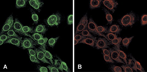
SKBr3 breast cancer cell line, stained with ( A ) anti-lamin B antibodies (revealed with fluorescein) and ( B ) anti-emerin antibodies (revealed with rhodamin). The two proteins linked to the nuclear ...
2
The arrangement of the nuclear membrane, as revealed by tagging membrane-associated lamin B, was observed by confocal microscopy, in cells from primary cultures of normal mammary epithelium ( A ) and ...
3
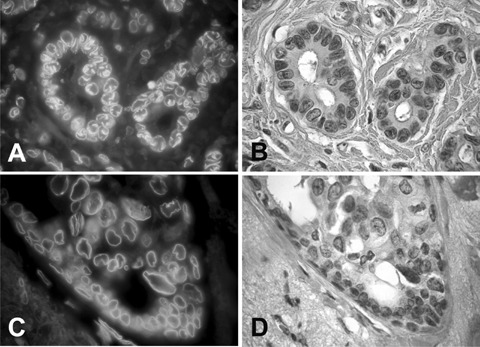
IF staining with anti-emerin antibody outlined high NEP ( A ) in a histologically low-grade ductal carcinoma (G1) as shown by staining with haematoxylin and eosin of a parallel section ( B ). Anti-eme...
Figure images are served from the NIH/NLM PubMed Central Open Access Subset or Europe PMC; copyright remains with the publishers and authors.
💬 Discussion
0 commentsNo comments yet. Be the first to start a discussion!
Leave a Comment