Abstract
Abstract Accurate histopathologic diagnosis is essential for providing optimal surgical management of pediatric brain tumors. Current methods for intraoperative histology are time- and labor-intensive and often introduce artifact that limit interpretation. Stimulated Raman histology (SRH) is a novel label-free imaging technique that provides intraoperative histologic images of fresh, unprocessed surgical specimens. Here we evaluate the capacity of SRH for use in the intraoperative diagnosis of pediatric type brain tumors. SRH revealed key diagnostic features in fresh tissue specimens collected from 33 prospectively enrolled pediatric type brain tumor patients, preserving tumor cytology and histoarchitecture in all specimens. We simulated an intraoperative consultation for 25 patients with specimens imaged using both SRH and standard hematoxylin and eosin histology. SRH-based diagnoses achieved near-perfect diagnostic concordance (Cohen's kappa, κ > 0.90) and an accuracy of 92% to 96%. We then developed a quantitative histologic method using SRH images based on rapid image feature extraction. Nuclear density, tumor-associated macrophage infiltration, and nuclear morphology parameters from 3337 SRH fields of view were used to develop and validate a decision-tree machine-learning model. Using SRH image features, our model correctly classified 25 fresh pediatric type surgical specimens into normal versus lesional tissue and low-grade versus high-grade tumors with 100% accuracy. Our results provide insight into how SRH can deliver rapid diagnostic histologic data that could inform the surgical management of pediatric brain tumors. Significance: A new imaging method simplifies diagnosis and informs decision making during pediatric brain tumor surgery. Cancer Res; 78(1); 278–89. ©2017 AACR.
🔬 Techniques
💻 Software
✨ Fluorophores
🧪 Sample Preparation
🔬 Cell Lines
🏭 Microscope Brands
💻 Software Details
💾 Data Repositories
🏛️ Research Organizations (ROR)
Affiliated research institutions:
📋 Methods
Study design
The study was approved by the University of Michigan Institutional Review Board (HUM00083059). Patient studies were conducted in accordance with the Declaration of Helsinki, International Ethical Guidelines for Biomedical Research Involving Human Subjects (CIOMS), Belmont Report and U.S. Common Rule. Patients were prospectively enrolled for 24 months with the following inclusion criteria: ( 1 ) male and female subjects undergoing brain tumor resection at the University of Michigan Health System, ( 2 ) subject or medical decision maker was able to provide informed written consent and ( 3 ) subjects in whom there was excess tumor tissue beyond what was needed for routine diagnosis. All patients 18 years or younger were included in the study preoperatively. Patients older than 18 years were enrolled postoperatively if they were diagnosed with pediatric type pathologies to increase study enrollment and ensure a patient cohort of representative pediatric type histology, including pilocytic astrocytoma, ependymoma, medulloblastoma and other embryonal tumors, ganglioglioma, diffuse midline glioma, hemangioblastoma, choroid plexus papilloma, chordoma, and germinoma. The list of pediatric type pathologies was provided by our expert panel of neuropathologists (S.C.P., A.P.L., K.A.M). Normal/non-neoplastic specimens were taken from a cohort of adult epilepsy and brain tumor patients. The primary goals of the investigation were to 1) establish SRH as a feasible method for obtaining histopathologic diagnosis in tumors common in the pediatric population and 2) develop a machine-learning method using quantitative SRH image features to provide rapid, automated detection of lesional tissue and tumor grade. Patients were recruited consecutively at a high-volume, tertiary-care hospital to obtain a representative sample of pediatric type brain tumors. All collected specimens were imaged immediately after removal with our clinical fiber-laser–based SRS microscope ( 13 ). A board-certified neuropathologist (A.P.L.) reviewed all images from both standard intraoperative pathology and SRH to determine adequacy and classify each specimen following the current World Health Organization (WHO) diagnostic classification criteria ( 14 ). We then implemented a web-based survey with three neuropathologists (S.C.P., K.A.M., M.S.) to determine the diagnostic concordance and accuracy of SRH compared to standard intraoperative H&E histology. To develop a quantitative histology, we used CellProfiler for image feature extraction ( 15 ). Image features were then used to develop and validate a random forest machine-learning method to provide automated classification of lesional tissue (i.e., normal versus lesional) and tumor grade (i.e., low grade versus high grade).
Show full methods section
Study design
The study was approved by the University of Michigan Institutional Review Board (HUM00083059). Patient studies were conducted in accordance with the Declaration of Helsinki, International Ethical Guidelines for Biomedical Research Involving Human Subjects (CIOMS), Belmont Report and U.S. Common Rule. Patients were prospectively enrolled for 24 months with the following inclusion criteria: ( 1 ) male and female subjects undergoing brain tumor resection at the University of Michigan Health System, ( 2 ) subject or medical decision maker was able to provide informed written consent and ( 3 ) subjects in whom there was excess tumor tissue beyond what was needed for routine diagnosis. All patients 18 years or younger were included in the study preoperatively. Patients older than 18 years were enrolled postoperatively if they were diagnosed with pediatric type pathologies to increase study enrollment and ensure a patient cohort of representative pediatric type histology, including pilocytic astrocytoma, ependymoma, medulloblastoma and other embryonal tumors, ganglioglioma, diffuse midline glioma, hemangioblastoma, choroid plexus papilloma, chordoma, and germinoma. The list of pediatric type pathologies was provided by our expert panel of neuropathologists (S.C.P., A.P.L., K.A.M). Normal/non-neoplastic specimens were taken from a cohort of adult epilepsy and brain tumor patients. The primary goals of the investigation were to 1) establish SRH as a feasible method for obtaining histopathologic diagnosis in tumors common in the pediatric population and 2) develop a machine-learning method using quantitative SRH image features to provide rapid, automated detection of lesional tissue and tumor grade. Patients were recruited consecutively at a high-volume, tertiary-care hospital to obtain a representative sample of pediatric type brain tumors. All collected specimens were imaged immediately after removal with our clinical fiber-laser–based SRS microscope ( 13 ). A board-certified neuropathologist (A.P.L.) reviewed all images from both standard intraoperative pathology and SRH to determine adequacy and classify each specimen following the current World Health Organization (WHO) diagnostic classification criteria ( 14 ). We then implemented a web-based survey with three neuropathologists (S.C.P., K.A.M., M.S.) to determine the diagnostic concordance and accuracy of SRH compared to standard intraoperative H&E histology. To develop a quantitative histology, we used CellProfiler for image feature extraction ( 15 ). Image features were then used to develop and validate a random forest machine-learning method to provide automated classification of lesional tissue (i.e., normal versus lesional) and tumor grade (i.e., low grade versus high grade).
Tissue collection and intraoperative SRH
Following standard operative procedures, neurosurgeons (D.A.O., C.O.M., H.J.L.G., K.M.M.) removed lesional tissue. Specimens were then split by the neurosurgeon with equal halves sent for intraoperative pathology and for SRH. Standard intraoperative pathology included cytologic preparation and frozen sectioning. To image fresh surgical specimens using the clinical SRS microscope, a small (approximately, 3 × 3 × 3 mm or 27 μL) unprocessed and unlabeled specimen was placed on a standard uncoated glass slide covered with a cover slip. Using custom imaging programs in μ-Manager and ImageJ software, 400 × 400-μm images from two SRS channels, 2845 cm -1 (CH 2 /lipid channel) and 2930 cm -1 (CH 3 /protein channel) Raman shift wave numbers, were obtained in a raster fashion. A mosaic image with automated image stitching was completed to obtain wider fields of view (FOV). In addition to 2845 cm -1 and 2930 cm -1 channel greyscale images, virtual hematoxylin and eosin (H&E) color scheme was used for histopathologic diagnosis ( Figure 1 ) ( 13 ). Survey methodology The web-based survey consisted of 25 cases, including 20 pediatric type brain tumors and 5 normal specimens from epilepsy operations. The survey was given to three blinded neuropathologists (S.C.P., K.A.M., M.S.). All cases included both SRH and conventional H&E histology (frozen sections and cytologic preparations) that were admixed and presented in random order. To simulate an intraoperative consultation, a short clinical narrative that included age group, sex, presenting symptoms, and tumor location accompanied each image. Responses were then scored for concordance and accuracy on the following three levels: 1) lesional versus non-lesional tissue for all specimens, 2) high-grade versus low-grade pathology for tumor specimens, and 3) diagnostic interpretation for all specimens. The clinical intra operative pathologic diagnosis provided at the time of surgery was considered the “ground truth”. Final WHO classification diagnoses using permanent sections were also recorded to document any discrepancies between intra operative and final pathologic diagnosis; none were identified upon the review of our supervising neuropathologist (A.P.L.). Diagnostic concordance was determined based on equivalent survey responses for H&E pathology and SRH images (survey-to-survey comparison). Diagnostic accuracy was determined by comparing the survey responses to the University of Michigan Health System diagnosis (survey-to-truth comparison).
Digital image processing of SRH images for quantitative histology
To extract histologic features from SRH images, we used Cell Profiler, an automated image analysis application for measuring cellular phenotypes in biological images ( 15 ). Three main histologic features were used for digital image analysis: 1) nuclear density, 2) tumor-associated macrophage (TAM) density, and 3) nuclear morphology. These image features were selected because they represent known histopathologic changes that occur in neoplastic tissues and because SRH is amenable to extracting these image features. A Cell Profiler pipeline was developed using the two SRS image channels for parallel processing of both tumor/normal cell nuclei (2930 cm -1 – 2845 cm -1 subtracted image) and TAM (2845 cm -1 image) segmentation. To glean information about nuclear anaplasia, a feature of neoplastic, aberrant differentiation and growth, we used 11 nuclear morphology parameters (area, perimeter, eccentricity, minimum feret diameter, maximum feret diameter, compactness, solidity, form factor, extent, orientation, maximum radius) to quantify the shape and size of segmented nuclei. Features were extracted from each 400 × 400-μm SRH field of view (FOV). Nuclear and TAM density were calculated as raw counts for each FOV. Nuclear morphology measures were calculated for each segmented cell, and then averaged over each field of view for further analysis. A detailed description of our Cell Profiler pipeline modules can be found in Supplementary Table 1 .
Machine-learning model for automated histopathologic classification
A random forest model was used to conduct decision tree-based supervised machine learning on SRH image features in order to rapidly identify residual tumor and malignant tissue( 16 ). A random forest machine-learning technique was chosen for model performance and interpretability. Random forest model was built and validated using R version 3.3.1. Package “randomForest” was used for rapid implementation of random forest and recursive partitioning algorithms. Model training and cross-validation was conducted using the “caret” package. Out-of-bag accuracy was used for model optimization and to select the highest performing mtry hyperparameter. Number of trees to grow was set at 500. Node impurity was measured by the Gini index. Twenty-five SRH mosaic images/specimens were selected by our supervising neuropathologist (A.P.L.) to be included for the development and validation of two random forest models: Model 1) differentiates normal versus lesional tissue and model 2) differentiates low-grade versus high-grade tissue. Image tiles within a mosaic that did not contain tissue were excluded. The same extracted image feature data were used for both random forest models as described above. Due to restricted sample size, model evaluation was achieved using ten-fold cross-validation completed independently for each model. Each model's performance was evaluated on two levels: 1) SRH FOV/tile (400 × 400-μm) level and 2) SRH mosaic level. Model 1 contained 1,780 SRH FOVs and model 2 contained 1,557 SRH FOVs. Because model predictions occurred at the SRH FOV level, we implemented a FOV-based modal approach to scale the model predictions to the mosaic level. The most common, or modal, predicted FOV class was assigned to the mosaic as a whole. A modal-predicted approach allows for the most represented histopathology within an SRH mosaic to provide the mosaic-level classification.
Statistical Analysis
For each pathologist, we calculated Cohen's kappa statistic for normal versus lesional, low-grade versus high-grade, and diagnostic class to determine concordance between SRH and H&E histology ( 17 ). This analysis provides information on how well SRH and H&E agree. Cohen's kappa was also calculated for SRH versus truth and for H&E versus truth. This analysis provides information on how well each pathologist was able to detect the truth from SRH and H&E histology (intrarater accuracy). Seven diagnostic classes were included for analysis: embryonal tumors ( 6 ), normal/non-neoplastic ( 5 ), pilocytic astrocytoma ( 5 ), circumscribed glioma/glioneuronal tumor (4, including ganglioglioma, pleomorphic xanthoastrocytoma, and angiocentric glioma), ependymoma ( 2 ), other (2, including germinoma and hemangioblastoma), and diffuse midline glioma ( 1 ). Lastly, we calculated the reliability among the three pathologists using Fleiss' kappa statistic (interrater accuracy) ( 18 ). For comparing the quantitative image features between normal tissue, low-grade tumors, and high-grade tumors, analysis of variance (ANOVA) testing was used to compare feature means. All statistical comparisons were made using an alpha of 0.05. Receiver operator characteristic (ROC) curves were generated and area under the curve (AUC) was calculated for random forest classifier using “pROC” and “ggplot2” packages. The R Environment of Statistical Computing (version 3.3.1; http://www.r-project.org ) was used for all statistical analyses.
Survey methodology The web-based survey consisted of 25 cases, including 20 pediatric type brain tumors and 5 normal specimens from epilepsy operations. The survey was given to three blinded neuropathologists (S.C.P., K.A.M., M.S.). All cases included both SRH and conventional H&E histology (frozen sections and cytologic preparations) that were admixed and presented in random order. To simulate an intraoperative consultation, a short clinical narrative that included age group, sex, presenting symptoms, and tumor location accompanied each image. Responses were then scored for concordance and accuracy on the following three levels: 1) lesional versus non-lesional tissue for all specimens, 2) high-grade versus low-grade pathology for tumor specimens, and 3) diagnostic interpretation for all specimens. The clinical intra operative pathologic diagnosis provided at the time of surgery was considered the “ground truth”. Final WHO classification diagnoses using permanent sections were also recorded to document any discrepancies between intra operative and final pathologic diagnosis; none were identified upon the review of our supervising neuropathologist (A.P.L.). Diagnostic concordance was determined based on equivalent survey responses for H&E pathology and SRH images (survey-to-survey comparison). Diagnostic accuracy was determined by comparing the survey responses to the University of Michigan Health System diagnosis (survey-to-truth comparison).
Supplementary Material 1 2 3 4
📊 Figures
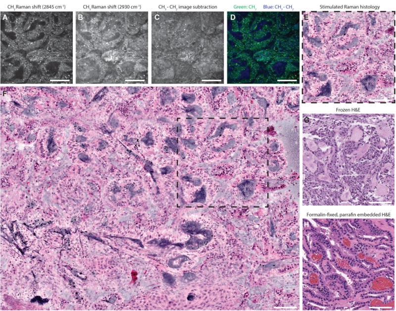
Figure 1
Label-free stimulated Raman histology (SRH) of fresh brain tumor tissue
A choroid plexus papilloma, WHO grade I, imaged at 2845 cm -1 (A) and 2930 cm -1 (B) Raman shift wave numbers with 400 u00d7 400-u03bcm fields of view at a rate of 2 seconds per frame. To highlight nu...
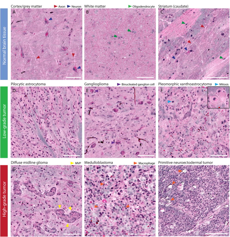
Figure 2
SRH histopathologic features of normal brain and pediatric brain tumors
(A) Normal neocortex shows large pyramidal neurons with lipofuscin cytoplasmic inclusions seen in bright pink. Axons are clearly visualized in neocortex as white lines. (B) Normal subcortical white ma...
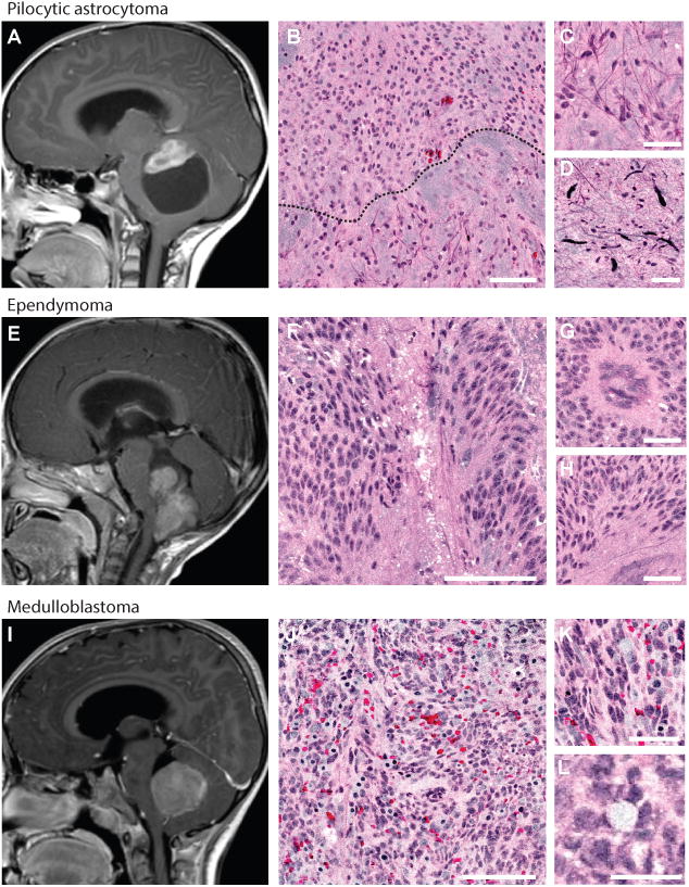
Figure 3
SRH identifies pediatric surgical lesions of the posterior fossa
Magnetic resonance images (midsagittal T1-weighted post-gadolinium) of the three most common surgical lesions of posterior fossa are shown: pilocytic astrocytoma (A), ependymoma (E), and medulloblasto...
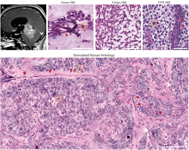
Figure 4
SRH preserves cytologic and histoarchitectural features of pediatric brain tumors
(A) Preoperative midsagittal T1-weighted post-gadolinium magnetic resonance image of posterior fossa germinoma. (B) Smear preparation shows the large germ cells with abundant foamy glycogen-rich cytop...
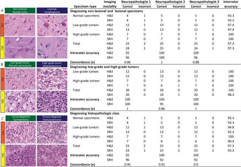
Figure 5
Evaluation of SRH via simulated intraoperative pathology consultation
Results from web-based survey shown in the table. SRH and standard H&E images from 25 patients were presented to three neuropathologists for evaluation. Free-text responses were evaluated on three lev...
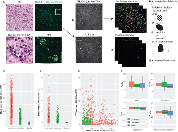
Figure 6
SRH feature extraction and quantitative histology
A) CellProfiler feature extraction pipeline was developed to split composite SRH images into 2845 cm -1 /CH 2 and 2930 u2013 2845 cm -1 /CH 3 -CH 2 images for nuclear and tumor-associated macrophage (...
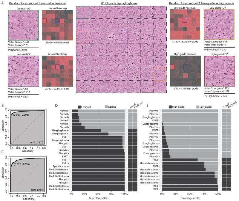
Figure 7
Validation of machine-learning model for classification of pediatric brain tumor specimens
A) SRH image mosaic (center) of a ganglioglioma, WHO grade I, is shown with individual FOV tiles demarcated with dashed black lines. Select color-coded tiles from the image mosaic are shown peripheral...
Figure images are served from the NIH/NLM PubMed Central Open Access Subset or Europe PMC; copyright remains with the publishers and authors.
💬 Discussion
0 commentsNo comments yet. Be the first to start a discussion!
Leave a Comment