Abstract
BACKGROUND: Transient receptor potential vanilloid subtype 1 (TRPV1) is activated by low pH/protons and is well known to be involved in hyperalgesia during inflammation. Tumor necrosis factor alpha (TNF-alpha), a proinflammatory cytokine, is involved in nociceptive responses causing hyperalgesia through TNF receptor type 1 (TNFR1) activation. Reactive oxygen species (ROS) production is also prominently increased in inflamed tissue. The present study investigated TNFR1 receptors in primary cultured mouse dorsal root ganglion (DRG) neurons after TRPV1 activation and the involvement of ROS. C57BL/6 mice, both TRPV1 knockout and wild type, were used for immunofluorescent and live cell imaging. The L4 and L5 DRGs were dissected bilaterally and cultured overnight. TRPV1 was stimulated with capsaicin or its potent analog, resiniferatoxin. ROS production was measured with live cell imaging and TNFR1 was detected with immunofluorescence in DRG primary cultures. The TRPV1 knockout mice, TRPV1 antagonist, capsazepine, and ROS scavenger, N-tert-Butyl-alpha-phenylnitrone (PBN), were employed to explore the functional relationship among TRPV1, ROS and TNFR1 in these studies. RESULTS: The results demonstrate that TRPV1 activation increases TNFR1 receptors and ROS generation in primary cultures of mouse DRG neurons. Activated increases in TNFR1 receptors and ROS production are absent in TRPV1 deficient mice. The PBN blocks increases in TNFR1 and ROS production induced by capsaicin/resiniferatoxin. CONCLUSION: TRPV1 activation increases TNFR1 in cultured mouse DRG neurons through a ROS signaling pathway, a novel sensitization mechanism in DRG neurons.
🔬 Techniques
💻 Software
✨ Fluorophores
🧪 Sample Preparation
🏭 Microscope Brands
🧪 Reagent Suppliers
💻 Software Details
🏛️ Research Organizations (ROR)
Affiliated research institutions:
📋 Methods
This study was performed in accordance with the Guide for the Care and Use of Laboratory Animals published by the National Institutes of Health and was approved by the University of Kentucky Institutional Animal Care and Use Committee.
Primary DRG neuronal cultures
Male and female TRPV1 knockout or wild type C57BL/6 mice (19 – 20 g, Jackson Lab, as a tissue sharing gift from Dr. Lu-Yuan Lee) were used in these experiments [ 42 ]. Mice were anesthetized and the vertebral columns were exposed, removed, and put into the ice-cold dissection solution (NaCl 135, KCl 5, KH 2 PO 4 2, HEPES 10, MgSO 4 6, CaCl 2 1.5, Glucose 10; in mM) bubbled with 100% O 2 . The L4, L5 DRGs were removed from the lumbar spinal roots and placed into the 30 mm dish with ice-cold dissection solution. After connective tissues were removed, DRGs were digested in fresh enzyme solution (10 mg trypsin+25 mg collagenase in 10 ml dissection solution) in 25 cm 2 flask saturated with O 2 . After shaking in a warm water bath (speed 150 rpm, 35°C) for 1 hour, 4 ml of culture medium (MEM with FBS, antibiotics) were added to the flask. The cells were transferred into a 15 ml centrifuge tube and spun at 800 rpm for 5 minutes. The supernatant was discarded and the cell pellet was re-suspended with culture medium. DRG cells were plated onto poly-L-Lysine pre-coated 12 mm glass-bottom home made tissue culture dish or 12 mm coverslips in 24 well culture plates (0.2 ml of cell suspension per well). Cultured DRG neurons were maintained in an incubator with 5% CO 2 , 37°C overnight and all experiments performed on the subsequent day.
Show full methods section
This study was performed in accordance with the Guide for the Care and Use of Laboratory Animals published by the National Institutes of Health and was approved by the University of Kentucky Institutional Animal Care and Use Committee.
Primary DRG neuronal cultures
Male and female TRPV1 knockout or wild type C57BL/6 mice (19 – 20 g, Jackson Lab, as a tissue sharing gift from Dr. Lu-Yuan Lee) were used in these experiments [ 42 ]. Mice were anesthetized and the vertebral columns were exposed, removed, and put into the ice-cold dissection solution (NaCl 135, KCl 5, KH 2 PO 4 2, HEPES 10, MgSO 4 6, CaCl 2 1.5, Glucose 10; in mM) bubbled with 100% O 2 . The L4, L5 DRGs were removed from the lumbar spinal roots and placed into the 30 mm dish with ice-cold dissection solution. After connective tissues were removed, DRGs were digested in fresh enzyme solution (10 mg trypsin+25 mg collagenase in 10 ml dissection solution) in 25 cm 2 flask saturated with O 2 . After shaking in a warm water bath (speed 150 rpm, 35°C) for 1 hour, 4 ml of culture medium (MEM with FBS, antibiotics) were added to the flask. The cells were transferred into a 15 ml centrifuge tube and spun at 800 rpm for 5 minutes. The supernatant was discarded and the cell pellet was re-suspended with culture medium. DRG cells were plated onto poly-L-Lysine pre-coated 12 mm glass-bottom home made tissue culture dish or 12 mm coverslips in 24 well culture plates (0.2 ml of cell suspension per well). Cultured DRG neurons were maintained in an incubator with 5% CO 2 , 37°C overnight and all experiments performed on the subsequent day.
Immunofluorescence study Dissociated
DRG neurons were cultured on coverslips in 24-well plates overnight. Cells were stimulated with capsaicin (1 μM) or resiniferatoxin (200 nM) for 30 minutes and then the media was replaced with Dulbecco’s phosphate buffered saline (DPBS) for 90 minutes. In some experiments, ROS scavenger PBN was applied for 90 minutes before capsaicin stimulation, or TRPV1 antagonist capsazepine (10 μM) was administered 10 minutes before capsaicin stimulation. At the end of incubation, cells were washed with 0.1 M phosphate buffered saline (PBS, pH7.4) 3 times × 5 minutes and fixed with 4% paraformaldehyde in 0.1 M PBS for 30 minutes. After washing with PBS, cells were incubated with 3% normal donkey serum in PBS plus 0.02% Triton X-100 for 30 minutes to block non-specific protein binding. Then, cells were incubated overnight with the primary antibody diluted in PBS TritonX-100 plus 1% normal donkey serum at 4°C. The anti-TRPV1 receptor antibody (VR1 P-19, Santa Cruz, and affinity purified goat polyclonal antibody against a peptide mapping near the N-terminus of VR1 of rat origin producing a single band in western blots) was diluted 1:100. The anti-TNFR1 receptor antibody (TNF-R1 H-5, Santa Cruz), a mouse monoclonal antibody raised against amino acids 30–301 mapping within the extracellular domain of TNF-R1 of human origin producing a single band in western blots, was diluted 1:50. After overnight incubation, the coverslips were rinsed in PBS 3 times × 5 minutes and incubated with FITC-donkey-anti-goat antibody, diluted 1:1000 and Texas Red-donkey-anti-mouse antibody, diluted 1:800 in PBS Triton-X plus 1% normal donkey serum for 1 hour at room temperature. For staining controls, primary antibodies were omitted. The coverslips were washed again 3 times × 5 minutes. Thereafter, the coverslips were mounted face down onto slides with mounting medium. Five different areas were picked randomly from each coverslip for pictures. MetaMorph offline software was used for digital image data analysis.
Determination of ROS production by H2DCFDA fluorescent probe
All experiments were done at room temperature. Cells were kept at room temperature for 30 min before replacing culture medium with DPBS, pH 7.4. On the day of the experiment, the ROS-sensitive fluorescent dye, 5-(and-6)-chloromethyl-2', 7'-dichlorodihydrofluorescein diacetate, acetyl ester (CM-H2DCFDA, Invitrogen) was freshly reconstituted. Cells were pre-loaded with CM-H2DCFDA in DPBS at a final concentration of 4 μM for 30 min. The loading buffer was removed and replaced with DPBS allowing 20 min to 2 h recovery time for cells to exhibit low levels of esterase activity. ROS production was monitored for 30 minutes in the live cell imaging experiment. Cells were stimulated with bath application of capsaicin (1 μM) or resiniferatoxin (200 nM) and in some cases neurons were pretreated 10 minutes with TRPV1 antagonist capsazepine (10 μM) before beginning the recording and capsaicin stimulation. ROS scavenger N-tert-Butyl-α-phenylnitrone (PBN, 2 mM) was administered for 90 minutes before capsaicin stimulation. The change in fluorescent intensity over time is proportional to DCF accumulation, an index of net oxidant activity in the cytoplasm. Fluorescent imaging was performed using an Eclipse 2000-S microscope (Nikon) equipped with a high sensitivity digital monochrome Cool SNAP ES camera (Photometrics, Tucson, AZ) controlled by MetaMorph software (Molecular Devices, Sunnyvale, CA). DCF was excited at 480 nm and emitted light was collected using a 520 nm long pass filter. The photos were captured under 10 ms exposure at 1 min intervals for 30 min. Emissions from DCFH-free buffer are not detectable under these conditions, and background correction is not required. Photooxidation artifact was controlled by conducting experiments in a darkened laboratory and by standardizing excitation parameters to minimize the cumulative delivered energy. The baseline fluorescent intensity was determined prior to exposing the cells to experimental inducements. MetaMorph offline software (Molecular Devices, Sunnyvale, CA) was used for image data analysis. Chemicals Trypsin, poly-L-Lysine, capsaicin, resiniferatoxin, capsazepine and N-tert-Butyl-α-phenylnitrone were obtained from Sigma-Aldrich (St. Louis, MO). Collagenase was purchased from Roche Applied Science (Indianapolis, IN). 5-(and-6)-chloromethyl-2', 7'-dichlorodihydrofluorescein diacetate, acetyl ester and DPBS were purchased from Invitrogen (Carlsbad, CA). Antibody against TRPV1, antibody against TNFR1, FITC-donkey-anti-goat antibody and Texas Red-donkey-anti-mouse antibody were obtained from Santa Cruz Biotechnology (Santa Cruz, CA). Mounting medium was purchased from Vector Laboratories (Burlingame, CA).
Statistics
All data were expressed as mean ± SEM. Comparisons among multiple means were analyzed with the one-way ANOVA. Post hoc test was analyzed with the Newman-Keuls Multiple Comparison test. A P < 0.05 was considered significant.
📊 Figures
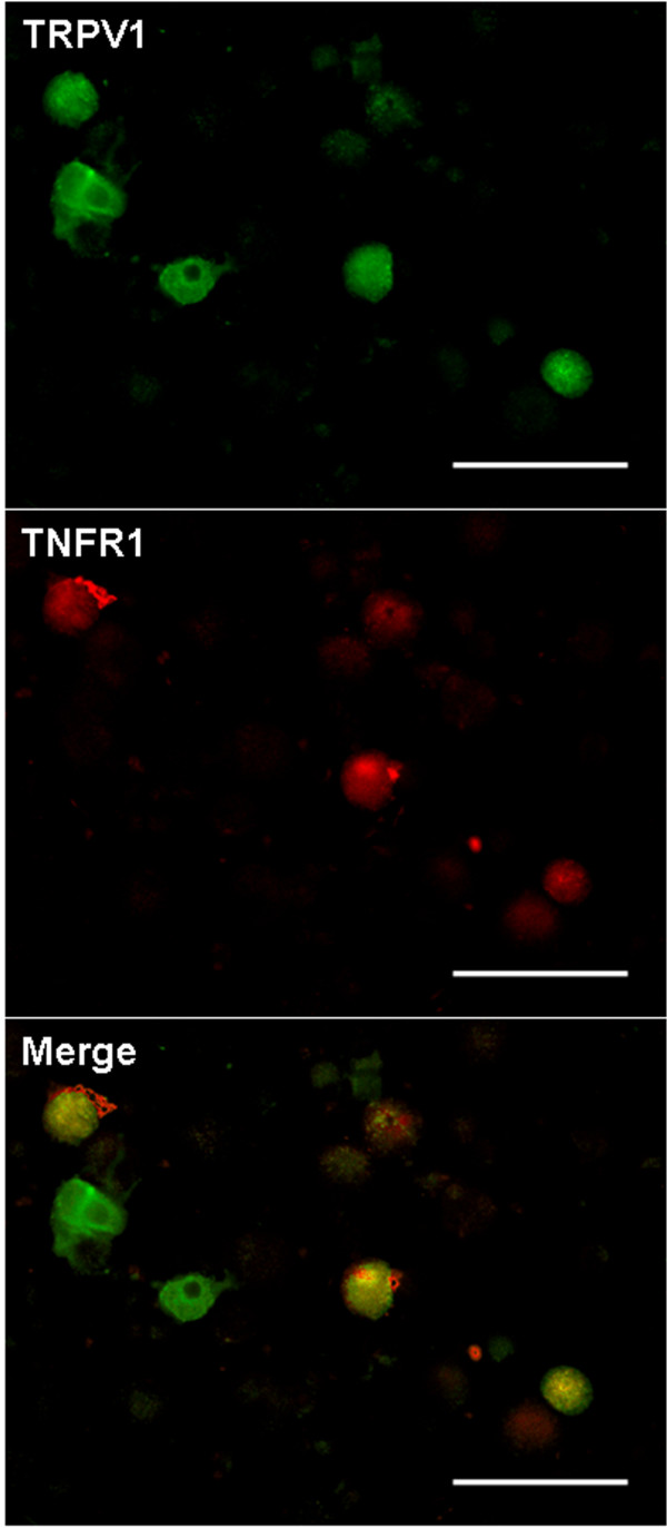
Figure 1
TRPV1 and TNFR1 in primary cultures of mouse DRG neurons . Photomicrographs show green TRPV1 immunofluorescence and red TNFR1 immunofluorescence in DRG neurons cultured overnight and then fixed for do...
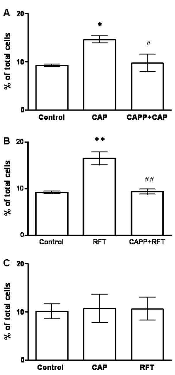
Figure 2
Capsaicin or resiniferatoxin activation of TRPV1 increases TNFR1 staining in cultured mouse DRG neurons . Histograms show the percentage of immunoreactive cells in DRG neuronal cultures. (A) Capsaicin...
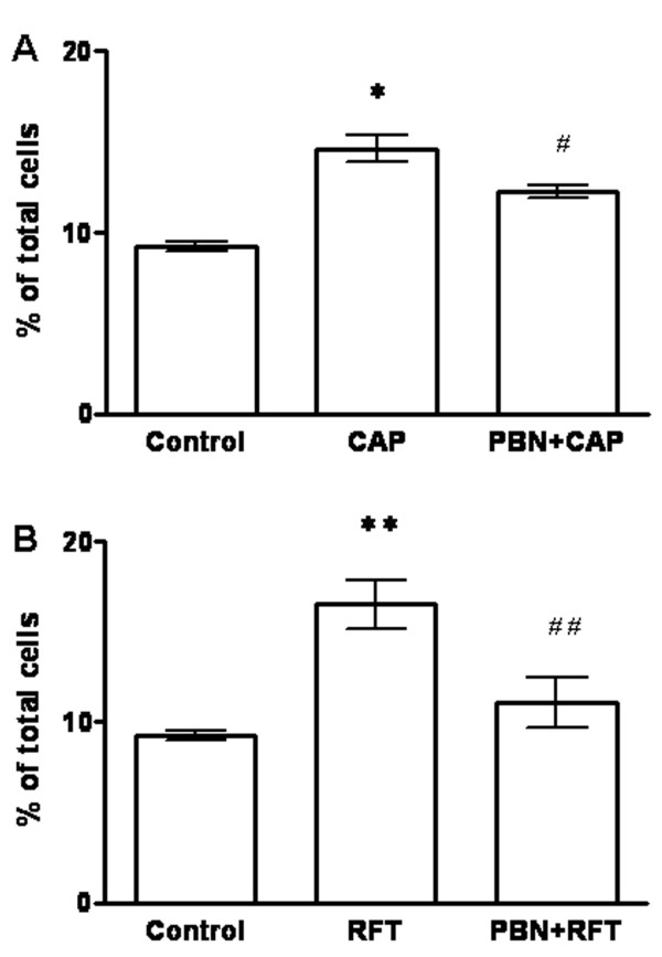
Figure 3
The ROS scavenger PBN blocks TNFR1 increases in cultured mouse DRG neurons induced by capsaicin or resiniferatoxin . Histograms showing the percentage of TNFR1 immunoreactive neurons in DRG neuronal c...
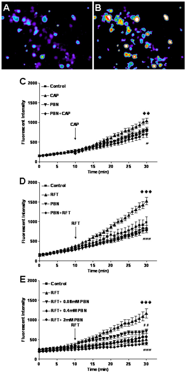
Figure 4
Capsaicin or resiniferatoxin increases ROS production in mouse primary DRG cultures . (A) Photomicrographs of ROS in mouse DRG in primary cultures at baseline. (B) ROS production increases after resin...
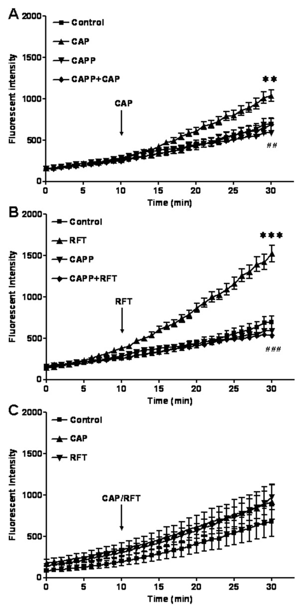
Figure 5
Capsaicin or resiniferatoxin induces ROS production in cultured mouse DRG neurons through TRPV1 activation . (A) Capsaicin significantly increases ROS production and capsazepine blocks capsaicin induc...
Figure images are served from the NIH/NLM PubMed Central Open Access Subset or Europe PMC; copyright remains with the publishers and authors.
💬 Discussion
0 commentsNo comments yet. Be the first to start a discussion!
Leave a Comment