Abstract
Metabolic sensing neurons in the ventromedial hypothalamus (VMH) alter their activity when ambient levels of metabolic substrates, such as glucose and fatty acids (FA), change. To assess the relationship between a high-fat diet (HFD; 60%) intake on feeding and serum and VMH FA levels, rats were trained to eat a low-fat diet (LFD; 13.5%) or an HFD in 3 h/day and were monitored with VMH FA microdialysis. Despite having higher serum levels, HFD rats had lower VMH FA levels but ate less from 3 to 6 h of refeeding than did LFD rats. However, VMH β-hydroxybutyrate (β-OHB) and VMH-to-serum β-OHB ratio levels were higher in HFD rats during the first 1 h of refeeding, suggesting that VMH astrocyte ketone production mediated their reduced intake. In fact, using calcium imaging in dissociated VMH neurons showed that ketone bodies overrode normal FA sensing, primarily by exciting neurons that were activated or inhibited by oleic acid. Importantly, bilateral inhibition of VMH ketone production with a 3-hydroxy-3-methylglutaryl-CoA synthase inhibitor reversed the 3- to 6-h HFD-induced inhibition of intake but had no effect in LFD-fed rats. These data suggest that a restricted HFD intake regimen inhibits caloric intake as a consequence of FA-induced VMH ketone body production by astrocytes.
🔬 Techniques
✨ Fluorophores
🧪 Sample Preparation
🔬 Cell Lines
🧪 Reagent Suppliers
💻 Software Details
🏛️ Research Organizations (ROR)
Affiliated research institutions:
📋 Methods
Research Design and Methods Animals
Animals were housed at 23–24°C on a reversed 12:12-h light-dark cycle (lights off at 1000) with ad libitum access to chow (13.5% kcal fat; Purina #5001) and water. Outbred male Sprague-Dawley rats were purchased from Charles River Laboratories. All work was in compliance with the East Orange Veterans Affairs Medical Center Institutional Animal Care and Use Committee. VMH Free FA and Food Intake Measurements Rats ( n = 8 per group; weight 300–400 g) were anesthetized with isoflurane (1.5% at 0.8 L/min), and a unilateral microdialysis probe with a 3-mm membrane length and 3,000 kDa pore size (MAB 5.15.3PE; Microbiotech/se AB, Stockholm, Sweden) was stereotaxically angled at 20° to the vertical at the junction between the arcuate and VMH nuclei (VMN) (VMH = VMN + arcuate; −2.9 mm bregma, ± 3.7 mm midline, and −8.5 mm dura). The next day at 0800 (2 h before food was presented), microdialysis was begun with probes perfused with artificial cerebrospinal fluid (aCSF; Harvard Apparatus, Holliston, MA) containing 3% FA-free BSA (Sigma-Aldrich, St. Louis, MO) at 1.0 μL/min. Eluates were collected every 30 min and were stored at −80°C until nonesterified FA assay. Probe placement was assessed terminally. A first set of rats was fasted for 24 h, and VMH probes were infused for 1 h at each of five concentrations (0, 20, 60, 150, 400 µmol/L) of OA, with monitoring of effluent OA concentrations at 20 min. Fasting VMH OA levels were calculated using the zero-net-flux method ( 19 ). A second set of rats was trained for 2 weeks to eat a 13.5% LFD (Purina #5001) or a 60% HFD (Research Diet D12492, New Brunswick, NJ) ( n = 8 per group) in 3 h each day from 1000 to 1300 in a BioDAQ food intake monitoring apparatus. After 1 week, jugular catheters were implanted, and rats were allowed 6 to 7 days to recover their preoperative body weight on their 3 h/day feeding schedule. On testing day, rats ate their respective diets for 6 h while their ongoing food intake was monitored, and they simultaneously underwent microdialysis and blood sampling at 30 min intervals over 6 h. VMH β-Hydroxybutyrate and Feeding Measurements Unilateral VMH guide cannulae were implanted in LFD rats ( n = 8 per group). After 1–2 weeks of recovery, they were fasted for 24 h for zero-net-flux assessment. The next day, at 0800, microdialysis probes (3-mm membrane length and 6-kDa pore size [CMA 11; Harvard Apparatus, Holliston, MA]) were inserted into the guide cannulae and perfused at 1.0 µL/min with β-hydroxybutyrate (β-OHB) diluted in aCSF for 40 min at each of four concentrations (0, 2, 30, 100 µmol/L), and effluent β-OHB concentrations were monitored. Next, additional rats were trained for 2 weeks to eat a LFD or HFD ( n = 8 per group) over 3 h/day and were implanted with jugular catheters and unilateral VMH guide cannulae. At 0800 on the test day, microdialysis probes were inserted, jugular catheters were connected, and the rats’ respective diets were returned from 1000 to 1300. At 1300, rats being fed the HFD were switched to the LFD, and all rats were allowed to eat for 3 h more, with monitoring of food intake, microdialysis eluates, and blood samples every 30 min over 6 h. A third set of rats ( n = 6–8 per group) was conditioned to eat the LFD or HFD in 3 h/day and implanted with bilateral VMH guide cannulae and jugular catheters. On test day, bilateral microdialysis probes were inserted at 0800, and an infusion with aCSF and 0.4% DMSO vehicle or 30 µmol/L hymeglusin, a 3-hydroxy-3-methylglutaryl–CoA (HMG-CoA) synthase inhibitor ( 20 ), in aCSF plus 0.4% DMSO at 1.0 µL/min ( n = 6–8 per group), along with simultaneous microdialysis for ketones was begun. The LFD or HFD was provided from 1000 to 1300 and the LFD from 1300 to 1600, with monitoring of food intake, VMH, and serum β-OHB levels. β-OHB and Acetoacetate Induced Changes in Intracellular Ca 2+ Oscillations in Dissociated VMN Neurons Postnatal day 21–28 (P21–28) rats were perfused, and neurons were dissociated from VMN punches, as previously described ( 18 , 21 , 22 ). Evaluation of glucose-, OA-, and ketone body-induced alterations in intracellular Ca 2+ [Ca 2+ ] i oscillations in individual VMN neurons were assessed using fura-2 AM (Invitrogen, Grand Island, NY), as previously described ( 18 , 21 , 22 ). Neurons were classified first as glucose excited (GE), glucose inhibited (GI), and non–glucose-sensing (NG), then as OA excited (OAE), OA inhibited (OAI), and OA nonresponsive (OAN), and as β-OHB and acetoacetate (AA) excited, inhibited, or nonresponsive using previously established criteria for changes in [Ca 2+ ] i area under the curve ( 21 , 22 ). Studies began with neurons held at 2.5 mmol/L or 0.5 mmol/L glucose, followed by 15 nmol/L OA and by 1–100 µmol/L β-OHB or 1 μmol/L AA. Also assessed was a combination of 1 μmol/L AA and 20 μmol/L β-OHB (their relative ratio in the brain). All neurons were incubated with 20 nmol/L glutamate terminally to assess viability. In Vitro Effects of FA on VMH Astrocyte-Induced Ketone Production The VMH was dissected from P21–28 rats and triturated in Neurobasal-A (Invitrogen) containing 5 mmol/L glucose, 0.23 mmol/L sodium pyruvate, 100 units/mL penicillin/streptomycin, 10 μg/mL gentamicin, and 10% FBS at pH 7.4. Dissociated cells were plated in 25-cm 2 flasks coated with poly- d -lysine (50 µg/mL) until confluence. Astrocytes were separated from remaining microglia by shaking at 250 rpm for 3 h at 37°C ( 23 ). Astrocytes attached to the flask were trypsinized with 0.05% trypsin-EDTA (Sigma-Aldrich) and centrifuged. The pellet was resuspended in Neurobasal-A and plated in 6-well plates. The day before the experiment, astrocytes were washed with PBS, and Neurobasal-A serum free was added overnight. Astrocytes were then treated with vehicle alone (0.4% DMSO), 150 μmol/L OA, palmitic acid (PA), or octanoic acid (OctA), or 30 µmol/L hymeglusin. Hymeglusin (synthesized by H.M.M.) is an HMG-CoA synthase inhibitor that is been demonstrated to inhibit ketone production in bacteria ( 20 , 24 ). Media were harvested at 0, 2, and 4 h, and total ketone levels were assayed. Assays of OA, β-OHB, and Total Ketones OA, total ketone, and β-OHB levels were analyzed using a colorimetric assay (Wako, Richmond, VA). Statistics Systat (Chicago, IL) and GraphPad Prism software (GraphPad, Inc., La Jolla, CA) were used to calculate one-way and two-way ANOVA and one-way ANOVA for repeated measures with post hoc Bonferonni corrections for the in vitro and in vivo studies. No more than two outliers per group were removed, if necessary, by using Systat software.
Show full methods section
Research Design and Methods Animals
Animals were housed at 23–24°C on a reversed 12:12-h light-dark cycle (lights off at 1000) with ad libitum access to chow (13.5% kcal fat; Purina #5001) and water. Outbred male Sprague-Dawley rats were purchased from Charles River Laboratories. All work was in compliance with the East Orange Veterans Affairs Medical Center Institutional Animal Care and Use Committee. VMH Free FA and Food Intake Measurements Rats ( n = 8 per group; weight 300–400 g) were anesthetized with isoflurane (1.5% at 0.8 L/min), and a unilateral microdialysis probe with a 3-mm membrane length and 3,000 kDa pore size (MAB 5.15.3PE; Microbiotech/se AB, Stockholm, Sweden) was stereotaxically angled at 20° to the vertical at the junction between the arcuate and VMH nuclei (VMN) (VMH = VMN + arcuate; −2.9 mm bregma, ± 3.7 mm midline, and −8.5 mm dura). The next day at 0800 (2 h before food was presented), microdialysis was begun with probes perfused with artificial cerebrospinal fluid (aCSF; Harvard Apparatus, Holliston, MA) containing 3% FA-free BSA (Sigma-Aldrich, St. Louis, MO) at 1.0 μL/min. Eluates were collected every 30 min and were stored at −80°C until nonesterified FA assay. Probe placement was assessed terminally. A first set of rats was fasted for 24 h, and VMH probes were infused for 1 h at each of five concentrations (0, 20, 60, 150, 400 µmol/L) of OA, with monitoring of effluent OA concentrations at 20 min. Fasting VMH OA levels were calculated using the zero-net-flux method ( 19 ). A second set of rats was trained for 2 weeks to eat a 13.5% LFD (Purina #5001) or a 60% HFD (Research Diet D12492, New Brunswick, NJ) ( n = 8 per group) in 3 h each day from 1000 to 1300 in a BioDAQ food intake monitoring apparatus. After 1 week, jugular catheters were implanted, and rats were allowed 6 to 7 days to recover their preoperative body weight on their 3 h/day feeding schedule. On testing day, rats ate their respective diets for 6 h while their ongoing food intake was monitored, and they simultaneously underwent microdialysis and blood sampling at 30 min intervals over 6 h. VMH β-Hydroxybutyrate and Feeding Measurements Unilateral VMH guide cannulae were implanted in LFD rats ( n = 8 per group). After 1–2 weeks of recovery, they were fasted for 24 h for zero-net-flux assessment. The next day, at 0800, microdialysis probes (3-mm membrane length and 6-kDa pore size [CMA 11; Harvard Apparatus, Holliston, MA]) were inserted into the guide cannulae and perfused at 1.0 µL/min with β-hydroxybutyrate (β-OHB) diluted in aCSF for 40 min at each of four concentrations (0, 2, 30, 100 µmol/L), and effluent β-OHB concentrations were monitored. Next, additional rats were trained for 2 weeks to eat a LFD or HFD ( n = 8 per group) over 3 h/day and were implanted with jugular catheters and unilateral VMH guide cannulae. At 0800 on the test day, microdialysis probes were inserted, jugular catheters were connected, and the rats’ respective diets were returned from 1000 to 1300. At 1300, rats being fed the HFD were switched to the LFD, and all rats were allowed to eat for 3 h more, with monitoring of food intake, microdialysis eluates, and blood samples every 30 min over 6 h. A third set of rats ( n = 6–8 per group) was conditioned to eat the LFD or HFD in 3 h/day and implanted with bilateral VMH guide cannulae and jugular catheters. On test day, bilateral microdialysis probes were inserted at 0800, and an infusion with aCSF and 0.4% DMSO vehicle or 30 µmol/L hymeglusin, a 3-hydroxy-3-methylglutaryl–CoA (HMG-CoA) synthase inhibitor ( 20 ), in aCSF plus 0.4% DMSO at 1.0 µL/min ( n = 6–8 per group), along with simultaneous microdialysis for ketones was begun. The LFD or HFD was provided from 1000 to 1300 and the LFD from 1300 to 1600, with monitoring of food intake, VMH, and serum β-OHB levels. β-OHB and Acetoacetate Induced Changes in Intracellular Ca 2+ Oscillations in Dissociated VMN Neurons Postnatal day 21–28 (P21–28) rats were perfused, and neurons were dissociated from VMN punches, as previously described ( 18 , 21 , 22 ). Evaluation of glucose-, OA-, and ketone body-induced alterations in intracellular Ca 2+ [Ca 2+ ] i oscillations in individual VMN neurons were assessed using fura-2 AM (Invitrogen, Grand Island, NY), as previously described ( 18 , 21 , 22 ). Neurons were classified first as glucose excited (GE), glucose inhibited (GI), and non–glucose-sensing (NG), then as OA excited (OAE), OA inhibited (OAI), and OA nonresponsive (OAN), and as β-OHB and acetoacetate (AA) excited, inhibited, or nonresponsive using previously established criteria for changes in [Ca 2+ ] i area under the curve ( 21 , 22 ). Studies began with neurons held at 2.5 mmol/L or 0.5 mmol/L glucose, followed by 15 nmol/L OA and by 1–100 µmol/L β-OHB or 1 μmol/L AA. Also assessed was a combination of 1 μmol/L AA and 20 μmol/L β-OHB (their relative ratio in the brain). All neurons were incubated with 20 nmol/L glutamate terminally to assess viability. In Vitro Effects of FA on VMH Astrocyte-Induced Ketone Production The VMH was dissected from P21–28 rats and triturated in Neurobasal-A (Invitrogen) containing 5 mmol/L glucose, 0.23 mmol/L sodium pyruvate, 100 units/mL penicillin/streptomycin, 10 μg/mL gentamicin, and 10% FBS at pH 7.4. Dissociated cells were plated in 25-cm 2 flasks coated with poly- d -lysine (50 µg/mL) until confluence. Astrocytes were separated from remaining microglia by shaking at 250 rpm for 3 h at 37°C ( 23 ). Astrocytes attached to the flask were trypsinized with 0.05% trypsin-EDTA (Sigma-Aldrich) and centrifuged. The pellet was resuspended in Neurobasal-A and plated in 6-well plates. The day before the experiment, astrocytes were washed with PBS, and Neurobasal-A serum free was added overnight. Astrocytes were then treated with vehicle alone (0.4% DMSO), 150 μmol/L OA, palmitic acid (PA), or octanoic acid (OctA), or 30 µmol/L hymeglusin. Hymeglusin (synthesized by H.M.M.) is an HMG-CoA synthase inhibitor that is been demonstrated to inhibit ketone production in bacteria ( 20 , 24 ). Media were harvested at 0, 2, and 4 h, and total ketone levels were assayed. Assays of OA, β-OHB, and Total Ketones OA, total ketone, and β-OHB levels were analyzed using a colorimetric assay (Wako, Richmond, VA). Statistics Systat (Chicago, IL) and GraphPad Prism software (GraphPad, Inc., La Jolla, CA) were used to calculate one-way and two-way ANOVA and one-way ANOVA for repeated measures with post hoc Bonferonni corrections for the in vitro and in vivo studies. No more than two outliers per group were removed, if necessary, by using Systat software.
📊 Figures
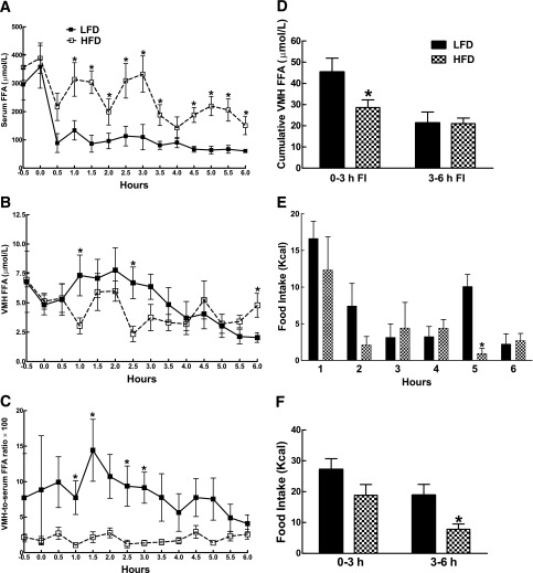
Figure 1
Rats were trained to eat all of an LFD (13.5% fat; n = 8) or an HFD (60% fat; n = 7) within 3 h of presentation at 0 h on the graph. On the day of testing, FFA levels were measured every 30 min in ser...
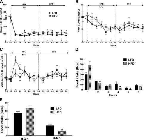
Figure 2
Rats were trained to eat all of an LFD (13.5% fat; n = 8) or HFD (60% fat; n = 7) within 3 h of presentation at 0 h on the graph. On the day of testing, u03b2-OHB levels were measured every 30 min in ...
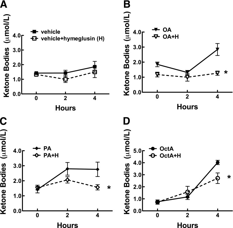
Figure 3
Ketone production in primary cultured VMH astrocytes from P21 rats. A : Effects of vehicle (0.4% DMSO) and vehicle plus 30 u00b5mol/L hymeglusin (H). Vehicle plus 30 u00b5mol/L hymeglusin in the prese...
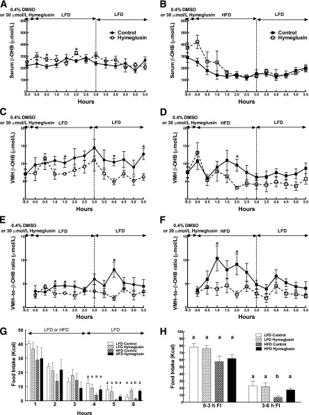
Figure 4
Rats were trained to eat all of their food (LFD, 13.5% fat [ n = 12]; HFD, 60% fat [ n = 16]) within 3 h of presentation at 0 h on the graph. On the day of testing, 0.4% DMSO (control; n = 6u20138) or...
Figure images are served from the NIH/NLM PubMed Central Open Access Subset or Europe PMC; copyright remains with the publishers and authors.
💬 Discussion
0 commentsNo comments yet. Be the first to start a discussion!
Leave a Comment