Abstract
Respiratory syncytial virus (RSV) nonstructural protein 1(NS1) attenuates type-I interferon (IFN) production during RSV infection; however the precise role of RSV NS1 protein in orchestrating the early host-virus interaction during infection is poorly understood. Since NS1 constitutes the first RSV gene transcribed and the production of IFN depends upon RLR (RIG-I-like receptor) signaling, we reasoned that NS1 may interfere with this signaling. Herein, we report that NS1 is localized to mitochondria and binds to mitochondrial antiviral signaling protein (MAVS). Live-cell imaging of rgRSV-infected A549 human epithelial cells showed that RSV replication and transcription occurs in proximity to mitochondria. NS1 localization to mitochondria was directly visualized by confocal microscopy using a cell-permeable chemical probe for His(6)-NS1. Further, NS1 colocalization with MAVS in A549 cells infected with RSV was shown by confocal laser microscopy and immuno-electron microscopy. NS1 protein is present in the mitochondrial fraction and co-immunoprecipitates with MAVS in total cell lysatesof A549 cells transfected with the plasmid pNS1-Flag. By immunoprecipitation with anti-RIG-I antibody, RSV NS1 was shown to associate with MAVS at an early stage of RSV infection, and to disrupt MAVS interaction with RIG-I (retinoic acid inducible gene) and the downstream IFN antiviral and inflammatory response. Together, these results demonstrate that NS1 binds to MAVS and that this binding inhibits the MAVS-RIG-I interaction required for IFN production.
🔬 Techniques
🔭 Microscopes
✨ Fluorophores
🧪 Sample Preparation
🔬 Cell Lines
🏭 Microscope Brands
🧪 Reagent Suppliers
💻 Software Details
🏛️ Research Organizations (ROR)
Affiliated research institutions:
📋 Methods
Cell culture Human embryonic lung fibroblast HEp-2
(CCL-23) cells, human alveolar epithelial A549 (CCL-185) cells and African green monkey kidney Vero (CCL-81) cells were all purchased from the American Type Culture Collection (ATCC) and were cultured in standard Dulbecco's MEM (DMEM) containing 5% heat-inactivated fetal bovine serum, L-glutamine, 100 IU/ml penicillin and 100 µg/ml streptomycin sulfate. Viruses Recombinant RSV strains rA2, rA2ΔNS1, rA2ΔNS2 and rA2-His 6 -NS1 have been previously described [25] . RgRSV, which expresses green fluorescent protein, was a gift from Dr. Mark Peeples. Viruses rA2-His 6 -NS1 and rA2 RSV were grown in HEp-2 cells and harvested when cytopathic effects became visible (2–3 days). RSV-infected cells were subjected to a single round of freeze-thaw cycles and viral supernatant was clarified by centrifugation at 3200 rpmat 4°C for 10 min. Viral titers were obtained through plaque assay with 0.8% methylcellulose overlay and immunostaining with monoclonal murine anti-RSV F antibody, followed by a horseradish peroxidase-conjugated secondary antibody and visualization with 4CN substrate (Kirkegaard and Perry Laboratories). The deletion mutant rA2ΔNS1 was cultured in Vero cells and viral titer was also determined in the same cells. For microscopy studies, A549 and HEp-2 cells were seeded to sub-confluency on glass-bottom culture dishes 24 hrs before infection. On the day of infection, growth media was replaced with Opti-MEM (Invitrogen) containing the appropriate amount of virus suspension for obtaining the indicated multiplicity of infection (MOI).
Show full methods section
Cell culture Human embryonic lung fibroblast HEp-2
(CCL-23) cells, human alveolar epithelial A549 (CCL-185) cells and African green monkey kidney Vero (CCL-81) cells were all purchased from the American Type Culture Collection (ATCC) and were cultured in standard Dulbecco's MEM (DMEM) containing 5% heat-inactivated fetal bovine serum, L-glutamine, 100 IU/ml penicillin and 100 µg/ml streptomycin sulfate. Viruses Recombinant RSV strains rA2, rA2ΔNS1, rA2ΔNS2 and rA2-His 6 -NS1 have been previously described [25] . RgRSV, which expresses green fluorescent protein, was a gift from Dr. Mark Peeples. Viruses rA2-His 6 -NS1 and rA2 RSV were grown in HEp-2 cells and harvested when cytopathic effects became visible (2–3 days). RSV-infected cells were subjected to a single round of freeze-thaw cycles and viral supernatant was clarified by centrifugation at 3200 rpmat 4°C for 10 min. Viral titers were obtained through plaque assay with 0.8% methylcellulose overlay and immunostaining with monoclonal murine anti-RSV F antibody, followed by a horseradish peroxidase-conjugated secondary antibody and visualization with 4CN substrate (Kirkegaard and Perry Laboratories). The deletion mutant rA2ΔNS1 was cultured in Vero cells and viral titer was also determined in the same cells. For microscopy studies, A549 and HEp-2 cells were seeded to sub-confluency on glass-bottom culture dishes 24 hrs before infection. On the day of infection, growth media was replaced with Opti-MEM (Invitrogen) containing the appropriate amount of virus suspension for obtaining the indicated multiplicity of infection (MOI).
Confocal microscopy
A549 cells were infected with rgRSV and imaged from the start of infection for 16 h using a Nikon T inverted microscope running the Perkin Elmer UltraVIEW®VoX 3D Live Cell Imaging System. In a different experiment, A549 cells were infected with rA2 or rA2ΔNS1 and 24 h later the cells were stained with the Image-iT Live Mitochondrial and Nuclear Labeling Kit (Molecular Probes, Invitrogen), using the mitochondrial stain CMXRos, according to the manufacturer's protocol. Cells were fixed with 4% paraformaldehyde, permeabilized with cold 0.2% Triton X-100 in PBS for 10 min, washed twice with PBS and incubated with primary rabbit IgG antibody conjugated to ZenonAlexa Fluor (Invitrogen). Alexa 488 was conjugated to anti-His 6 -NS1antibody and Alexa 647 was conjugated to anti-MAVS antibody (Bethyl Laboratories). Stained cells were viewed under a Leica TCS SP2 laser scanning confocal microscope. Fluorescent imaging of histidine-tagged protein using cell permeable Ni 2+ NTA-BM dye in live cells HEp-2 cells were seeded in 35 mm glass-bottom confocal culture dishes and infected with rA2-His 6 -NS1 at an MOI of 2 for 10 h. Ni 2+ -NTA 2 -dibromobimane (Ni 2+ -NTA 2 -BM) was prepared as previously described [26] , [27] and used to label rA2-His 6 -NS1-infected live HEp-2 cells 10 hrs post-infection. 50 µM NTA 2 -BM was complexed with a 10-fold molar excess of NiSO 4 and incubated with ammonium bicarbonate buffer-washed RSV-infected cells, together with a mitochondrial labeling probe, CMXRos and nuclear (DRAQ5, Cell Signaling) stain, following manufacturers' instructions. Infection with rA2 was used as a control to test target specificity. Fluorescence was visualized by laser confocal microscopy at wavelengths of 405, 563 and 633 nm. Cells were also photographed using differential interference contrast (DIC). A series of 0.5 µm Z-stack images was collected of each specimen and experiments were performed in triplicate. Semi-quantitative analysis with the JACoP provided Pearson's and Manders' overlap coefficients ( http://rsb.info.nih.gov/ij/plugins/track/jacop.html ).
Immunogold electron microscopy
A549 cells were grown to near confluence in T-75 flasks (USA Scientific), infected with 0.5 MOI of rA2-His 6 -NS1 and harvested at 12 and 24 h post-infection. The cells were pelleted and fixed in 1% paraformaldehyde, 0.5% glutaraldehyde, 0.05% sodium cacodylate, pH 7.1, for 10 min at 4°C, then in 2% paraformaldehyde, 2.5% glutaraldehyde, 0.05% sodium cacodylate, pH 7.1, for 30 min at 4°C. After washing three times (10 min each time) with 0.05 M sodium cacodylate, the cells were dehydrated with a series of ethanol concentrations (50%, 70%, 85%, 95%, and 3× with 100%) for 30 min for each step at 4°C. Cells were then infiltrated with ethanol∶LR White Resin using ratios of 1∶1 (2 h at 4°C), and 1∶3 (overnight at 4°C) followed by pure LR White (24 h at 4°C, with a resin change after 8 h). The cells were then embedded in gelatin capsules, and resin was polymerized at 4°C for 48 h under UV light. The blocks were sectioned at the Lisa Muma Weitz Advanced Microscopy & Cell Imaging Core Laboratory (University of South Florida) and the sections were laid on nickel grids. Gold labeling of the nickel grids was carried out as described previously [28] . Grids were treated with 25 µl of TBS-supplemented buffer (0.05 M Tris, 0.85% NaCl, pH 8.3–8.5, 0.5% normal goat serum, 0.5% normal pig serum and 0.5% BSA) with 3% nonfat dry milk for 2 h at room temperature. Grids with thin sections were incubated with 50 µl of anti-MAVS rabbit antibody (1∶100 dilution) diluted in TBS-supplemented buffer with 3% nonfat dry milk for 3 h at room temperature, washed and stained with the anti-rabbit antibody conjugated with 15 nm gold (Ted Pella, Inc.) for 1 h at room temperature. The grids were washed thoroughly and treated with 1∶20 dilution of 5 nm gold-Ni-NTA (Molecular Probes) that binds to His 6 tags.The grids were washed three times with TBS buffer, and incubated with 25 µl of 1∶100 dilution of goat anti-rabbit antibody conjugated with 10 nm gold particles (Ted Pella Inc.) for 1 h at room temperature. After stream washing and three washes with distilled water (10 min each), the grids were dried and stained with 2% aqueous uranyl acetate for 5 min and examined on a JOEL 1200 EX scanning/transmission electron microscope at 80 kV.
Plasmids
The plasmids used in the study were pNS1-Flag, pFlag vector control for the mitochondrial fraction isolation, pVAX, pNS1, pRIG-I (Invivogen) and pMAVS (Invivogen). Cells were transfected with the specified amounts of DNA using Lipofectamine 2000 (Invitrogen). Plasmid pNS1 was constructed by subcloning a codon-optimized RSVNS1 sequence with restriction enzyme digestion into the parental pVAX vector. pNS1-Flag was similarly constructed by restriction enzyme digest into pFlag vector, with Flag on the amino terminus of NS1. Constructs were verified with sequencing and molecular weight of pNS1-Flag was ∼17 kDa when probed with an anti-Flag antibody. Mitochondrial isolation 20×10 6 cells were either transfected with pNS1-Flag or pFlag vector alone, then the mitochondrial fraction was isolated using a mitochondrial isolation kit (ThermoFisher Scientific). The mitochondrial protein was estimated using the BCA protein assay (ThermoFisher Scientific) and subjected to western blot analysis as described below. Immunoprecipitation 30×10 6 cells were either transfected with the indicated plasmids or infected with RSV at the specified MOI. At specified times, the cells were lysed, and 500 µg aliquots of each protein sample were incubated with the indicated antibody and immunoprecipitated using protein A-agarose beads (Invitrogen). Immunoprecipitation (protocols from eBioScience) was performed using anti-Flag antibody (Sigma), anti-MAVS antibody (Bethyl Laboratories), or anti-RIG-I (Cell Signaling) antibodies and the antigen-antibody complexes were evaluated by western blot analysis.
Western blot analysis of proteins
Aliquots of the mitochondrial proteins (100 µg) or the immunoprecipitated complexes were boiled for 5 min in 2× protein denaturation buffer (2.3% SDS, 10% glycerol, 5% 2-mercaptoethanol, 62.5 mMTris-HCl and 0.01% bromophenol blue, pH 6.8) and were resolved by electrophoresis on 4–20% SDS-PAGE gels (Bio-Rad Laboratories). Proteins were transferred for 30 min with Towbin buffer (10 mMTris base, 96 mM glycine in 10% methanol) to nitrocellulose membranes (Bio-Rad). Blots were blocked for 30 min at room temperature with 5% nonfat dry milk and 0.1% Tween-20 in Tris-buffered saline (TBS). Blots were separately probed with rabbit anti-Flag antibody (Sigma), mouse anti-MAVS (Santa Cruz), anti-COX-IV (Cell Signaling), anti-RIG-I antibody (Cell Signaling) or polyclonal rabbit antiserum against recombinant His 6 -NS1 [25] , and finally, the protein bands were detected by ECL (ThermoFischer Scientific, SuperSignal West Pico).
📊 Figures
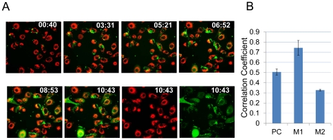
Figure 1
Live cell imaging of Rg-RSV-infected cells.
( A ). A549 cells were seeded in 35 mm glass-bottom fluorodishes (Fisher Scientific) and stained with CMXRos, live mitochondrial labeling kit (Molecular Probes) for 15 min at 37u00b0C, washed and then...
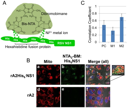
Figure 2
Co-localization of His 6 -NS1 with mitochondria in infected cells using Ni 2+ -NTA 2 -BM.
( A ) Structure of Ni 2+ -NTA 2 -BM [26] . ( B ) rA2-His 6 -NS1 or rA2 were used to infect HEp-2 cells at a MOI of 2. At 10h p.i., infected cells were labeled with CMXRos (red), DRAQ5 (blue), and 50 u...
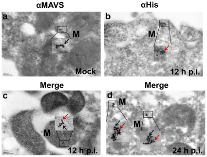
Figure 3
Colocalization of NS1 with MAVS by using immunogold electron microscopy.
A549 cells were mock-infected or infected with 0.5 MOI rA2-His 6 -NS1 for 1 h. Cells were harvested at 12 and 24 h post-infection and processed for immunogoldelectron microscopy. Thin sections on nick...
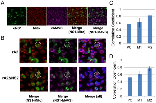
Figure 4
Co-localization of NS1 with MAVS in mitochondria during RSV infection.
A549 cells were infected with rA2 or rA2u0394NS2 (0.1 MOI).Twelve hours after infection, cells were stained with the mitochondrial stain CMXRos (red). Cells were then fixed and stained with Alexa 488-...
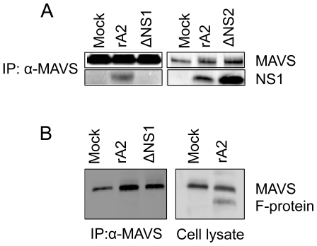
Figure 5
MAVS co-immunoprecipitates with NS1 in epithelial cells infected with rA2- RSV or rA2.
( A ) HEp-2 cells were eithermock-infectedor infected with rA2 or rA2u0394NS1or rA2u0394NS2 at an MOI of 1. At 24 h p.i., total cell extracts were immunoprecipitated with anti-MAVS followed by western...
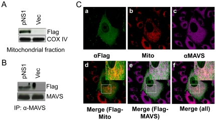
Figure 6
RSV NS1 is present on the mitochondria and co-immunoprecipitates with MAVS in NS1-transfected cells.
( A ). A549 cells were transfected with pNS1-Flag (NS1) or pFlag (Vec). Mitochondria were isolated and the proteins were analyzed by western blot for Flag and Cox-IV ( B ). Total cell extracts were im...
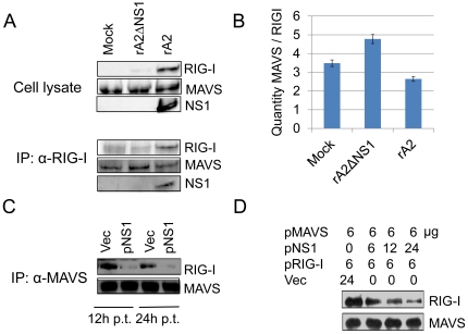
Figure 7
NS1 protein prevents RIG-I from binding to MAVS.
( A ) A549 cells were mock-infected or infected with rA2 or rA2u0394NS1 (MOIu200a=u200a1). Whole-cell lysates were subjected to western blot analysis with anti-RIG-I, anti-MAVS, and anti-NS1antibodies...
Figure images are served from the NIH/NLM PubMed Central Open Access Subset or Europe PMC; copyright remains with the publishers and authors.
💬 Discussion
0 commentsNo comments yet. Be the first to start a discussion!
Leave a Comment