Abstract
PURPOSE: To characterize the concordance/symmetry of each retinal layers in individuals without macular pathology and to further characterize the localization of inner retinal thinning in eyes receiving silicone oil-based endotamponade. METHODS: Retinal layers of one hundred eyes of 50 individuals without macular pathology were imaged using spectral domain optical coherence tomography (SD-OCT) and manually segmented using ImageJ software (developed by Wayne Rasband, NIH, Bethesda, MD, USA). In the second part of the study, retrospective analysis of 3028 cases of pars plana vitrectomy in University Eye Hospital Cologne, Germany, was conducted, retrieving nine patients with silicone oil-based endotamponade with no macular condition interfering retinal layers measurements. These patients had retinal detachment not involving the macula due to various conditions. In these patients, retinal layer segmentation was performed and compared with the fellow eye. RESULTS: There is a moderate-to-high concordance for all retinal layers between the right and the left eye of the same individual. In eyes receiving silicone oil-based endotamponade, the inner retinal layers become subsequently thinner. Ganglion cell and inner plexiform layers contribute most to this thinning, that is, 0.537 ± 0.096 mm(3) compared with 0.742 ± 0.117 mm(3) ; p = 0.006. Outer retinal layers were not affected by silicone oil-based endotamponade (p = 0.439 for the differences of calculated outer retinal layers). CONCLUSION: Ganglion cell and inner retinal layers become subsequently thinner after the use of silicone oil-based endotamponade. This study advocates the use of spectral domain optical coherence tomography for patient management with silicone oil endotamponade to early detect subsequent retinal thinning.
🔬 Techniques
🧪 Sample Preparation
🧪 Reagent Suppliers
💻 Software Details
🏛️ Research Organizations (ROR)
Affiliated research institutions:
📋 Methods
Retinal layers of one hundred eyes of 50 individuals without macular pathology were imaged using spectral domain optical coherence tomography (SD-OCT) and manually segmented using ImageJ software (developed by Wayne Rasband, NIH, Bethesda, MD, USA). In the second part of the study, retrospective analysis of 3028 cases of pars plana vitrectomy in University Eye Hospital Cologne, Germany, was conducted, retrieving nine patients with silicone oil-based endotamponade with no macular condition interfering retinal layers measurements. These patients had retinal detachment not involving the macula due to various conditions. In these patients, retinal layer segmentation was performed and compared with the fellow eye.
Patients and Methods
Retinal layers concordance in eyes without macular pathologies We chose 100 eyes of 50 random subjects over 60 years of age with no macular pathologies (epiretinal gliosis, glaucoma, age-related macular degenerations, macular oedema, etc.) and with best-corrected visual acuity better than 20/25 from the European Genetic Database (EuGenDa). It is a German–Dutch genetic database studying age-related macular degeneration. Subjects without macular pathologies were included as control patients in the database. The patients chosen for this study are part of a previous project, studying the variability of central retinal thickness (Caramoy et al. 2012 ). The main inclusion criteria were no macular pathologies as assessed using SD-OCT and visual acuity better than 20/25. Main exclusion criterion was insufficient SD-OCT image quality that might interfere retinal layer segmentation. Demographic characteristics of these patients are shown in Table 1 . All eyes examined were phakic. Patients selected for this study were the first 50 patients recruited in the year 2009. The research adhered to the tenets of the Declaration of Helsinki. Table 1 Demographic characteristics of subjects without macular pathologies evaluated in this study Variables Value (mean ± standard deviation) Age (years) 67.34 ± 5.04 (range 60–78) Gender 21 men, 29 women Medical history * n (%) Arterial hypertension 22 (45.8) Type 2 diabetes mellitus 7 (14) Conditions after stroke 3 (6) Coronary heart disease, myocardial infarction, stents or bypass surgery 2 (4) Asthma or other allergies 18 (36) Hyper- or hypothyreosis 7 (14) Oesophageal reflux 1 (2) Migraine 3 (6) Psoriasis 1 (2) Rheumatoid arthritis 2 (4) Hypercholesterinaemia 1 (2) Kidney stone 1 (2) Other malignancies 3 (6) Other surgeries 4 (8) Right eye Left eye Spherical refractive error (dioptre) 0.80 ± 2.09 (range −5.0 to +6.0) 0.80 ± 2.30 (range −6.5 to +5.5) BCVA (LogMAR) 0.02 ± 0.05 (range −0.1 to 0.1) 0.03 ± 0.06 (range −0.1 to 0.1) * Multiple diagnosis for one subject possible. Eyes with silicone oil-based endotamponade A retrospective database search of patients receiving silicone oil-based endotamponade was conducted in University Eye Hospital of Cologne, Germany, from the year 2008 (the first use of SD-OCT in our clinic) to 2011. Main inclusion criteria were patients receiving silicone oil-based endotamponade in one eye with subsequent SD-OCT imaging on both eyes. All eyes receiving silicone oil tamponade due to various diseases were included (Table 2 ). These eyes had retinal detachment not involving the macula. Main exclusion criterion was other condition on the retina affecting retinal layers measurement, for example macular pucker, retinal detachment involving the macula or internal limiting membrane peeling. Patients included in this study were called for a follow-up examinations using SD-OCT protocol described below. SD-OCT measurements were performed after silicone oil removal. Table 2 Demographic of patients receiving silicone oil-based endotamponade Case no. Sex/Age Preop BCVA (LogMAR) Clock hours detached Indication for endotamponade Endotamponade Tamponade duration (days) Final BCVA (LogMAR) 1 F/55 0.7 4 Recurrent retinal detachment 5000 cSt SiO 93 0.6 2 M/42 2.0 2 IOFB 5000 cSt SiO 211 1.3 3 M/57 0 5 Giant retinal tears 5000 cSt SiO 311 1.0 4 F/19 0 3 IOFB 5000 cSt SiO 127 0.5 5 M/60 0.2 6 PVR HSO 65 0.4 6 M/54 0.4 6 Giant retinal tears 2000 cSt SiO 74 0.2 7 F/54 0.4 4 Giant retinal tears 2000 cSt SiO 99 0.5 8 F/40 0 6 PVR 2000 cSt SiO 125 0.4 9 M/62 1.5 2 Multiple tears and vitreous haemorrhage 2000 cSt SiO 106 0.7 F = female, M = male, BCVA = best-corrected visual acuity, LogMAR = logarithm of the minimum angle of resolution, cSt = centistokes, SiO = silicone oil, HSO = heavy silicone oil, IOFB = intraocular foreign body, PVR = proliferative vitreoretinopathy. Spectral domain optical coherence tomography and measurements using ImageJ Spectral domain optical coherence tomography was performed using the Spectralis HRA + OCT (Heidelberg Engineering, Heidelberg, Germany). A standard protocol comprising a scan of 37 B-scans in 20°×15° field was used. Each B-scan consisted of 40 000 A-scans. For the measurement of retinal layers, the segmentation misalignment of the Heidelberg Eye Explorer software (version 1.7.1.0, Heidelberg Engineering, Heidelberg, Germany) was corrected manually. A clinically significant macular edema (CSME) grid (Campbell et al. 2007 ) was placed on the macula and manually centred on the fovea. This was performed to neutralize the effect of possible eccentric fixation and/or measurements. The 37 images from the 37 B-scans were downloaded. Using the ImageJ software, these images were configurated into stacks. The volume of each retinal layers was calculated using the formula: where m is the first image and n is the last image showing the marking line in the image stack. The pixel area is represented by p. x and z represent the scaling of the SD-OCT images in the x - and z -axis. b represents the distance between B-scans. The values of x , z and b can be seen in the ‘additional information’ button in the Heidelberg Eye Explorer software on the upper left-hand side for each SD-OCT image. The retinal layers measured in this study are the following: RNFL = retinal nerve fibre layer, GCLIPL = ganglion cell layer and inner plexiform layer, INL = inner nuclear layer, OPLONLPIS = outer plexiform layer, Henle's fibre layer, outer nuclear layer and inner part of the photoreceptor layer, POSRPEBM = posterior part of the photoreceptor layer, retinal pigment epithelium and Bruch's membrane, MACVOL = macular volume measured as total of the above mentioned retinal layers (Fig. 1 ). Figure 1 Morphology of the macula (case no. 4) of the silicone oil-filled eye (upper) shows thinning of inner retinal layers in comparison with the fellow eye (bottom). Cf. the methods section for the abbreviation used. These layers were chosen, because of their visibility. For example, using the current technology, it is not always possible to differentiate the ganglion cell layer (GCL) and the inner plexiform layer (IPL). Another example is to sum the outer plexiform layer (OPL), the Henle's fibre layer (HFL), the outer nuclear layer (ONL) and the inner part of the photoreceptor layer (PIS) into one single layer OPLONLPIS, because the thickness of HFL depends on the directionality of the SD-OCT (Lujan et al. 2011 ). To test the concurrence validity, the measured MACVOL using this ImageJ algorithm was compared with the measured MACVOL using the Heidelberg Eye Explorer for all patients. Measurement repeatability was tested using 20 eyes of 10 patients. For this purpose, all retinal layers from RNFL to POSRPEBM were measured twice and compared. Statistical analysis was performed using SPSS (IBM, version 21.0, Armonk, NY, USA) and MedCalc (Mariakerke, Belgium). Demographic characteristics of the population are described with summary statistics, including frequency and percentage for categorical data. Continuous data are presented with mean ± standard deviation. Testing for normality was carried out either using Shapiro–Wilk test for sample size
Show full methods section
Retinal layers of one hundred eyes of 50 individuals without macular pathology were imaged using spectral domain optical coherence tomography (SD-OCT) and manually segmented using ImageJ software (developed by Wayne Rasband, NIH, Bethesda, MD, USA). In the second part of the study, retrospective analysis of 3028 cases of pars plana vitrectomy in University Eye Hospital Cologne, Germany, was conducted, retrieving nine patients with silicone oil-based endotamponade with no macular condition interfering retinal layers measurements. These patients had retinal detachment not involving the macula due to various conditions. In these patients, retinal layer segmentation was performed and compared with the fellow eye.
Patients and Methods
Retinal layers concordance in eyes without macular pathologies We chose 100 eyes of 50 random subjects over 60 years of age with no macular pathologies (epiretinal gliosis, glaucoma, age-related macular degenerations, macular oedema, etc.) and with best-corrected visual acuity better than 20/25 from the European Genetic Database (EuGenDa). It is a German–Dutch genetic database studying age-related macular degeneration. Subjects without macular pathologies were included as control patients in the database. The patients chosen for this study are part of a previous project, studying the variability of central retinal thickness (Caramoy et al. 2012 ). The main inclusion criteria were no macular pathologies as assessed using SD-OCT and visual acuity better than 20/25. Main exclusion criterion was insufficient SD-OCT image quality that might interfere retinal layer segmentation. Demographic characteristics of these patients are shown in Table 1 . All eyes examined were phakic. Patients selected for this study were the first 50 patients recruited in the year 2009. The research adhered to the tenets of the Declaration of Helsinki. Table 1 Demographic characteristics of subjects without macular pathologies evaluated in this study Variables Value (mean ± standard deviation) Age (years) 67.34 ± 5.04 (range 60–78) Gender 21 men, 29 women Medical history * n (%) Arterial hypertension 22 (45.8) Type 2 diabetes mellitus 7 (14) Conditions after stroke 3 (6) Coronary heart disease, myocardial infarction, stents or bypass surgery 2 (4) Asthma or other allergies 18 (36) Hyper- or hypothyreosis 7 (14) Oesophageal reflux 1 (2) Migraine 3 (6) Psoriasis 1 (2) Rheumatoid arthritis 2 (4) Hypercholesterinaemia 1 (2) Kidney stone 1 (2) Other malignancies 3 (6) Other surgeries 4 (8) Right eye Left eye Spherical refractive error (dioptre) 0.80 ± 2.09 (range −5.0 to +6.0) 0.80 ± 2.30 (range −6.5 to +5.5) BCVA (LogMAR) 0.02 ± 0.05 (range −0.1 to 0.1) 0.03 ± 0.06 (range −0.1 to 0.1) * Multiple diagnosis for one subject possible. Eyes with silicone oil-based endotamponade A retrospective database search of patients receiving silicone oil-based endotamponade was conducted in University Eye Hospital of Cologne, Germany, from the year 2008 (the first use of SD-OCT in our clinic) to 2011. Main inclusion criteria were patients receiving silicone oil-based endotamponade in one eye with subsequent SD-OCT imaging on both eyes. All eyes receiving silicone oil tamponade due to various diseases were included (Table 2 ). These eyes had retinal detachment not involving the macula. Main exclusion criterion was other condition on the retina affecting retinal layers measurement, for example macular pucker, retinal detachment involving the macula or internal limiting membrane peeling. Patients included in this study were called for a follow-up examinations using SD-OCT protocol described below. SD-OCT measurements were performed after silicone oil removal. Table 2 Demographic of patients receiving silicone oil-based endotamponade Case no. Sex/Age Preop BCVA (LogMAR) Clock hours detached Indication for endotamponade Endotamponade Tamponade duration (days) Final BCVA (LogMAR) 1 F/55 0.7 4 Recurrent retinal detachment 5000 cSt SiO 93 0.6 2 M/42 2.0 2 IOFB 5000 cSt SiO 211 1.3 3 M/57 0 5 Giant retinal tears 5000 cSt SiO 311 1.0 4 F/19 0 3 IOFB 5000 cSt SiO 127 0.5 5 M/60 0.2 6 PVR HSO 65 0.4 6 M/54 0.4 6 Giant retinal tears 2000 cSt SiO 74 0.2 7 F/54 0.4 4 Giant retinal tears 2000 cSt SiO 99 0.5 8 F/40 0 6 PVR 2000 cSt SiO 125 0.4 9 M/62 1.5 2 Multiple tears and vitreous haemorrhage 2000 cSt SiO 106 0.7 F = female, M = male, BCVA = best-corrected visual acuity, LogMAR = logarithm of the minimum angle of resolution, cSt = centistokes, SiO = silicone oil, HSO = heavy silicone oil, IOFB = intraocular foreign body, PVR = proliferative vitreoretinopathy. Spectral domain optical coherence tomography and measurements using ImageJ Spectral domain optical coherence tomography was performed using the Spectralis HRA + OCT (Heidelberg Engineering, Heidelberg, Germany). A standard protocol comprising a scan of 37 B-scans in 20°×15° field was used. Each B-scan consisted of 40 000 A-scans. For the measurement of retinal layers, the segmentation misalignment of the Heidelberg Eye Explorer software (version 1.7.1.0, Heidelberg Engineering, Heidelberg, Germany) was corrected manually. A clinically significant macular edema (CSME) grid (Campbell et al. 2007 ) was placed on the macula and manually centred on the fovea. This was performed to neutralize the effect of possible eccentric fixation and/or measurements. The 37 images from the 37 B-scans were downloaded. Using the ImageJ software, these images were configurated into stacks. The volume of each retinal layers was calculated using the formula: where m is the first image and n is the last image showing the marking line in the image stack. The pixel area is represented by p. x and z represent the scaling of the SD-OCT images in the x - and z -axis. b represents the distance between B-scans. The values of x , z and b can be seen in the ‘additional information’ button in the Heidelberg Eye Explorer software on the upper left-hand side for each SD-OCT image. The retinal layers measured in this study are the following: RNFL = retinal nerve fibre layer, GCLIPL = ganglion cell layer and inner plexiform layer, INL = inner nuclear layer, OPLONLPIS = outer plexiform layer, Henle's fibre layer, outer nuclear layer and inner part of the photoreceptor layer, POSRPEBM = posterior part of the photoreceptor layer, retinal pigment epithelium and Bruch's membrane, MACVOL = macular volume measured as total of the above mentioned retinal layers (Fig. 1 ). Figure 1 Morphology of the macula (case no. 4) of the silicone oil-filled eye (upper) shows thinning of inner retinal layers in comparison with the fellow eye (bottom). Cf. the methods section for the abbreviation used. These layers were chosen, because of their visibility. For example, using the current technology, it is not always possible to differentiate the ganglion cell layer (GCL) and the inner plexiform layer (IPL). Another example is to sum the outer plexiform layer (OPL), the Henle's fibre layer (HFL), the outer nuclear layer (ONL) and the inner part of the photoreceptor layer (PIS) into one single layer OPLONLPIS, because the thickness of HFL depends on the directionality of the SD-OCT (Lujan et al. 2011 ). To test the concurrence validity, the measured MACVOL using this ImageJ algorithm was compared with the measured MACVOL using the Heidelberg Eye Explorer for all patients. Measurement repeatability was tested using 20 eyes of 10 patients. For this purpose, all retinal layers from RNFL to POSRPEBM were measured twice and compared. Statistical analysis was performed using SPSS (IBM, version 21.0, Armonk, NY, USA) and MedCalc (Mariakerke, Belgium). Demographic characteristics of the population are described with summary statistics, including frequency and percentage for categorical data. Continuous data are presented with mean ± standard deviation. Testing for normality was carried out either using Shapiro–Wilk test for sample size
📊 Figures
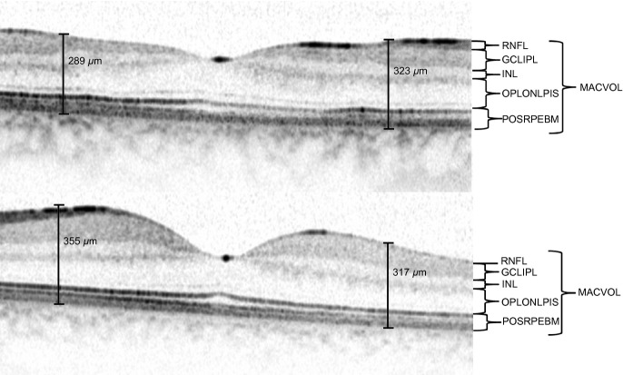
Figure 1
Morphology of the macula (case no. 4) of the silicone oil-filled eye (upper) shows thinning of inner retinal layers in comparison with the fellow eye (bottom). Cf. the methods section for the abbrevia...
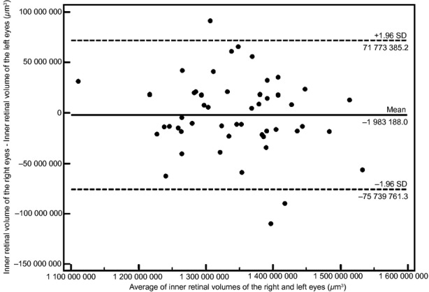
Figure 2
Bland-Altmann plot of inner retinal volume of the right and left eyes of patients without macular pathology.
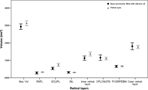
Figure 3
Retinal layer volumes in eyes previously received silicone oil tamponade ( n = 9) are reduced in comparison with the fellow eyes.
Figure images are served from the NIH/NLM PubMed Central Open Access Subset or Europe PMC; copyright remains with the publishers and authors.
💬 Discussion
0 commentsNo comments yet. Be the first to start a discussion!
Leave a Comment