Abstract
African swine fever virus (ASFV), a devastating pathogen to the worldwide swine industry, mainly targets macrophage/monocyte lineage, but how the virus enters host cells has remained unclear. Here, we report that ASFV utilizes apoptotic bodies (ApoBDs) for infection and cell-cell transmission. We show that ASFV induces cell apoptosis of primary porcine alveolar macrophages (PAMs) at the late stage of infection to productively shed ApoBDs that are subsequently swallowed by neighboring PAMs to initiate a secondary infection as evidenced by electron microscopy and live-cell imaging. Interestingly, the virions loaded within ApoBDs are exclusively single-enveloped particles that are devoid of the outer layer of membrane and represent a predominant form produced during late infection. The in vitro purified ApoBD vesicles are capable of mediating virus infection of naive PAMs, but the transmission can be significantly inhibited by blocking the "eat-me" signal phosphatidyserine on the surface of ApoBDs via Annexin V or the efferocytosis receptor TIM4 on the recipient PAMs via anti-TIM4 antibody, whereas overexpression of TIM4 enhances virus infection. The same treatment however did not affect the infection by intracellular viruses. Importantly, the swine sera to ASFV exert no effect on the ApoBD-mediated transmission but can partially act on the virions lacking the outer layer of membrane. Thus, ASFV has evolved to hijack a normal cellular pathway for cell-cell spread to evade host responses.
🔬 Techniques
🔭 Microscopes
✨ Fluorophores
🧪 Sample Preparation
🔬 Cell Lines
🏭 Microscope Brands
💻 Software Details
🏛️ Research Organizations (ROR)
Affiliated research institutions:
📋 Methods
Reagents. Primary PAMs from 1-mo-old SPF piglets and WSL cells were maintained in RPMI-1640 medium. Type II ASFV strain CADC_HN09 (GenBank accession no: MZ614662.1 ) was used a model organism in this study. The commercial antibodies and chemicals are from various sources, and the plasmids were engineered by standard recombinant DNA procedures. ApoBD Isolation. ApoBDs were purified from ASFV-infected PAMs via differential centrifugation. Briefly, the samples were centrifuged at 300 g to obtain the ApoCells-enriched fraction, whereas the supernatant containing ApoBDs was centrifuged at 3,000 g to pellet ApoBDs. Purified-ApoBD samples were validated by DIC microscopy and transmission electron microscopy. Blocking Assay. The isolated ApoBDs were incubated with Annexin V or swine sera for 1 h prior to infection of recipient PAMs. To block the function of efferocytosis receptor on recipient PAMs, the cells were incubated with anti-TIM4 antibody prior to incubation with ApoBDs. The unbound viruses or ApoBDs were washed off with serum-free RPMI-1640, and the cells were cultured in maintenance medium containing 2% FBS. ASFV replication was measured at 12 or 24 hpi by western analysis and IFA. Annexin V Conjugates for Apoptosis Detection. PAMs were washed with annexin-binding buffer and incubated with the Alexa Fluor @ 568 conjugated Annexin V for 15 min before being washed three times and observed with a fluorescence microscope. Construction of Recombinant ASFV. The GFP was inserted to replace the loci MGF360-18R under the control of p72 promoter. The recombinant virus ASFV-GFP was generated via homologous recombination, followed by successive rounds of plaque assay in PAMs, and confirmed by PCR. Live-Cell Imaging. PAMs on coverslip-bottomed dishes were infected with ASFV-GFP or purified ApoBDs and imaged for time-lapse and DIC microscopy with a Nikon A1 confocal microscope. Transmission Electron Microscopy. PAMs or isolated ApoBDs were fixed with 0.1 M phosphate buffer containing 4% paraformaldehyde and 2% glutaraldehyde. The pellet was then enrobed in low melting point agarose and post-fixed in 1% osmium tetroxide in cacodylate buffer and en bloc stained with 1% uranyl acetate. Following dehydration with acetone, it was embedded in epoxy (TAAB 812 resin). After polymerization, 80-nm-thick (ultrathin) sections were obtained and stained with uranyl acetate and lead citrate. Imaging was performed in a HITACHI HT7700 electron microscope. Statistical Analysis. Statistical significance was analyzed by two-tailed unpaired Student’s t test. Significance symbols are defined as follows: NS, no significance; * P < 0.05; ** P < 0.01; *** P < 0.001. Error bars indicate means ± SD. Detailed descriptions are provided in SI Appendix , Materials and Methods .
Show full methods section
Reagents. Primary PAMs from 1-mo-old SPF piglets and WSL cells were maintained in RPMI-1640 medium. Type II ASFV strain CADC_HN09 (GenBank accession no: MZ614662.1 ) was used a model organism in this study. The commercial antibodies and chemicals are from various sources, and the plasmids were engineered by standard recombinant DNA procedures. ApoBD Isolation. ApoBDs were purified from ASFV-infected PAMs via differential centrifugation. Briefly, the samples were centrifuged at 300 g to obtain the ApoCells-enriched fraction, whereas the supernatant containing ApoBDs was centrifuged at 3,000 g to pellet ApoBDs. Purified-ApoBD samples were validated by DIC microscopy and transmission electron microscopy. Blocking Assay. The isolated ApoBDs were incubated with Annexin V or swine sera for 1 h prior to infection of recipient PAMs. To block the function of efferocytosis receptor on recipient PAMs, the cells were incubated with anti-TIM4 antibody prior to incubation with ApoBDs. The unbound viruses or ApoBDs were washed off with serum-free RPMI-1640, and the cells were cultured in maintenance medium containing 2% FBS. ASFV replication was measured at 12 or 24 hpi by western analysis and IFA. Annexin V Conjugates for Apoptosis Detection. PAMs were washed with annexin-binding buffer and incubated with the Alexa Fluor @ 568 conjugated Annexin V for 15 min before being washed three times and observed with a fluorescence microscope. Construction of Recombinant ASFV. The GFP was inserted to replace the loci MGF360-18R under the control of p72 promoter. The recombinant virus ASFV-GFP was generated via homologous recombination, followed by successive rounds of plaque assay in PAMs, and confirmed by PCR. Live-Cell Imaging. PAMs on coverslip-bottomed dishes were infected with ASFV-GFP or purified ApoBDs and imaged for time-lapse and DIC microscopy with a Nikon A1 confocal microscope. Transmission Electron Microscopy. PAMs or isolated ApoBDs were fixed with 0.1 M phosphate buffer containing 4% paraformaldehyde and 2% glutaraldehyde. The pellet was then enrobed in low melting point agarose and post-fixed in 1% osmium tetroxide in cacodylate buffer and en bloc stained with 1% uranyl acetate. Following dehydration with acetone, it was embedded in epoxy (TAAB 812 resin). After polymerization, 80-nm-thick (ultrathin) sections were obtained and stained with uranyl acetate and lead citrate. Imaging was performed in a HITACHI HT7700 electron microscope. Statistical Analysis. Statistical significance was analyzed by two-tailed unpaired Student’s t test. Significance symbols are defined as follows: NS, no significance; * P < 0.05; ** P < 0.01; *** P < 0.001. Error bars indicate means ± SD. Detailed descriptions are provided in SI Appendix , Materials and Methods .
Supplementary Material Appendix 01 (PDF) Click here for additional data file. Movie S1. Live-cell-imaging of the dynamics of ApoBDs formation in ASFV-GFP-infected PAMs. PAMs were seeded into 35 mm dishes and infected with ASFV-GFP at an MOI of 0.01. At 36 hpi, cells were imaged for time-lapse GFP and DIC microscopy with a Nikon A1 confocal microscope. Oil objective: 100 X; zoom in 1 X. Movie S2.
Live-cell-imaging of the infection dynamics of ASFV-GFP-infected
PAMs and GFP-positive ApoBDs. PAMs were seeded into 35 mm dishes and infected with ASFVGFP at an MOI of 0.01. At 36 hpi, cells were imaged for time-lapse GFP and DIC microscopy with a Nikon A1 confocal microscope. Oil objective: 100 X; zoom in 1 X. Movie S3.
Live-cell-imaging of the infection dynamics of the purified
ApoBDs from ASFVGFP-infected PAMs. PAMs were seeded into 35 mm dishes and infected with ApoBDs from ASFV-GFP-infected PAMs. At 0 hpi, cells were imaged for time-lapse GFP and DIC microscopy with a Nikon A1 confocal microscope. Oil objective: 100 X; zoom in 1 X. Movie S4.
Live-cell-imaging of the infection dynamics of A5-alexa568-labeled
ApoBDs that were from ASFV-GFP-infected PAMs. PAMs in 35 mm dishes were infected with A5-alexa-labled ApoBDs, and the cells were imaged from 0 hpi for time-lapse GFP and DIC microscopy with a Nikon A1 confocal microscope. Oil objective: 100 X; zoom in 1 X.
Data, Materials, and Software Availability All study data are included in the article and/or supporting information .
📊 Figures
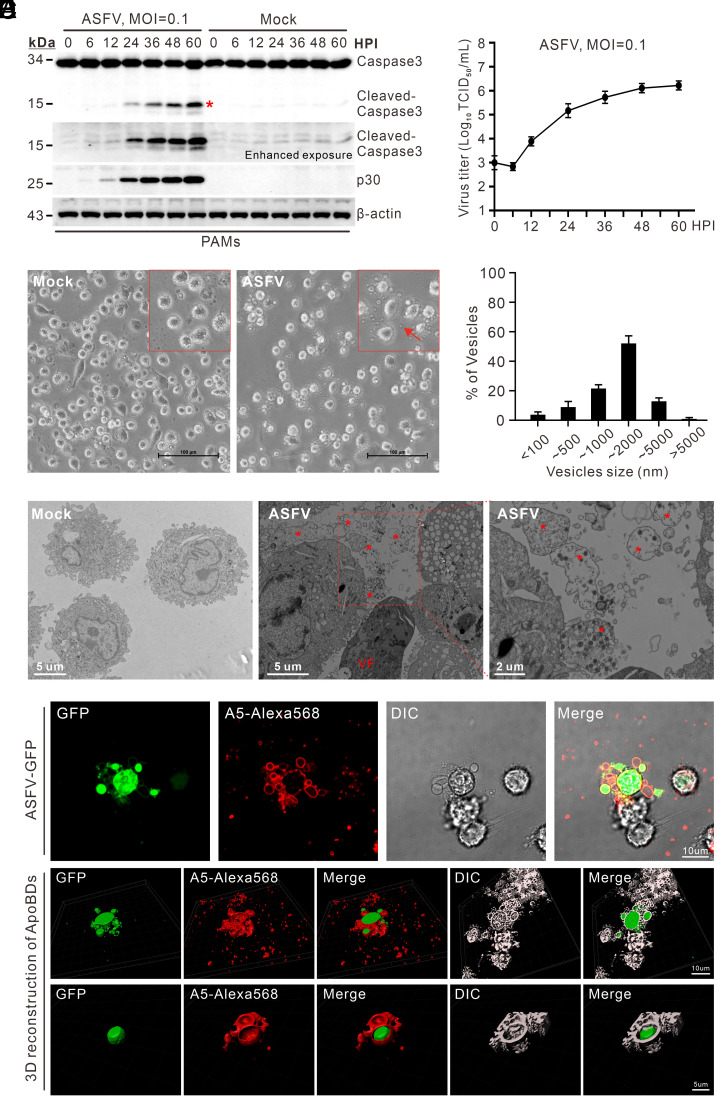
Fig. 1.
ASFV induces apoptosis of PAMs to shed ApoBDs. ( A ) Analysis of cellular apoptosis of ASFV-infected cells. PAMs were mock-infected or infected with ASFV strain HN09 at an MOI of 0.1 and harvested at ...
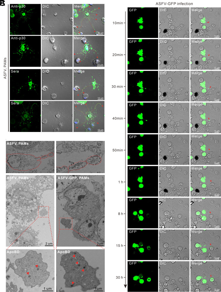
Fig. 2.
ApoBDs derived from ASFV-infected PAMs can be swallowed by neighboring PAMs to establish a secondary infection. ( A ) Distribution pattern of the ASFV-positive ApoBDs. PAMs on coverslips in six-well p...
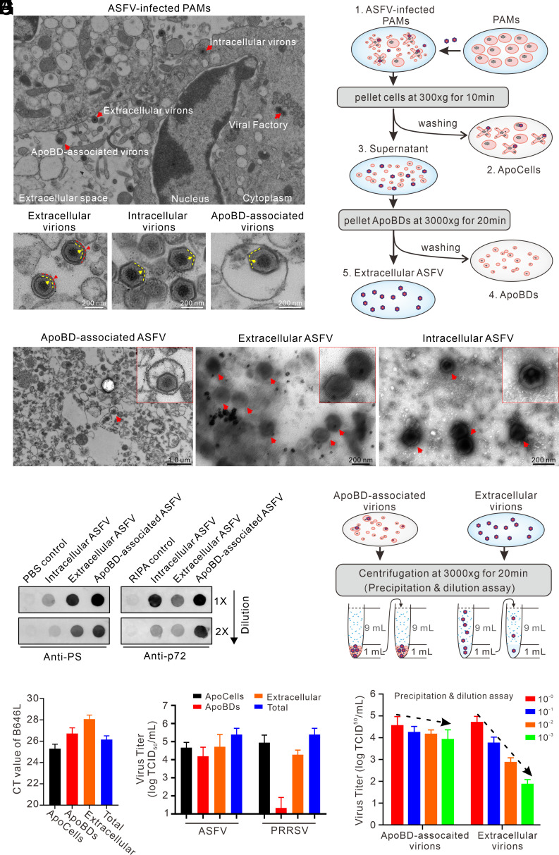
Fig. 3.
Purified ApoBDs from ASFV-infected PAMs contain infectious virions. ( A ) Presence of different forms of virions in ASFV-infected PAMs. ( B ) Schematic diagram of the procedure for purifying ApoBDs vi...
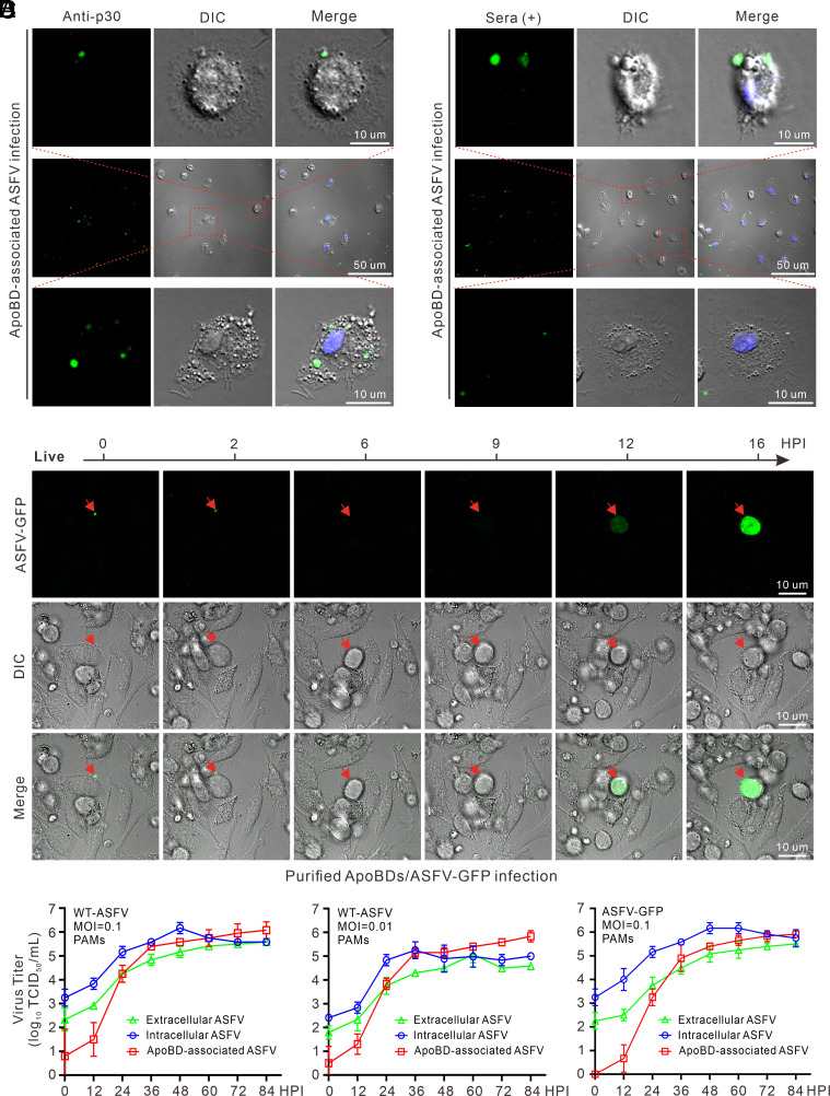
Fig. 4.
The purified ASFV-containing ApoBDs can transmit ASFV and establish a productive infection. PAMs grown on coverslips in six-well plates were incubated with the purified ApoBDs from WT ASFV-infected PA...
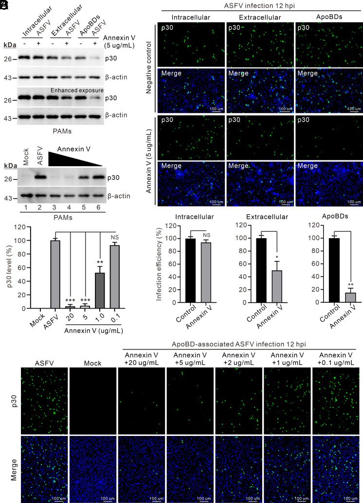
Fig. 5.
Blocking PS lipids suppresses ApoBD-mediated ASFV infection. Different forms of ASFV were isolated at 48 hpi from PAMs infected with ASFV strain HN09 at an MOI of 0.1 and then incubated, respectively,...
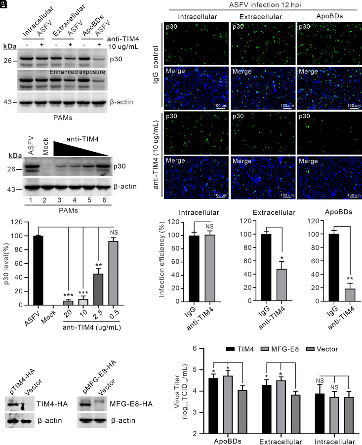
Fig. 6.
ApoBD-mediated ASFV infection is dependent on the efferocytosis receptor TIM4. ( A ) PAMs in 24-well plates were incubated with antibodies to TIM4 (10 u00b5g/mL) or isotype IgG for 1 h and then expose...
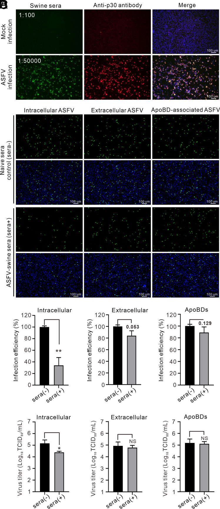
Fig. 7.
ApoBD-mediated viral transmission is fully resistant to swine sera to ASFV. ( A ) IFA analysis of the titer of swine anti-ASFV serum in PAMs with the antibody to p30 as a control. ( B u2013 D ) Intrac...
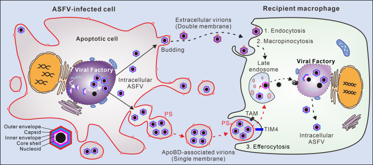
Fig. 8.
A proposed model for ASFV transmission. ASFV induces cell apoptosis to shed the virion-loaded ApoBDs at late stage of infection via caspase-3 activation. The PS-positive ApoBDs carrying single-membran...
Figure images are served from the NIH/NLM PubMed Central Open Access Subset or Europe PMC; copyright remains with the publishers and authors.
💬 Discussion
0 commentsNo comments yet. Be the first to start a discussion!
Leave a Comment