Abstract
Phosphatidylserine, a phospholipid with a negatively charged head-group, is an important constituent of eukaryotic cellular membranes. On the plasma membrane, rather than being evenly distributed, phosphatidylserine is found preferentially in the inner leaflet. Disruption of this asymmetry, leading to the appearance of phosphatidylserine on the surface of the cell, is known to play a central role in both apoptosis and blood clotting. Despite its importance, comparatively little is known about phosphatidylserine in cells: its precise subcellular localization, transmembrane topology and intracellular dynamics are poorly characterized. The recent development of new, genetically-encoded probes able to detect phosphatidylserine within live cells, however, is leading to a more in-depth understanding of the biology of this phospholipid. This review aims to give an overview of the current methods for phosphatidylserine detection within cells, and some of the recent realizations derived from their use.
🔬 Techniques
✨ Fluorophores
🧪 Sample Preparation
🔬 Cell Lines
🏛️ Research Organizations (ROR)
Affiliated research institutions:
📋 Methods
Used to Date for Phosphatidylserine Detection and Characterization 2.1.
PS-Binding Probes
One of the first methods used to detect the presence of PS involved the chemical 2,4,6-trinitrobenzenesulfonate (TNBS), which covalently reacts with amines; in biological membranes it therefore preferentially labels the aminophospholipids, phosphatidylethanolamine (PE) and PS. Because TNBS is membrane-impermeant and since the labelled lipids can be distinguished from the non-labelled PE and PS by thin layer chromatography (TLC) or mass spectrometry, TNBS has been used to assess the transmembrane topology of the aminophospholipids. Indeed, this is the approach used to establish that PS is present almost exclusively in the cytoplasmic leaflet of the plasma membrane of most mammalian cells [ 10 , 11 ]. TNBS has also been used to determine the sidedness of PS in intracellular organelles, such as mitochondria and the sarcoplasmic reticulum [ 12 , 13 ]. Despite its selectivity for aminophospholipids, TNBS is of limited use in live cells. It fails to react with PS in unperturbed, healthy cells and, even when PS is exposed during stimulation or apoptosis, long incubation times (15–60 minutes) are required for the reaction to become manifest, preventing meaningful dynamic studies on a timescale faster than hourly. Even in the case of fractionated cells, the probe is far from perfect: homogenization procedures and separation of organelles prior to labelling can potentially alter lipid sidedness and may breach the permeability barrier on which the sidedness determinations rely [ 14 ]. The use of antibodies to specifically recognize PS has also been considered. Such antibodies have been used successfully to detect PS on the outer leaflet of the membrane of apoptotic cells or activated platelets [ 15 ]. However, while anti-PS antibodies can be specific, there is also a significant risk of cross-reactivity with other phospholipids [ 16 ], so batch-specific characterization must be undertaken. More importantly, to immunostain PS in organellar membranes the cells must be fixed and permeabilized, in order to grant the antibodies access to the cell interior. Because permeabilization involves treatment with detergents or organic solvents that disrupt lipid architecture, it is not suitable for reliable PS detection. Additionally, the need for fixation precludes dynamic PS studies. While microinjection can in principle be used to deliver antibodies into live cells, the inability to wash the excess (unbound) antibodies prevents definition of the fraction bound to PS. Currently, the most commonly used probe for PS detection is annexin A5, a member of the annexin family of Ca 2+ -dependent, non-covalent lipid-binding proteins. Annexin A5 binds all negatively charged lipids with relatively high affinity; however as PS is by far the most abundant negatively charged lipid (particularly on the external surface of apoptotic cells), annexin binding is generally associated with PS. Annexin A5 is readily available for purchase as a fluorescently-tagged protein, and is used extensively for detection of exofacial PS by flow cytometry or microscopy. As in the case of antibodies, delivery of exogenous annexin to the cell interior is limited by the need for fixation and permeabilization. However, because the sequence of annexin is known, it is in principle possible to generate an annexin A5-GFP (green fluorescent protein) chimeric construct [ 17 ] for endogenous expression in transfected cells. Unfortunately, annexin is capable of binding PS only when free calcium is comparatively high: concentrations >10 −5 M are required for proper recognition of PS [ 18 ]. Such concentrations are at least two orders of magnitude higher than the prevailing cytosolic [Ca 2+ ] [ 19 ]. While it is possible to manipulate intracellular [Ca 2+ ] by means such as the application of ionophores, the physiological consequences of such manipulations are deleterious and can alter the native PS distribution. Indeed, the concentrations of [Ca 2+ ] required to facilitate annexin binding are known to stimulate scramblases that disrupt lipid asymmetry [ 20 ]. These considerations rule out the use of annexin A5 as a reliable probe for detection of internal PS in intact live cells. 2.2.
Show full methods section
Used to Date for Phosphatidylserine Detection and Characterization 2.1.
PS-Binding Probes
One of the first methods used to detect the presence of PS involved the chemical 2,4,6-trinitrobenzenesulfonate (TNBS), which covalently reacts with amines; in biological membranes it therefore preferentially labels the aminophospholipids, phosphatidylethanolamine (PE) and PS. Because TNBS is membrane-impermeant and since the labelled lipids can be distinguished from the non-labelled PE and PS by thin layer chromatography (TLC) or mass spectrometry, TNBS has been used to assess the transmembrane topology of the aminophospholipids. Indeed, this is the approach used to establish that PS is present almost exclusively in the cytoplasmic leaflet of the plasma membrane of most mammalian cells [ 10 , 11 ]. TNBS has also been used to determine the sidedness of PS in intracellular organelles, such as mitochondria and the sarcoplasmic reticulum [ 12 , 13 ]. Despite its selectivity for aminophospholipids, TNBS is of limited use in live cells. It fails to react with PS in unperturbed, healthy cells and, even when PS is exposed during stimulation or apoptosis, long incubation times (15–60 minutes) are required for the reaction to become manifest, preventing meaningful dynamic studies on a timescale faster than hourly. Even in the case of fractionated cells, the probe is far from perfect: homogenization procedures and separation of organelles prior to labelling can potentially alter lipid sidedness and may breach the permeability barrier on which the sidedness determinations rely [ 14 ]. The use of antibodies to specifically recognize PS has also been considered. Such antibodies have been used successfully to detect PS on the outer leaflet of the membrane of apoptotic cells or activated platelets [ 15 ]. However, while anti-PS antibodies can be specific, there is also a significant risk of cross-reactivity with other phospholipids [ 16 ], so batch-specific characterization must be undertaken. More importantly, to immunostain PS in organellar membranes the cells must be fixed and permeabilized, in order to grant the antibodies access to the cell interior. Because permeabilization involves treatment with detergents or organic solvents that disrupt lipid architecture, it is not suitable for reliable PS detection. Additionally, the need for fixation precludes dynamic PS studies. While microinjection can in principle be used to deliver antibodies into live cells, the inability to wash the excess (unbound) antibodies prevents definition of the fraction bound to PS. Currently, the most commonly used probe for PS detection is annexin A5, a member of the annexin family of Ca 2+ -dependent, non-covalent lipid-binding proteins. Annexin A5 binds all negatively charged lipids with relatively high affinity; however as PS is by far the most abundant negatively charged lipid (particularly on the external surface of apoptotic cells), annexin binding is generally associated with PS. Annexin A5 is readily available for purchase as a fluorescently-tagged protein, and is used extensively for detection of exofacial PS by flow cytometry or microscopy. As in the case of antibodies, delivery of exogenous annexin to the cell interior is limited by the need for fixation and permeabilization. However, because the sequence of annexin is known, it is in principle possible to generate an annexin A5-GFP (green fluorescent protein) chimeric construct [ 17 ] for endogenous expression in transfected cells. Unfortunately, annexin is capable of binding PS only when free calcium is comparatively high: concentrations >10 −5 M are required for proper recognition of PS [ 18 ]. Such concentrations are at least two orders of magnitude higher than the prevailing cytosolic [Ca 2+ ] [ 19 ]. While it is possible to manipulate intracellular [Ca 2+ ] by means such as the application of ionophores, the physiological consequences of such manipulations are deleterious and can alter the native PS distribution. Indeed, the concentrations of [Ca 2+ ] required to facilitate annexin binding are known to stimulate scramblases that disrupt lipid asymmetry [ 20 ]. These considerations rule out the use of annexin A5 as a reliable probe for detection of internal PS in intact live cells. 2.2.
Fluorescent Analogues Another method commonly used to examine
PS within the cellular environment is the use of fluorescently-labelled PS analogues. 7-nitro-2-1,3-benzoxadiazol-4-yl (NBD) is an aromatic fluorescent compound that, when attached to the end of a shortened (usually 6 or 12 carbon) sn -2 acyl chain, can be used to monitor the distribution and dynamics of PS without altering the unique structure of the PS head group, or having to contend with the promiscuous binding of the probes mentioned above. NDB-PS analogues are also easy to use, as they are readily loaded into cellular membranes, either by liposome transfer or using albumin as a carrier. NBD-PS has been useful in determining specific features of the physiology of PS, such as the process whereby it is translocated (flipped) across the plasma membrane, and has aided in the identification of the responsible proteins [ 21 – 23 ]. On the other hand, there are a number of undesirable features that preclude the use of NBD-PS for long-term cellular imaging and thorough characterization of PS dynamics. Firstly, NBD photobleaches extremely rapidly, thus severely limiting the imaging period [ 24 ]. Additionally, the strong dipole moment of the NBD moiety forces the acyl chain to which it is attached to loop back towards the aqueous interphase [ 25 – 27 ]. The resulting distortion of the labelled PS-like molecule alters the packing of the endogenous annular lipids, modifying the behaviour of the bilayer. This likely explains why NBD-PS is readily removed from membranes by albumin, while native PS and other natural lipids are refractory to extraction. How well NBD-PS mimics the behaviour of endogenous PS is therefore contentious. Its altered behaviour may explain why studies of intracellular trafficking using NBD-PS have been inconclusive, suggesting the existence of cellular transport-independent processes [ 22 ]. Another fluorophore, BODIPY, can also be used to label lipids without disruption of the chemical nature of the head group. The greater photostability and hydrophobicity of BODIPY hold promise for the study of PS though, to our knowledge, the use of acyl-labelled BODIPY derivatives of PS has not been reported to date.
📊 Figures
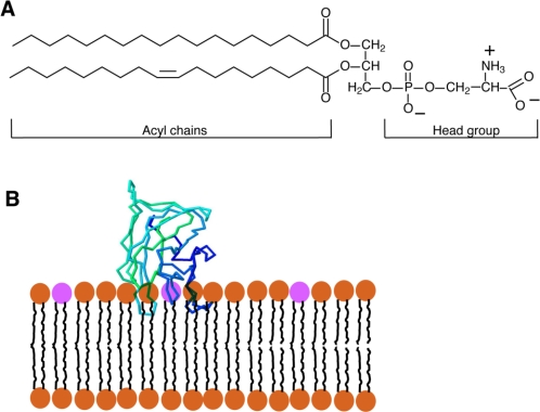
Figure 1.
(A) Diagram of the structure of a prototypical phosphatidylserine (PS), a glycerophospholipid, with saturated and unsaturated fatty acyl chains. Note that at physiological pH the head-group bears one ...
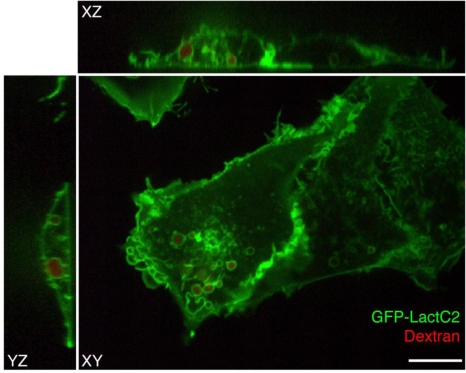
Figure 2.
Distribution of GFP-LactC2, a PS-specific probe, in mammalian cells. COS-7 cells expressing GFP-LactC2 were pulsed with rhodamine-conjugated dextran followed by a 15 minute chase and were imaged live ...
Figure images are served from the NIH/NLM PubMed Central Open Access Subset or Europe PMC; copyright remains with the publishers and authors.
💬 Discussion
0 commentsNo comments yet. Be the first to start a discussion!
Leave a Comment