Abstract
Squamous cell carcinoma (SCC) accounts for most nonmelanoma skin cancer-related metastatic disease and deaths. Histopathology and correct surgical excision remain the gold standard for the diagnosis and treatment of SCC; however, new diagnostic imaging techniques such as dermoscopy and reflectance confocal microscopy have increased the diagnostic accuracy in terms of early recognition, better differential diagnosis, more precise selection of areas to biopsy, and noninvasive monitoring of treatments. The therapeutic intervention in patients with severe actinic damage and multiple in situ/low-risk SCC, and the development of innovative treatments such as epidermal growth factor receptor inhibitors and immune checkpoint inhibitors for locally advanced and metastatic SCC, are improving considerably the approach to the disease. This review summarizes the up-to-date knowledge in the field of detection, treatment, and monitoring of cutaneous SCC.
🔬 Techniques
🧪 Sample Preparation
🔬 Cell Lines
🏛️ Research Organizations (ROR)
Affiliated research institutions:
📊 Figures
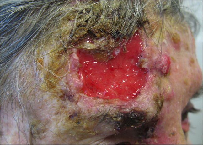
Figure 1
Advanced and recurrent squamous cell carcinoma with nodal involvement in an elderly woman with dementia and chronic myeloid leukemia. Large ulcerative and keratotic tumor with numerous actinic keratos...
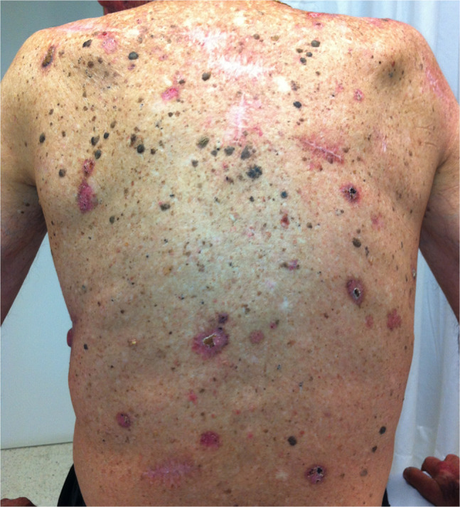
Figure 2
Multiple skin cancers, mainly squamous cell carcinomas, in a solid organ transplant patient.
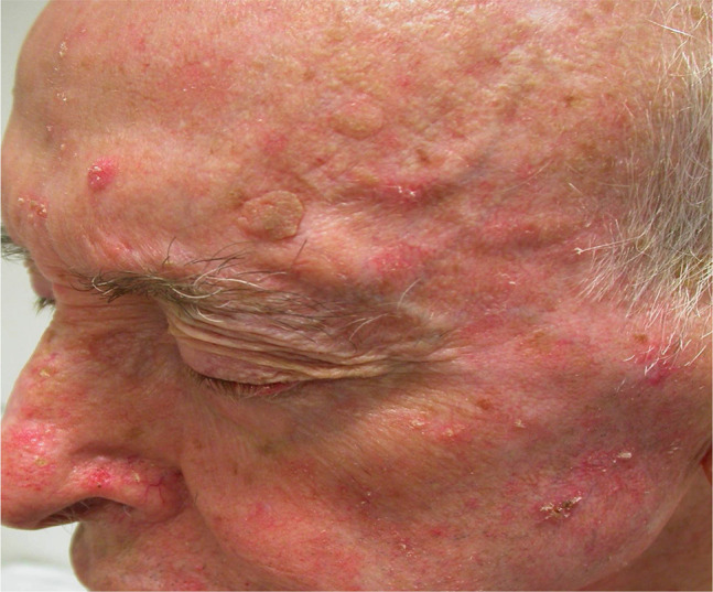
Figure 3
Example of cancerization field. Multiple actinic keratoses in a patient with marked sun-damaged skin.
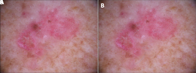
Figure 4
(A) Clinical image of Bowen disease. (B) Dermoscopic image of Bowen disease where clusters of glomerular and dotted vessels and whitish scales can be identified.
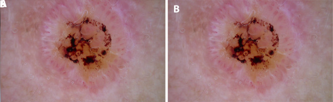
Figure 5
(A) Clinical image of a well-differentiated squamous cell carcinoma (keratoacanthoma-like) on the nose of a patient with sun-damaged skin. (B) Dermoscopy shows a symmetric lesion with a central ulcera...
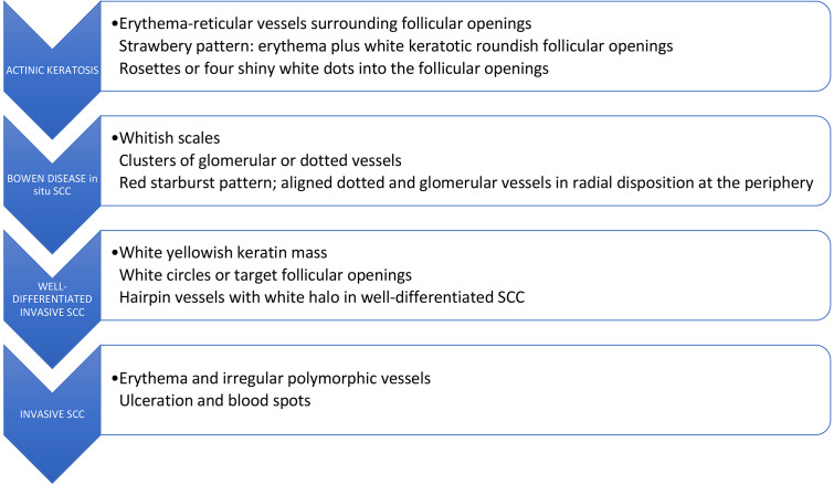
Figure 6
Dermoscopic features found in the progression model of actinic keratosis into squamous cell carcinoma (SCC) in situ and invasive based on the article of Zalaudek et al [ 6 ].
Figure images are served from the NIH/NLM PubMed Central Open Access Subset or Europe PMC; copyright remains with the publishers and authors.
💬 Discussion
0 commentsNo comments yet. Be the first to start a discussion!
Leave a Comment