Abstract
Glutamate receptors are ligand-gated tetrameric ion channels that mediate synaptic transmission in the central nervous system. They are instrumental in vertebrate cognition and their dysfunction underlies diverse diseases. In both the resting and desensitized states of AMPA and kainate receptor subtypes, the ion channels are closed, whereas the ligand-binding domains, which are physically coupled to the channels, adopt markedly different conformations. Without an atomic model for the desensitized state, it is not possible to address a central problem in receptor gating: how the resting and desensitized receptor states both display closed ion channels, although they have major differences in the quaternary structure of the ligand-binding domain. Here, by determining the structure of the kainate receptor GluK2 subtype in its desensitized state by cryo-electron microscopy (cryo-EM) at 3.8 Å resolution, we show that desensitization is characterized by the establishment of a ring-like structure in the ligand-binding domain layer of the receptor. Formation of this 'desensitization ring' is mediated by staggered helix contacts between adjacent subunits, which leads to a pseudo-four-fold symmetric arrangement of the ligand-binding domains, illustrating subtle changes in symmetry that are important for the gating mechanism. Disruption of the desensitization ring is probably the key switch that enables restoration of the receptor to its resting state, thereby completing the gating cycle.
🔬 Techniques
🧬 Organisms
✨ Fluorophores
🧪 Sample Preparation
🔬 Cell Lines
🏭 Microscope Brands
🧪 Reagent Suppliers
💻 Software Details
💾 Data Repositories
🏛️ Research Organizations (ROR)
Affiliated research institutions:
📋 Methods
Protein expression and purification
The full-length rat GluK2 subunit cDNA sequence (UniProt ID: P42260 ) was cloned into the pFastBac1 vector for protein expression in insect cells. GluK2 EM was created by introducing four mutations in the LBD (A487T, A658S, N690S and F704L) which convert the sequence to that found in GluK1, creating a high affinity binding site for the GluK1 selective antagonist LY466195 16 . The construct was RNA edited at position 536 (I to V) and had two mutations (C545V (M1) and C564S (M1-M2 loop) which increased yield and tetramer stability. For fluorescence detection and affinity purification, a thrombin recognition site and linker sequence (GLVPRGSAAAA) was inserted between GluK2 EM and the coding sequence for the A207K dimerization suppressed EGFP mutant, with a C-terminal SGLRHis8 affinity tag. Membranes isolated from Sf9 cells were solubilized and purified as described previously using n-dodecyl-β-D-maltopyranoside (DDM) and cholesterol hemisuccinate (CHS) 5 . The GluK2 EM S1S2 LBD construct encoding residues S398-K513 and P636-E775 connected by a GT dipeptide linker was expressed in Escherichia coli and purified as reported previously for wild type GluK2 26 .
Specimen preparation and cryo-electron microscopy
Vitrified samples of GluK2 solublized in DDM-CHS were prepared with protein concentrated to 4.2 mg/mL. A volume of 3 μL was added to R1.2/1.3 holey carbon grids (Quantifoil) rendered hydrophilic with self-assembled monolayer functionalization 27 , and grids were frozen with a Vitrobot Mk IV robot (FEI Company, Hillsboro, OR, USA) or a Leica EM GP (Leica Microsystems Inc., Buffalo Grove, IL, USA). Data was collected using a Titan Krios, operated at 300 kV, aligned for parallel illumination, and equipped with a GIF Quantum Energy Filter (Gatan, Inc.) operated in zero-energy-loss mode with a slit width of 20 eV ( Extended Data Table 2 ). Images were acquired manually on a K2 Summit camera (Gatan, Inc.), at 105,000 X nominal magnification corresponding to a 1.324 Å physical pixel size. Each exposure was recorded in super-resolution mode as a 38-frame movie, with dose rate and exposure time of 3 e Å −1 s −1 and 15 s, respectively.
Show full methods section
Protein expression and purification
The full-length rat GluK2 subunit cDNA sequence (UniProt ID: P42260 ) was cloned into the pFastBac1 vector for protein expression in insect cells. GluK2 EM was created by introducing four mutations in the LBD (A487T, A658S, N690S and F704L) which convert the sequence to that found in GluK1, creating a high affinity binding site for the GluK1 selective antagonist LY466195 16 . The construct was RNA edited at position 536 (I to V) and had two mutations (C545V (M1) and C564S (M1-M2 loop) which increased yield and tetramer stability. For fluorescence detection and affinity purification, a thrombin recognition site and linker sequence (GLVPRGSAAAA) was inserted between GluK2 EM and the coding sequence for the A207K dimerization suppressed EGFP mutant, with a C-terminal SGLRHis8 affinity tag. Membranes isolated from Sf9 cells were solubilized and purified as described previously using n-dodecyl-β-D-maltopyranoside (DDM) and cholesterol hemisuccinate (CHS) 5 . The GluK2 EM S1S2 LBD construct encoding residues S398-K513 and P636-E775 connected by a GT dipeptide linker was expressed in Escherichia coli and purified as reported previously for wild type GluK2 26 .
Specimen preparation and cryo-electron microscopy
Vitrified samples of GluK2 solublized in DDM-CHS were prepared with protein concentrated to 4.2 mg/mL. A volume of 3 μL was added to R1.2/1.3 holey carbon grids (Quantifoil) rendered hydrophilic with self-assembled monolayer functionalization 27 , and grids were frozen with a Vitrobot Mk IV robot (FEI Company, Hillsboro, OR, USA) or a Leica EM GP (Leica Microsystems Inc., Buffalo Grove, IL, USA). Data was collected using a Titan Krios, operated at 300 kV, aligned for parallel illumination, and equipped with a GIF Quantum Energy Filter (Gatan, Inc.) operated in zero-energy-loss mode with a slit width of 20 eV ( Extended Data Table 2 ). Images were acquired manually on a K2 Summit camera (Gatan, Inc.), at 105,000 X nominal magnification corresponding to a 1.324 Å physical pixel size. Each exposure was recorded in super-resolution mode as a 38-frame movie, with dose rate and exposure time of 3 e Å −1 s −1 and 15 s, respectively.
Image processing and structure analysis Movie frames from the K2
Summit were aligned using the UCSF drift correction software 28 . Data was 2 × 2 binned, yielding a pixel size of 1.324 Å. Particles were manually identified and selected using the program e2boxer within the EMAN2 program suite 29 . Integrated and unintegrated multi-frame images were processed in the framework of either Relion version 1.3 or 1.4 30 . The integrated images were used for CTF estimation with CTFFIND3 31 as implemented in the Relion workflow. Single particles were subjected to 2D (T = 2) and 3D (T = 4) classifications with C2 symmetry imposed (or C1 symmetry where specified) and low-population or poorly-defined classes were discarded at both stages. Data was subjected to particle polishing as implemented in Relion 32 , and resulting “shiny” particles refined to give final structures that were B-factor corrected in Relion. “Gold standard” FSC resolution plots were calculated using the EMAN2 program e2proc3d 29 with a soft shape mask applied to independent unfiltered half maps from Relion. Three-dimensional classification was bootstrapped using previous GluK2 cryo-EM maps filtered to 60 Å as initial models. Extended Data Table 2 contains numbers of micrographs and particles used at all stages of image processing. Graphics for figures were prepared using UCSF Chimera 33 and PyMOL 34 . The ion channel profiles in Figs. 3d and 3e were determined using the HOLE program 35 . Local resolution visualizations in Extended Data Figs. 1e and 3e were generated using the blocres utility in the Bsoft package 36 . LBD crystallization and atomic model building of full-length receptor Crystals for the GluK2 EM LBD complex with (2S,4R)-4-methylglutamate were grown using a reservoir containing 18% PEG 8K, with a buffer solution of 20 mM NaCl, 1 mM EDTA, 5 mM 2S,4R-4-methylglutamate and 2 mM Tris pH 8.0. Crystals for the GluK2 EM LBD complex with LY466195 were grown using a reservoir containing 2M Li 2 SO 4 , 3% PEG 4K, 0.1 M MgSO 4 , 5 mM LY466195 and 0.1 M Na Acetate pH 5.5. The structures were solved by molecular replacement, using the upper and lower lobes of the wild type GluK2 LBD (PDB ID: 3G3F) glutamate complex as search probes, and refined to good statistics using PHENIX 37 and COOT 38 ( Extended Data Table 3 ). The GluK2 EM LBD complex with 2S,4R-4-methylglutamate crystallized with one protomer in the asymmetric unit. Due in large part to the N690S and F704L mutations the solvent accessible volume of the ligand binding pocket increased from 270 Å 3 for wild type GluK2 to 310 Å 3 for GluK2 EM , comparable to the value of 305 Å 3 for GluK1 26 . Identical to the structure of GluK1 an additional trapped water molecule entered the binding pocket, forming a hydrogen bond network made by the Asn690 side chain carbonyl oxygen atom in the wild type protein. A dimer assembly essentially identical to that for wild type GluK2 (PDB ID: 3G3F) was created by least squares superposition using domain one coordinates (RMSD 0.23 Å) to position two GluK2 EM protomers. The GluK2 EM LBD complex with LY466195 crystallized as a dimer, but surprisingly one protomer contained a bound glutamate molecule while the second protomer bound LY466195 . The glutamate bound protomer adopted a closed cleft conformation, while the LY466196 bound protomer adopted an open cleft conformation produced by a 27° rotation of domain 2. The structure revealed hydrogen bonds with the bound LY466195 ligand formed by the hydroxyl groups of the A487T and A658S mutant side chains, while the N690S mutation relieved a bad steric clash that prevents binding of LY466195 to wild type GluK2. A dimer with two open cleft protomers was created by least squares superposition using domain one coordinates (RMSD 0.31 Å) to position a copy of the LY466195 bound protomer in place of the glutamate bound protomer. The GluK2 EM desensitized state atomic model was built using rigid body fitting in UCSF Chimera 33 of two copies of a GluK2 ATD dimer crystal structure (PDB ID: 3H6G) and four copies of a 2S,4R-4-methylglutamate-bound GluK2 EM LBD protomer crystal structure (PDB ID: 5CMM) to the cryo-EM density map, followed by rebuilding in COOT. The ion channel and linkers to the LBD were also built in COOT using the GluA2 resting state crystal structure (PDB ID: 3KG2) as a guide, followed by real space refinement of the complete model using PHENIX. The ATD-LBD linker regions spanning residues 385-398 were then modeled using RosettaCM 39 with C2 symmetry and the real space refined GluK2 EM model as input. The antagonist-bound resting state model of GluK2 EM was built from rigid body fits of two copies of a GluK2 ATD dimer crystal structure (PDB ID: 3H6G) and two copies of the manually generated dimer corresponding to the LY466195 -bound GluK2 EM LBD (PDB ID: 5CMK). Electrophysiological experiments Outside-out patch recordings from HEK cells transfected with wild type and mutant GluK2 constructs, with fast solution exchange achieved using four-bore glass tubing mounted on a P245.30 piezoelectric stack driven by a P-270 HVA amplifier (Physik Instrumente), were performed at room temperature using a Axopatch 200A amplifier as described previously 40 . The external solution contained (in mM) 145 NaCl, 2.5 KCl, 1.8 CaCl 2 , 1 MgCl 2 , 5 HEPES (pH 7.3), and 10 glucose, with 10 mM L-glutamate used for activation. The internal solution contained (in mM) 105 NaCl, 20 NaF, 5 Na 4 BAPTA, 0.5 CaCl 2 , 5 HEPES (pH 7.3), and 10 mM Na 2 ATP. Two electrode voltage clamp recordings at a holding potential of −60 mV, with 3M KCl agarose tipped electrodes of resistance 0.1 – 0.8 MΩ, were performed using stage 5–6 Xenopus oocytes, 2–3 days after injection of cRNA for either GluK2 EM or wild type GluK2. The bath solution contained 100 mM NaCl, 1 mM KCl, 5 mM HEPES pH 7.5, 0.8 mM BaCl 2 and 1 mM MgCl 2 , with concanavalin A (Sigma Type IV) 0.6 mg/ml used to block desensitization. The initial response to 100 μM glutamate following preincubation with 1.5 – 300 nM LY466195 was recorded as a step response before the slow increase in current due to dissociation of antagonist and fit with the Hill equation.
Supplementary Material supp_guide supp_video
📊 Figures
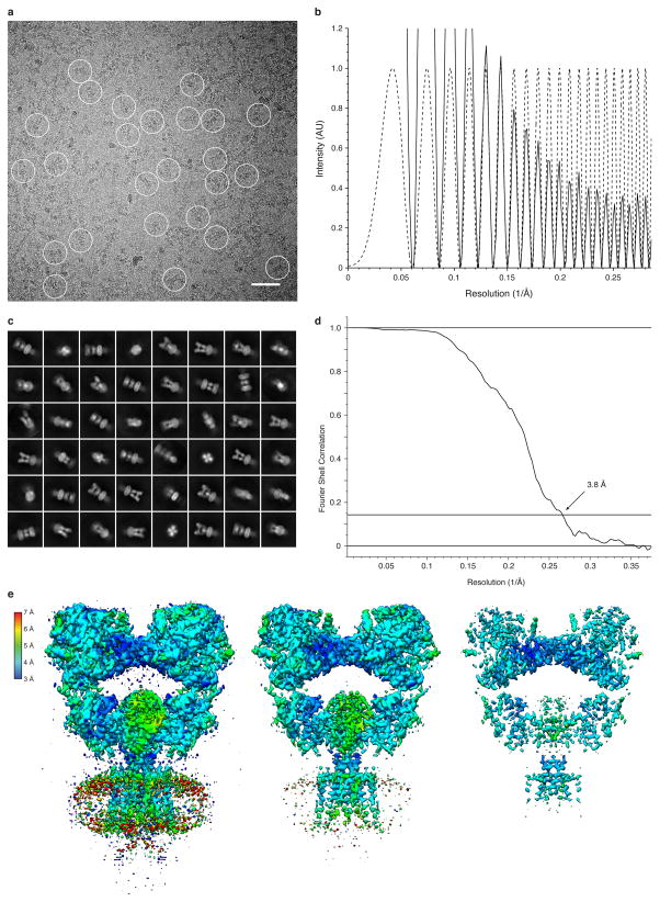
Extended Data Figure 1
Desensitized GluK2 imaging and structure determination
a, b, Representative cryo-EM image of GluK2 EM solubilized in DDM-CHS and bound by 2S,4R-4-methylglutamate (a), with the corresponding image power spectrum and CTF estimate showing signal beyond 3 u00...

Extended Data Figure 2
Desensitized GluK2 transmembrane and glycosylation features
a, b, Cryo-EM density for resolved S1-M1 (a) and M3-S2 (b) linkers and TM helices, displayed with Cu03b1 trace of the atomic model. c, Representative sites with densities for complex glycans at Asn244...

Extended Data Figure 3
Reconstruction of desensitized GluK2 without computational symmetry
a, Cryo-EM density map for the reconstruction of agonist bound GluK2 without imposition of computational symmetry, and colored according to local resolution. b, FSC curve with reported resolution of 4...

Extended Data Figure 4
Inhibition of GluK2 EM by LY466195 and LBD crystal structures for agonist and antagonist complexes
a, Crystal structure for the GluK2 EM isolated LBD dimer assembly complex with 2S,4R-4-methylglutamate; the upper/lower lobes for the two subunits are colored orange/pale yellow and teal/pale cyan res...

Extended Data Figure 5
Imaging and structure of GluK2 EM bound by antagonist LY466195
a, b, Representative cryo-EM image of GluK2 EM solubilized in DDM-CHS and bound by LY466195 (a), with the corresponding image power spectrum and CTF estimate showing signal beyond 6 u00c5 resolution (...

Extended Data Figure 6
ATD-LBD interface of desensitized GluK2
a, Desensitized GluK2 shown in surface representation with ATD-LBD interfaces highlighted. b, Top down view of LBD layer shown with perspective indicated by eye icon in (a). c, Underside of the ATD la...

Extended Data Figure 7
Desensitization ring residues that influence recovery kinetics
au2013d, Rate of recovery from desensitization measured using twin pulse applications of 10 mM glutamate (data points show mean u00b1 SD; fits are shown in red). a, Wild type GluK2 fit with a single e...

Figure 1
Desensitized kainate receptor at 3.8 u00c5 resolution
a, b, Cryo-EM density map (a) and atomic model (b) of 2S,4R-4-methylglutamate bound GluK2 EM with each chain colored uniquely. Panel (a) has features shown from two map contours. c, Side and top view ...

Figure 2
Desensitization ring
a, Top view of LBD layer highlighting desensitization ring helices E and G colored magenta for AC subunits and cyan for BD subunits. Residues S669 and D672, mutated in functional experiments (gu2013i)...

Figure 3
Desensitized state ion channel
au2013c, Selected sidechain densities with atomic model for M1 (a), M3 (b), and M4 (c) helices. d, The ion channel with surface modeling of the channel. Regions with pore radius less than 1.15 u00c5 a...
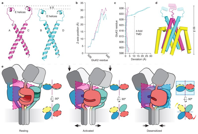
Figure 4
LBD-TM linkers mediate channel closing and LBD reorganization
a, Comparison of the regions encompassing the M3, M3-S2 linkers, and E helices for AC (magenta) and BD (cyan) chains of desensitized state GluK2 EM . b, The vertical rise in Cu03b1 position as a funct...
Figure images are served from the NIH/NLM PubMed Central Open Access Subset or Europe PMC; copyright remains with the publishers and authors.
💬 Discussion
0 commentsNo comments yet. Be the first to start a discussion!
Leave a Comment