Abstract
Steroid receptors activate gene transcription by recruiting coactivators to initiate transcription of their target genes. For most nuclear receptors, the ligand-dependent activation function domain-2 (AF-2) is a primary contributor to the nuclear receptor (NR) transcriptional activity. In contrast to other steroid receptors, such as ERα, the activation function of androgen receptor (AR) is largely dependent on its ligand-independent AF-1 located in its N-terminal domain (NTD). It remains unclear why AR utilizes a different AF domain from other receptors despite that NRs share similar domain organizations. Here, we present cryoelectron microscopy (cryo-EM) structures of DNA-bound full-length AR and its complex structure with key coactivators, SRC-3 and p300. AR dimerization follows a unique head-to-head and tail-to-tail manner. Unlike ERα, AR directly contacts a single SRC-3 and p300. The AR NTD is the primary site for coactivator recruitment. The structures provide a basis for understanding assembly of the AR:coactivator complex and its domain contributions for coactivator assembly and transcriptional regulation.
🔬 Techniques
🔭 Microscopes
🧬 Organisms
💻 Software
✨ Fluorophores
🧪 Sample Preparation
🔬 Cell Lines
🏭 Microscope Brands
🧪 Reagent Suppliers
💻 Software Details
💾 Data Repositories
🏛️ Research Organizations (ROR)
Affiliated research institutions:
📋 Methods
KEY RESOURCES TABLE
REAGENT or RESOURCE SOURCE IDENTIFIER Antibodies ERα Santa Cruz Cat#Sc-8002 SRC-3 Monoclonal Antibody/Recombinant Protein Expression Core at BCM N/A AR-Ab1 Monoclonal Antibody/Recombinant Protein Expression Core at BCM AR Ab clone #705 AR-Ab2 Monoclonal Antibody/Recombinant Protein Expression Core at BCM AR Ab clone #818 Flag M2 beads Sigma Cat#F2426 AR Santa Cruz Cat#Sc-816 P300 Santa Cruz Cat#Sc-584 Flag-HRP Sigma Cat#A8592 Chemicals, Peptides, and Recombinant Proteins ERα Thermo Fisher Cat#A15674 SRC-3 Monoclonal Antibody/Recombinant Protein Expression Core at BCM N/A P300 Monoclonal Antibody/Recombinant Protein Expression Core at BCM N/A AR Monoclonal Antibody/Recombinant Protein Expression Core at BCM N/A Dynabeads™ M-280 Streptavidin Thermo Fisher Cat#11205D R1881 Sigma Cat#R0908 Glutathione Sepharose 4B GE Life Sciences Cat#17075601 Pierce Fab Micro Preparation Kit Thermo Fisher Cat#44685 Tri-reagent Molecular Research Center Cat#TR118 Turbo DNase Thermo Fisher Cat#AM2239 17β-estradiol Sigma Cat#E2758 Experimental Models: Cell Lines HEK 293T/17 Tissue Culture Core (BCM) Cat# ATCC® CRL-11268™ Sf9 Monoclonal Antibody/recombinant Protein Expression Core (BCM) Cat# ATCC® CRL-1711™ Bacterial and Virus Strains E.coli BL21 (DE3) Agilent Cat#200131 Recombinant DNA pCDNA3.1-flag-AR WT/Mut This study N/A pCMV-flag-SRC-3 WT/Mut This study N/A pGEX-SRC-3 fragments Yi et al. 2015 Mol. Cell N/A 3XARE-E4 This study N/A Oligonucleotides ARE III PCR forward primer Thermo Fisher N/A ARE III PCR reverse primer Thermo Fisher N/A ARE oligonucleotide Santa Cruz Cat#sc-2551 Deposited Data CryoEM maps of AR complexes This study EMD-22079 and 22080 Software and Algorithms EMAN2.2 https://cryoem.bcm.edu/cryoem/downloads/view_eman2_versions N/A Relion3.0 https://www3.mrc-lmb.cam.ac.uk/relion/index.php/Download_%26_install N/A gctf https://www2.mrc-lmb.cam.ac.uk/research/locally-developed-software/zhang-software/ N/A Chimera https://www.cgl.ucsf.edu/chimera/download.htm l N/A ChimeraX https://www.rbvi.ucsf.edu/chimerax/download.html N/A Other SerialEM http://bio3d.colorado.edu/SerialEM/ N/A RESOURCE AVAILABILITY Lead Contact Further information and requests for resources and reagents should be directed to and will be fulfilled by the Lead Contact, Bert O’Malley ( berto@bcm.edu ).
Show full methods section
KEY RESOURCES TABLE
REAGENT or RESOURCE SOURCE IDENTIFIER Antibodies ERα Santa Cruz Cat#Sc-8002 SRC-3 Monoclonal Antibody/Recombinant Protein Expression Core at BCM N/A AR-Ab1 Monoclonal Antibody/Recombinant Protein Expression Core at BCM AR Ab clone #705 AR-Ab2 Monoclonal Antibody/Recombinant Protein Expression Core at BCM AR Ab clone #818 Flag M2 beads Sigma Cat#F2426 AR Santa Cruz Cat#Sc-816 P300 Santa Cruz Cat#Sc-584 Flag-HRP Sigma Cat#A8592 Chemicals, Peptides, and Recombinant Proteins ERα Thermo Fisher Cat#A15674 SRC-3 Monoclonal Antibody/Recombinant Protein Expression Core at BCM N/A P300 Monoclonal Antibody/Recombinant Protein Expression Core at BCM N/A AR Monoclonal Antibody/Recombinant Protein Expression Core at BCM N/A Dynabeads™ M-280 Streptavidin Thermo Fisher Cat#11205D R1881 Sigma Cat#R0908 Glutathione Sepharose 4B GE Life Sciences Cat#17075601 Pierce Fab Micro Preparation Kit Thermo Fisher Cat#44685 Tri-reagent Molecular Research Center Cat#TR118 Turbo DNase Thermo Fisher Cat#AM2239 17β-estradiol Sigma Cat#E2758 Experimental Models: Cell Lines HEK 293T/17 Tissue Culture Core (BCM) Cat# ATCC® CRL-11268™ Sf9 Monoclonal Antibody/recombinant Protein Expression Core (BCM) Cat# ATCC® CRL-1711™ Bacterial and Virus Strains E.coli BL21 (DE3) Agilent Cat#200131 Recombinant DNA pCDNA3.1-flag-AR WT/Mut This study N/A pCMV-flag-SRC-3 WT/Mut This study N/A pGEX-SRC-3 fragments Yi et al. 2015 Mol. Cell N/A 3XARE-E4 This study N/A Oligonucleotides ARE III PCR forward primer Thermo Fisher N/A ARE III PCR reverse primer Thermo Fisher N/A ARE oligonucleotide Santa Cruz Cat#sc-2551 Deposited Data CryoEM maps of AR complexes This study EMD-22079 and 22080 Software and Algorithms EMAN2.2 https://cryoem.bcm.edu/cryoem/downloads/view_eman2_versions N/A Relion3.0 https://www3.mrc-lmb.cam.ac.uk/relion/index.php/Download_%26_install N/A gctf https://www2.mrc-lmb.cam.ac.uk/research/locally-developed-software/zhang-software/ N/A Chimera https://www.cgl.ucsf.edu/chimera/download.htm l N/A ChimeraX https://www.rbvi.ucsf.edu/chimerax/download.html N/A Other SerialEM http://bio3d.colorado.edu/SerialEM/ N/A RESOURCE AVAILABILITY Lead Contact Further information and requests for resources and reagents should be directed to and will be fulfilled by the Lead Contact, Bert O’Malley ( berto@bcm.edu ).
Materials Availability
Plasmids generated in this study will be available upon request.
Data and Code Availability
All the cryo-EM maps have been deposited to EMDB under accession code EMD-22079 and EMD-22080.
EXPERIMENTAL MODEL AND SUBJECT DETAILS Cell line culture 293T
Cells were cultured at 37°C in Dulbecco’s Modified Eagle Medium (DMEM, Gibco) supplemented with 10% Fetal Bovine Serum (Gibco) and 100 U/ml Penicillin-Streptomycin (Gibco).
Materials Availability
Plasmids generated in this study will be available upon request.
EXPERIMENTAL MODEL AND SUBJECT DETAILS Cell line culture 293T
Cells were cultured at 37°C in Dulbecco’s Modified Eagle Medium (DMEM, Gibco) supplemented with 10% Fetal Bovine Serum (Gibco) and 100 U/ml Penicillin-Streptomycin (Gibco).
METHOD DETAILS Protein purification
Sf9 insect cells were infected with flag-AR expressing baculoviruses (that were produced in BCM Monoclonal Antibody/recombinant Protein Expression Core Facility). Cells were cultured in the presence of 1 μM R1881 24 hours post-infection. Cells were harvested 48 hours post-infection. Cells were washed and pelleted down at 5,000 rpm for 10 minutes at 4°C. Sf9 cells were further resuspended in lysis buffer (50 mM Tris-HCl, pH8.0; 150 mM NaCl; 100 nM R1881; 0.5% NP40) and lysed using a homogenizer. After 40 minutes centrifugation at 15,000 rpm, the cleared lysate was incubated with Ni-NTA (Qiagen) resins, washed three times with wash buffer (50 mM Tris-HCl, pH8.0; 150 mM NaCl; 100 nM R1881; 0.05% NP40; 25 mM imidazole) and finally eluted with elution buffer (50 mM Tris-HCl, pH8.0; 150 mM NaCl; 100 nM R1881; 0.05% NP40; 300 mM imidazole). Eluted protein was further applied on a gel-filtration column (Superdex 200 Increase 10/300 GL, GE Healthcare) pre-equilibrated with gel filtration buffer (50 mM Tris-HCl, pH8.0; 150 mM NaCl; 100 nM R1881; 0.05% NP40). Purified protein fractions were pooled together (~0.5 mg/ml) and for all the studies.
Cell-free transcription The 3xARE3–E4
DNA template contains three copies of the ARE-III from the human PSA/KLK3 gene enhancer ( Cleutjens et al., 1997 ) fused to the minimal Adenovirus E4 gene promoter. It was assembled into chromatin using HeLa core histone using a salt dilution method as described ( Panigrahi et al., 2018 ). Each in vitro transcription reaction (50 μL) contained 0.2 pmoles of chromatinized 3xARE3–E4 in 12 mM HEPES-KOH (pH7.9), 12% glycerol, 60 mM KCl, 12 mM MgCl2, 0.12 mM EDTA, 0.3 mM DTT, 1 mM ATP, 0.9 mM acetyl CoA, 2% Poly(vinyl alcohol) diluted in DEPC-treated H 2 O, 100 nM R1881, and 50 μg of HeLa S3 nuclear extract (NE). The reactions were run in duplicate and the HeLa S3 NE was prepared as described ( Foulds et al., 2013 ). Where indicated, reactions included recombinant AR (1 ng), p300 (6 ng), and/or SRC-3 (3 ng) protein. After incubating at room temperature for 25 min to allow for preinitiation complex assembly, 5 μL of 5 mM NTPs was added to allow for active transcription and the samples were transferred to 30°C for 50 min. 250 μL of Tri-reagent was added to each reaction, mixed by vortexing, and then 15 μL of BAN (4-bromoanisole) was added to phase separate the RNA from the DNA and protein. The RNA was then isolated and purified following the manufacturer’s protocol (Molecular Research Center). The RNA precipitates were dissolved in 35 μL DEPC-treated H 2 O and then treated with Turbo DNase (Ambion) following manufacturer’s protocol to remove any DNA contamination; 2 μL of each RNA sample (5% of 40 μL final volume) were then used in One-step RT-qPCR reactions (Bioline) utilizing an E4 gene primer pair. An additional qPCR reaction containing 10 fmoles of the template DNA was included to normalize Ct values. Results were normalized to the transcript levels from the reactions without any recombinant protein added (i.e. basal transcription) to determine relative E4 mRNA fold change. Statistical test used was the Student’s t test and p
📊 Figures
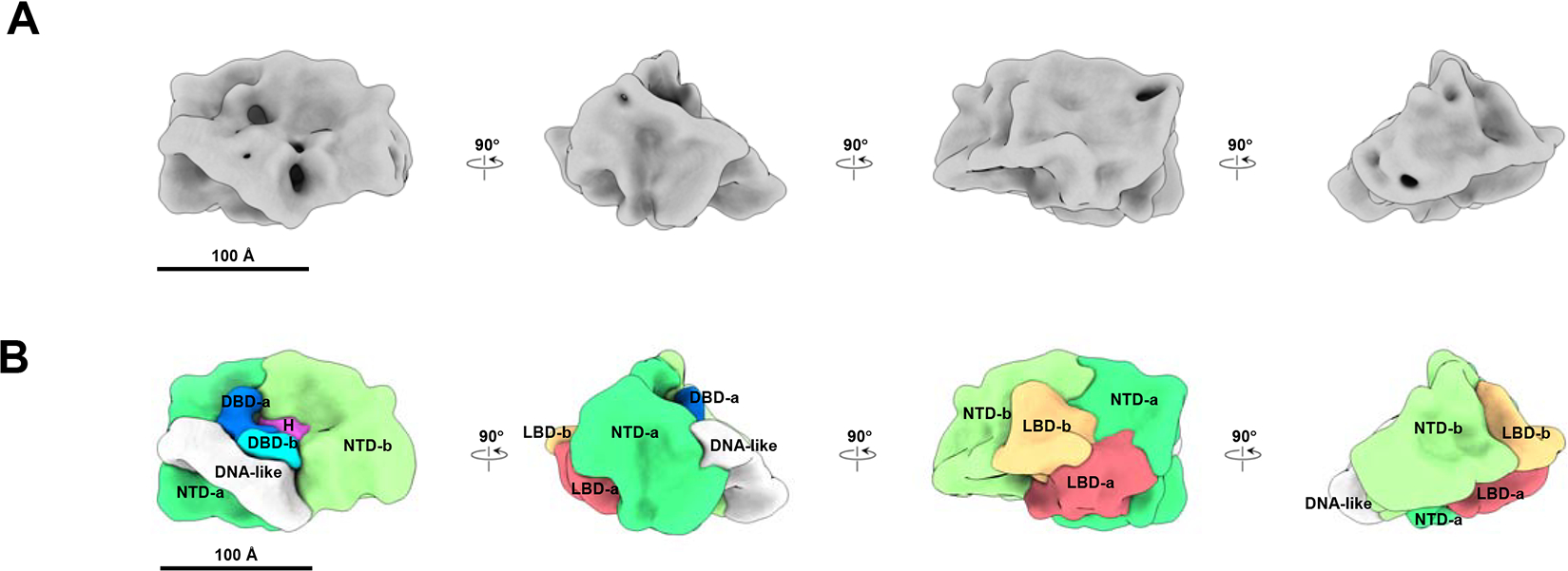
Figure 1.
ARE DNA bound AR density map and segmentation.
(A) Cryo-EM density of ARE-DNA/AR at resolution ~12.6 u00c5 viewed from different orientation rotating in vertical direction. (B) Segmentation result of ARE-DNA/AR. The segments of two ARs are shown a...
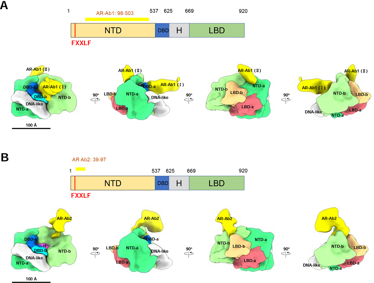
Figure 2.
NTD and LBD annotation validation by antibody labeling.
(A) AR-Ab1 labels the NTDs. Top panel, schematic representation of AR functional domains. The long yellow bar represents the region (residues 98u2013503) recognized by the AR-Ab1. The vertical red bar...
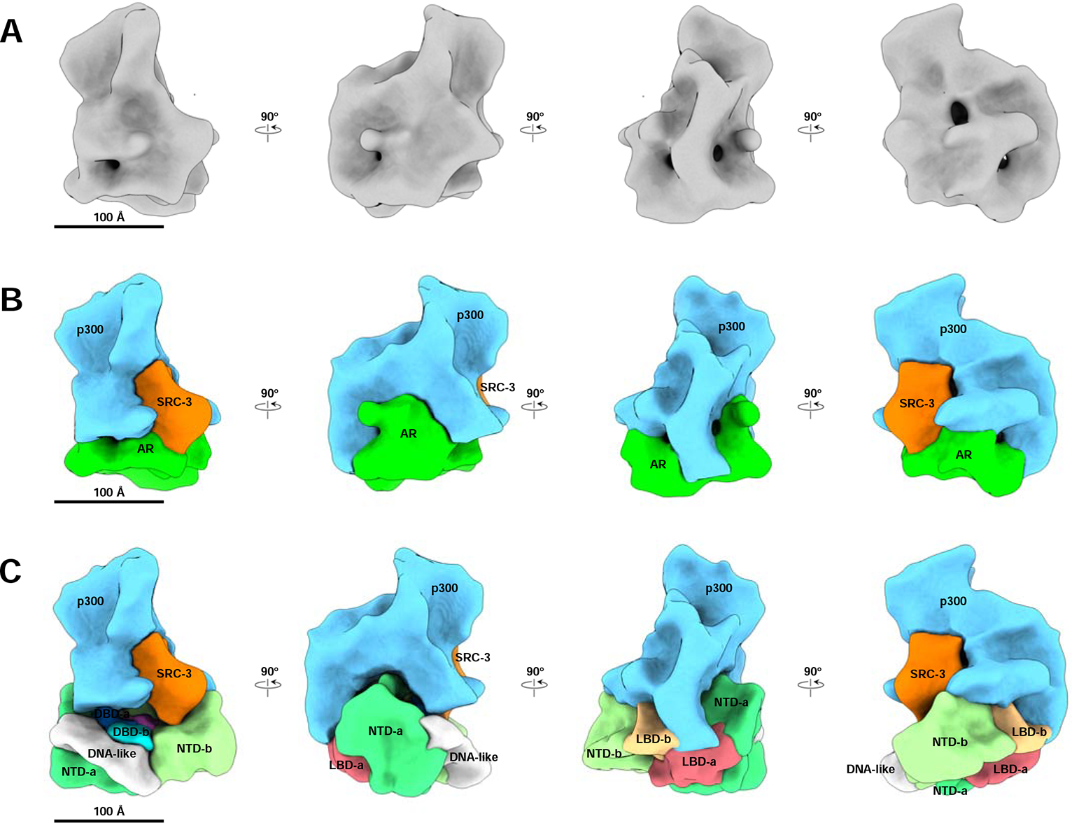
Figure 3.
The ARE DNA-bound AR/SRC-3/p300 complex density map and segmentation.
(A) Cryo-EM density map of the ARE DNA-bound AR/SRC-3/p300 complex at resolution ~20 u00c5. Shown are 4 different angles of the map rotating every 90 degrees. (B) Segmentation of ARE-DNA/AR/SRC-3/p300...
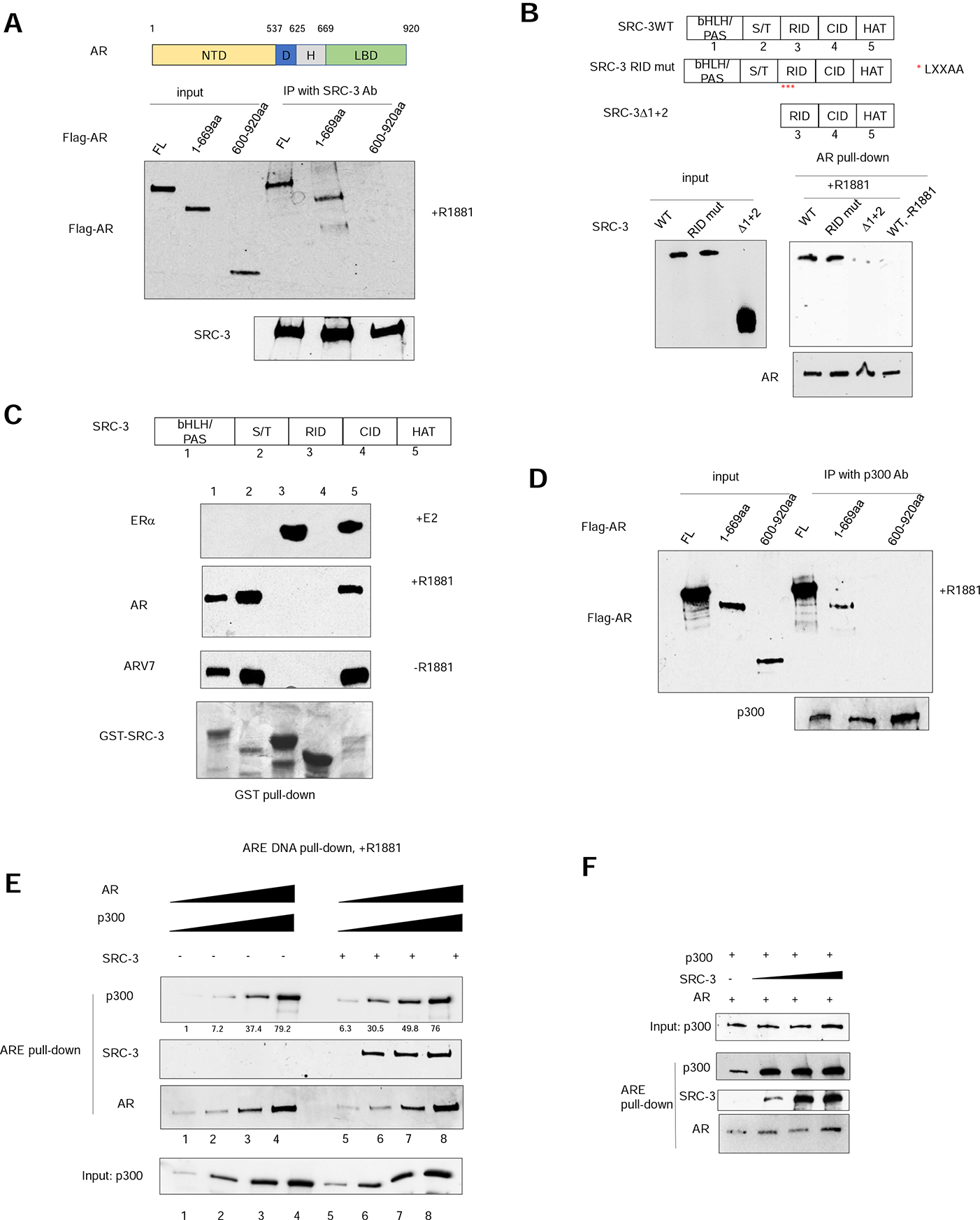
Figure 4.
The AR NTD is essential for SRC-3 and p300 coactivator recruitment.
(A) AR interacts with SRC-3 through its N-terminal domain (NTD) in vitro. Purified flag-tagged full-length AR, AR(1u2013669aa) or AR (600u2013920aa) was incubated with purified, recombinant SRC-3 in t...
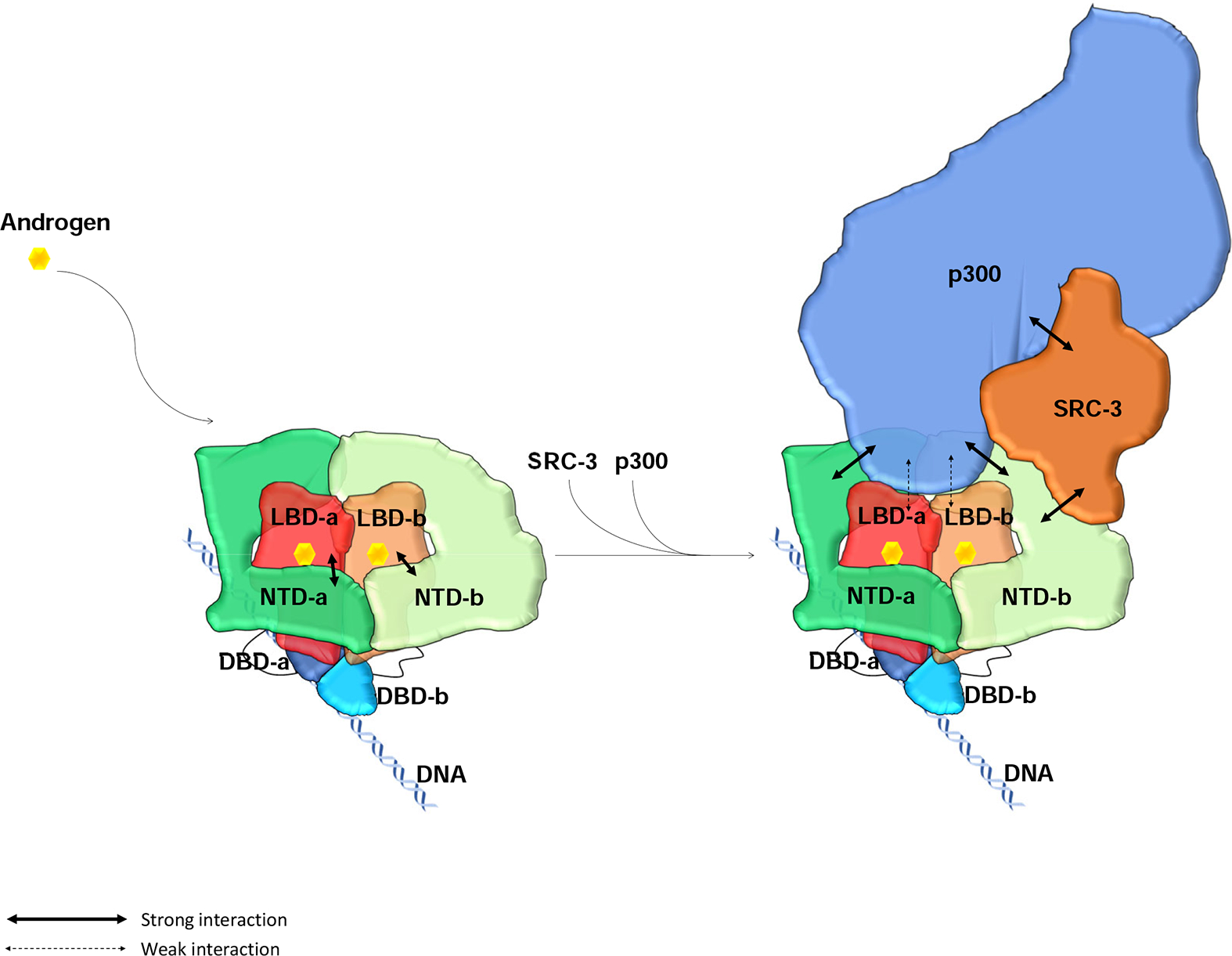
Figure 5:
Model of full-length AR domain organization and its NTD-mediated SRC-3 and p300 coactivator recruitment.
AR dimerizes upon binding to its ligand androgen. Its LBD and DBD are located at the center to form a tight dimerization interface. The two NTDs in the dimer adopt slightly different conformations, wr...
Figure images are served from the NIH/NLM PubMed Central Open Access Subset or Europe PMC; copyright remains with the publishers and authors.
💬 Discussion
0 commentsNo comments yet. Be the first to start a discussion!
Leave a Comment