Abstract
The uptake of peptides in mammals plays a crucial role in nutrition and inflammatory diseases. This process is mediated by promiscuous transporters of the solute carrier family 15, which form part of the major facilitator superfamily. Besides the uptake of short peptides, peptide transporter 1 (PepT1) is a highly abundant drug transporter in the intestine and represents a major route for oral drug delivery. PepT2 also allows renal drug reabsorption from ultrafiltration and brain-to-blood efflux of neurotoxic compounds. Here, we present cryogenic electron microscopy (cryo-EM) structures of human PepT1 and PepT2 captured in four different states throughout the transport cycle. The structures reveal the architecture of human peptide transporters and provide mechanistic insights into substrate recognition and conformational transitions during transport. This may support future drug design efforts to increase the bioavailability of different drugs in the human body.
🔬 Techniques
💻 Software
✨ Fluorophores
🧪 Sample Preparation
🔬 Cell Lines
🏭 Microscope Brands
🧪 Reagent Suppliers
📷 Detectors
💻 Software Details
💾 Data Repositories
🏛️ Research Organizations (ROR)
Affiliated research institutions:
📋 Methods
Expression and purification of Hs PepT2 The N528Q-N587Q Hs
PepT2 gene was cloned into a pXLG vector containing an expression cassette composed of an N-terminal Twin-Streptavidin tag followed by the human rhinovirus 3C (HRV-3C) protease recognition sequence. The double mutation in Hs PepT2 was introduced to decrease sample heterogeneity caused by glycosylation and to increase expression levels. HEK293F cells were collected 48 hours after transient transfection as previously described ( 60 ) and stored at −80°C until further use. Frozen cell pellets were resuspended in 300 mM NaCl, 20 mM NaPi (pH 7.5), 0.5 mM tris(2-carboxyethyl)phosphine) (TCEP), and 5% glycerol, supplemented with cOmplete EDTA-free protease inhibitors, and were disrupted using an Avestin Emulsiflex homogenizer. The lysate was centrifuged for 10 min at 10,000 g , and the supernatant was centrifuged for 90 min at 95,000 g (Optima XE-90, Beckman Coulter). The pellet containing the membrane fraction was solubilized in 1% N -dodecyl-β- d -maltopyranoside (DDM; Anatrace) and 0.1% cholesteryl hemisuccinate (CHS; Tris Salt, Anatrace) for 1 hour at 4°C. The sample was centrifuged for 50 min at 70,000 g , and the supernatant was applied to Strep-TactinXT beads (IBA). After 20 min of incubation on a rotating wheel, the suspension was transferred to a gravity column. Following two wash steps with 300 mM NaCl, 20 mM Hepes (pH 7.5), 0.03% DDM, and 0.003% CHS, Hs PepT2 was eluted with 0.03% DDM, 0.003% CHS, 150 mM NaCl, 20 mM Hepes (pH 7.5), and 10 mM desthiobiotin (Sigma-Aldrich). 3C cleavage was performed in 30 min, and the protease was separated from Hs PepT2 by gel filtration using Superose 6 Increase 10/300 (Sigma-Aldrich). The top fraction was concentrated to 10 mg/ml using a 100-kDa cutoff concentrator (Corning Spin-X UF concentrators) and stored at −80°C until further use.
Show full methods section
Expression and purification of Hs PepT2 The N528Q-N587Q Hs
PepT2 gene was cloned into a pXLG vector containing an expression cassette composed of an N-terminal Twin-Streptavidin tag followed by the human rhinovirus 3C (HRV-3C) protease recognition sequence. The double mutation in Hs PepT2 was introduced to decrease sample heterogeneity caused by glycosylation and to increase expression levels. HEK293F cells were collected 48 hours after transient transfection as previously described ( 60 ) and stored at −80°C until further use. Frozen cell pellets were resuspended in 300 mM NaCl, 20 mM NaPi (pH 7.5), 0.5 mM tris(2-carboxyethyl)phosphine) (TCEP), and 5% glycerol, supplemented with cOmplete EDTA-free protease inhibitors, and were disrupted using an Avestin Emulsiflex homogenizer. The lysate was centrifuged for 10 min at 10,000 g , and the supernatant was centrifuged for 90 min at 95,000 g (Optima XE-90, Beckman Coulter). The pellet containing the membrane fraction was solubilized in 1% N -dodecyl-β- d -maltopyranoside (DDM; Anatrace) and 0.1% cholesteryl hemisuccinate (CHS; Tris Salt, Anatrace) for 1 hour at 4°C. The sample was centrifuged for 50 min at 70,000 g , and the supernatant was applied to Strep-TactinXT beads (IBA). After 20 min of incubation on a rotating wheel, the suspension was transferred to a gravity column. Following two wash steps with 300 mM NaCl, 20 mM Hepes (pH 7.5), 0.03% DDM, and 0.003% CHS, Hs PepT2 was eluted with 0.03% DDM, 0.003% CHS, 150 mM NaCl, 20 mM Hepes (pH 7.5), and 10 mM desthiobiotin (Sigma-Aldrich). 3C cleavage was performed in 30 min, and the protease was separated from Hs PepT2 by gel filtration using Superose 6 Increase 10/300 (Sigma-Aldrich). The top fraction was concentrated to 10 mg/ml using a 100-kDa cutoff concentrator (Corning Spin-X UF concentrators) and stored at −80°C until further use.
Expression and purification of Hs PepT1
The wild-type Hs PepT1 gene was cloned into a pXLG vector containing an expression cassette composed of an N-terminal Twin-Streptavidin tag followed by the HRV-3C protease recognition sequence. HEK293F cells were collected 48 hours after transient transfection and resuspended in 300 mM NaCl, 20 mM NaPi (pH 7.5), 0.5 mM TCEP, and 5% glycerol, supplemented with cOmplete EDTA-free protease inhibitors. Whole cells were solubilized overnight in 1% lauryl maltose neopentyl glycol (LMNG; Anatrace) and 0.2% CHS (Tris Salt Anatrace). The sample was then centrifuged for 60 min at 70,000 g , and the supernatant was applied to Strep-TactinXT beads (IBA). After 30-min incubation on a rotating wheel, the suspension was transferred to a gravity column. Following two wash steps with 300 mM NaCl and 20 mM Hepes (pH 7.5) supplied with 0.03% DDM, 0.003% LMNG, and 0.006% CHS, Hs PepT1 was eluted with 0.03% DDM, 0.003% LMNG, 0.0006% CHS, 150 mM NaCl, 20 mM Hepes (pH 7.5), and 10 mM desthiobiotin (Sigma-Aldrich). The sample was concentrated to 100 μl using a 100-kDa cutoff concentrator (Corning Spin-X UF concentrators) and run directly on a Superdex Increase 200 5/150 gel filtration column for vitrification in 0.015% DDM, 0.0015% LMNG, 0.003% CHS, 150 mM NaCl, 50 mM Hepes (pH 7.5), and 0.5 mM TCEP. The top fraction reached a concentration of 2 mg/ml and was stored at −80°C until further use.
Whole-cell uptake assays
The wild-type Hs PepT2 gene was cloned into a pXLG vector ( 61 ) containing an expression cassette composed of an N-terminal hexa-histidine tag followed by enhanced green fluorescent protein and a tobacco etch virus protease cleavage site. HEK293F cells grown in suspension in FreeStyle medium were transfected with wild-type Hs PepT2 using a mass ratio of 2:1 polyethyleneimine:DNA. Hs PepT2 was expressed for 48 hours at 37°C, 8% CO 2 at 220 rpm. For competition assays, 4 × 10 6 cells/ml resuspended in phosphate-buffered saline buffer at pH 6.0 supplemented with 5 mM glucose were incubated in 96-well plates, with 50 μM β-Ala-Lys-AMCA in the absence or presence of dipeptides, tripeptides, or drugs for 10 min at 37°C. The reaction was stopped by adding 200 μl of ice-cold buffer, and the cells were then washed three times with the same buffer. Last, the cells were resuspended in 200 μl of buffer, and the fluorescence was measured in an M1000 microplate reader (TECAN) with excitation at 350 nm and emission at 450 nm. All experiments were performed in triplicates. The results were normalized by the fluorescence value of the control (cells overexpressing Hs PepT2 incubated with AK-AMCA in the absence of inhibitor) and plotted as AK-AMCA uptake rate percentage. For concentration-dependent uptake experiments, IC 50 values were processed in GraphPad Prism 9.0 (GraphPad Software) using sigmoidal four-parameter curve fitting.
Thermal stability measurements
The differential scanning fluorimetry method was used to follow the thermal unfolding event of Hs PepT2 and Hs PepT1 with a Prometheus NT.48 device (NanoTemper Technologies, Munich, Germany). Purified Hs PepT2 was diluted to 0.3 mg/ml and supplemented with decreasing amounts of Ala-Phe dipeptide in a dilution series of 13 points, starting at 10 mM down to 2.4 μM. Purified Hs PepT1 was diluted to 0.3 mg/ml and supplemented with 0, 1, or 10 mM Ala-Phe. The fluorescence at 330 and 350 nm was recorded over a temperature gradient scan from 15° to 95°C and processed in GraphPad Prism 9.0 (GraphPad Software).
Cryo-EM sample preparation and data collection on apo Hs
PepT1 in the outward-facing open state Four microliters of purified Hs PepT1 at 2 mg/ml was applied to a glow-discharged gold holey carbon 2/1 300-mesh grid (Quantifoil). The grid was blotted for 4 s at 0 force before being plunge-vitrified in liquid propane using Mark IV Vitrobot (Thermo Fisher Scientific). The blotting chamber was maintained at 4°C and 100% humidity during freezing. Movies were collected using a Titan Krios (Thermo Fisher Scientific) equipped with a K3 camera and BioQuantum energy filter (Gatan) set to 20 eV. A total of 22,537 movies were collected at a nominal magnification of ×105,000 and a physical pixel size of 0.85 Å, with a 70-μm C2 aperture and 100-μm objective aperture at a dose rate of 16 e − /pixel per second. A total dose of 66 e − /Å 2 was collected with 3-s exposure as movies of 50 frames. Data were collected using EPU (Thermo Fisher Scientific).
Cryo-EM sample preparation and data collection on Hs
PepT2 bound to Ala-Phe in the inward-facing partially occluded state One hour before vitrification, purified N528Q-N587Q Hs PepT2 was thawed on ice and run on a Superdex Increase 200 5/150 column in 0.015% DDM, 0.0015% CHS, 100 mM NaCl, 10 mM Hepes (pH 7.5), and 0.5 mM TCEP. The top fraction reached a concentration of 1 mg/ml, and 3.6 μl supplemented with 5 mM of the dipeptide alanine-phenylalanine (Bachem) was applied to glow-discharged gold holey carbon 2/1 300-mesh grids (Quantifoil). Grids were blotted for 4 s at 0 force and 2-s wait time before being plunge-vitrified in liquid propane using Mark IV Vitrobot (Thermo Fisher Scientific). The blotting chamber was maintained at 4°C and 100% humidity during freezing. Movies were collected using Titan Krios (Thermo Fisher Scientific) outfitted with a K3 camera and BioQuantum energy filter (Gatan) set to 10 eV. A total of 34,712 movies were collected at a nominal magnification of ×105,000 and a physical pixel size of 0.85 Å, with a 70-μm C2 aperture and 100-μm objective aperture at a dose rate of 19.5 e − /pixel per second. A total dose of 81 e − /Å 2 was collected with 3-s exposure as movies of 45 frames. Data were collected using EPU (Thermo Fisher Scientific).
Cryo-EM sample preparation and data collection on Hs
PepT1 bound to Ala-Phe in the outward-facing open state and in the outward-facing occluded state Four microliters of purified Hs PepT1 at 2 mg/ml supplemented with 20 mM Ala-Phe was applied to a glow-discharged gold holey carbon 2/1 300-mesh grid (Quantifoil). The grid was blotted for 4.0 s at 0 force before being plunge-vitrified in liquid propane using Mark IV Vitrobot (Thermo Fisher Scientific). The blotting chamber was maintained at 4°C and 100% humidity during freezing. Movies were collected using Titan Krios (Thermo Fisher Scientific) equipped with a K3 camera and BioQuantum energy filter (Gatan) set to 15 eV. A total of 37,822 movies were collected in two separate sessions (16,522 in the first session and 21,300 in the second session) using EPU (Thermo Fisher Scientific) at a nominal magnification of ×130,000 and a physical pixel size of 0.67 Å, with a 70-μm C2 aperture and 100-μm objective aperture at a dose rate of 15 e − /pixel per second. A total dose of 55 e − /Å 2 was collected with 1.7-s exposure as movies of 40 frames.
Cryo-EM image processing of apo Hs
PepT1 in the outward-facing open state Movies were motion-corrected using Relion-3.1 ( 62 ) own implementation of MotionCor2 ( 63 ). Contrast transfer function parameters were calculated using CTFFIND4 ( 64 ). A total of 2,091,726 coordinates were extracted from 22,537 micrographs using CrYOLO ( 65 ), with a 200-pixel box and binning to 50 pixels, and were subjected to multiple rounds of 2D classification in Relion-3.1. A total of 1,459,348 particles were selected and reextracted with a 200-pixel box size without binning. A 3D ab initio reconstruction was generated in CryoSPARCv2 with all particles and low pass–filtered at 30 Å for 3D classification in Relion-3.1. After multiple rounds of 3D classification using T = 10 and K = 4, the best classes were selected for additional 3D classification without image alignment in Relion-3.1, focusing on the protein and excluding the micelle. The selection of 593,757 particles from 3D classes with strong signal inside the micelle and in the extracellular domain led to a reconstruction of 4.6 Å in Relion-3.1. CTF refine (per particle defocus and beam tilt) and Bayesian polishing (using optimized trained parameters on a subset of 20,000 particles) were performed in Relion-3.1. The shiny particles were then imported in CryoSPARCv3 for nonuniform refinement ( 66 ) that led to a 3.9-Å reconstruction estimated in cryoSPARCv3 using the Fourier shell correlation (FSC) = 0.143 cutoff. Local resolution estimations were calculated in CryoSPARCv3 using 0.5 FSC cutoff. Postprocessing in DeepEMhancer ( 67 ) using the two half maps as input and the default tightTarget model resulted in a more interpretable map, which was used only for illustration purposes in Fig. 1B , while the model was build and refined in a map postprocessed using default parameters in the Phenix Autosharpen utility ( 68 ).
Cryo-EM image processing of Hs
PepT2 bound to Ala-Phe in the inward-facing partially occluded state Movies were motion-corrected using Relion-3.1 ( 62 ) own implementation of MotionCor2 ( 63 ). Contrast transfer function parameters were calculated using CTFFIND4 ( 64 ). A total of 4,388,314 coordinates were extracted from 34,712 micrographs using CrYOLO ( 65 ), with a 200-pixel box and binning to 50 pixels, and were subjected to multiple rounds of 2D classification in Relion-3.1. A total of 2,944,737 particles were selected and reextracted with a 200-pixel box size without binning. A 3D ab initio reconstruction was generated in CryoSPARCv2 with a subset of particles and low pass–filtered at 30 Å for 3D refinement in Relion-3.1 on the 2,944,737 particles, yielding a 7.5-Å reconstruction. After multiple rounds of 3D classification without image alignment in Relion-3.1, using T = 4, 8, 10, 20, 30, and 40 focusing on the protein and excluding the micelle, the selection of 454,149 particles from 3D classes with strong signal inside the micelle and in the extracellular domain led to a reconstruction of 4.3 Å in Relion-3.1 using SIDESPLITTER ( 69 ) and a soft mask covering the micelle and the extracellular domain. Bayesian polishing was performed in Relion-3.1 using optimized trained parameters on a subset of 20,000 particles. The shiny particles were then imported in CryoSPARCv3 for CTF Refinement per particle (defocus and beamtilt). Nonuniform refinement ( 66 ) led to a 3.8-Å reconstruction estimated in cryoSPARCv3 using the FSC = 0.143 cutoff. Local resolution estimations were calculated in CryoSPARCv3 using 0.5 FSC cutoff. Postprocessing in DeepEMhancer ( 67 ) using the two half maps as input and the default tightTarget model resulted in a more interpretable map, which was used only for illustration purposes in Fig. 1C , while the model was build and refined in a map postprocessed using default parameters in the Phenix Autosharpen utility ( 68 ).
Cryo-EM image processing of Hs
PepT1 bound to Ala-Phe in the outward-facing open and occluded states Movies were motion-corrected using Relion-3.1 ( 62 ) own implementation of MotionCor2 ( 63 ). Contrast transfer function parameters were calculated using CTFFIND4 ( 64 ). A total of 6,046,602 coordinates were extracted from 37,822 micrographs using CrYOLO ( 65 ), with a 200-pixel box and binning to 50 pixels, and were subjected to multiple rounds of 2D classification in Relion-3.1. A total of 4,247,238 particles were selected and reextracted with a 200-pixel box size without binning. The 3D volume of apo Hs PepT1 in its outward-facing open conformation was low pass–filtered at 40 Å for 3D classification in Relion-3.1, resulting in a reconstruction at 4.1 Å with 486,562 particles for the first dataset, and 4.5 Å with 599,754 particles for the second, after performing Bayesian polishing using individually optimized trained parameters on a subset of 30,000 particles for each dataset. In both datasets, further 3D classifications allowed to separate two distinct conformational states, including the substrate-bound outward-facing open state and the substrate-bound outward-facing occluded state differing by the opening or closure of TM2. Focused 3D classification without alignment on the transmembrane domain in Relion-3.1 allowed to improve the resolution of the substrate-bound outward-facing open state from 3.7 to 3.5 Å after nonuniform refinement ( 66 ) in cryoSPARCv3. The substrate-bound outward-facing occluded state reached a modest resolution of 4.1 Å after nonuniform refinement in cryoSPARCv3 but led to a clear resolution of all TMs, including the closed TM2. For the 3.5-Å resolution substrate-bound outward-facing open reconstruction, postprocessing was done in Phenix using default parameters in the Autosharpen utility ( 68 ). This map was subsequently used for model building and refinement. For illustration purposes in Fig. 5E only, the half maps were also subjected to postprocessing in DeepEMhancer ( 67 ) using the two half maps as input and the default tightTarget model. For the 4.1-Å resolution substrate-bound outward-facing occluded reconstruction, postprocessing was done in cryoSPARCv3.
Model building and refinement
The transmembrane domain of Hs PepT2 was built manually in Coot ( 70 ), guided by secondary structure predictions from PSIPRED ( 71 ). The resolution in the extracellular domain of Hs PepT2 did not allow de novo model building. Instead, the structure of Hs PepT2-ECD predicted by AlphaFold ( 50 ) was docked in the cryo-EM density and linked to the transmembrane domain. The model was iteratively adjusted and refined using Isolde ( 72 ) and Phenix real-space refine ( 73 ). An inward open partially occluded model of Hs PepT1 was generated in SWISSMODEL ( 74 ), using Hs PepT2 as a reference template. The model was then subjected to multiple rounds of refinement in NAMDINATOR ( 75 ), Coot, and Phenix real-space refine ( 73 ) to fit in the apo outward-facing open reconstruction. Once the model fitted the density, the extracellular domain was replaced by the AlphaFold Hs PepT1-ECD prediction, which improved the quality of the model significantly. This model was then subjected to adjustments in Isolde and a final refinement using Phenix real-space refine. The substrate-bound outward-facing open and occluded models were generated using the final apo outward-facing open model, and manual adjustments in Coot and Isolde before real-space refinement in Phenix. Validation of the models was performed using MolProbity in Phenix.
Supplementary Materials This PDF file includes: Figs. S1 to S16 Table S1 Legends for movie S1 Click here for additional data file. Other Supplementary Material for this manuscript includes the following: Movie S1 Click here for additional data file.
Other Supplementary Material for this manuscript includes the following: Movie S1 Click here for additional data file.
📊 Figures
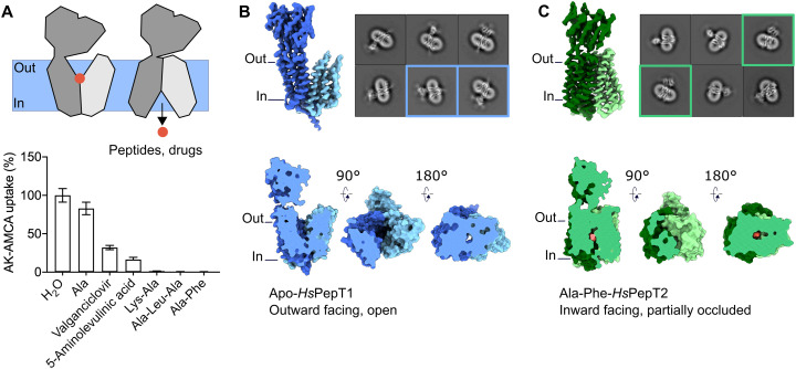
Fig. 1.
Cryo-EM structures of apo Hs PepT1 and Hs PepT2 bound to Ala-Phe.
( A ) Whole-cell transport competition assays of the u03b2-Ala-Lys peptide coupled to the fluorescent AMCA moiety (AK-AMCA) in Hs PepT2-transfected HEK293 cells showing reduced AK-AMCA uptake in the p...
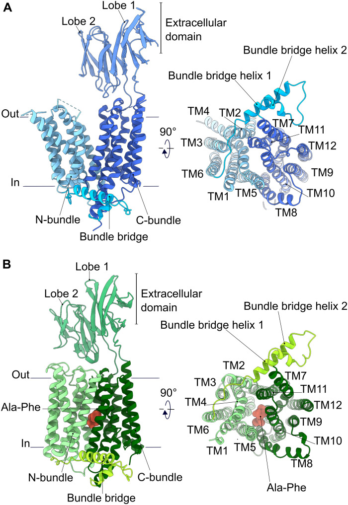
Fig. 2.
Overall architecture of human POTs.
( A ) Apo- Hs PepT1 and ( B ) substrate-bound Hs PepT2 models shown as cartoon representation. The different architectural elements are labeled. Loops that could not be modeled because of poor density...
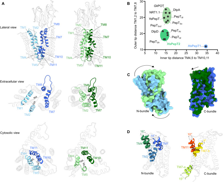
Fig. 3.
Structural comparison between the outward- and inward-facing states observed in apo Hs PepT1 and substrate-bound Hs PepT2.
( A ) Opening and closing of the substrate binding site to the extracellular and intracellular milieu observed in Hs PepT1 (blue) and Hs PepT2 (green). ( B ) The distances between C u03b1 atoms of the...
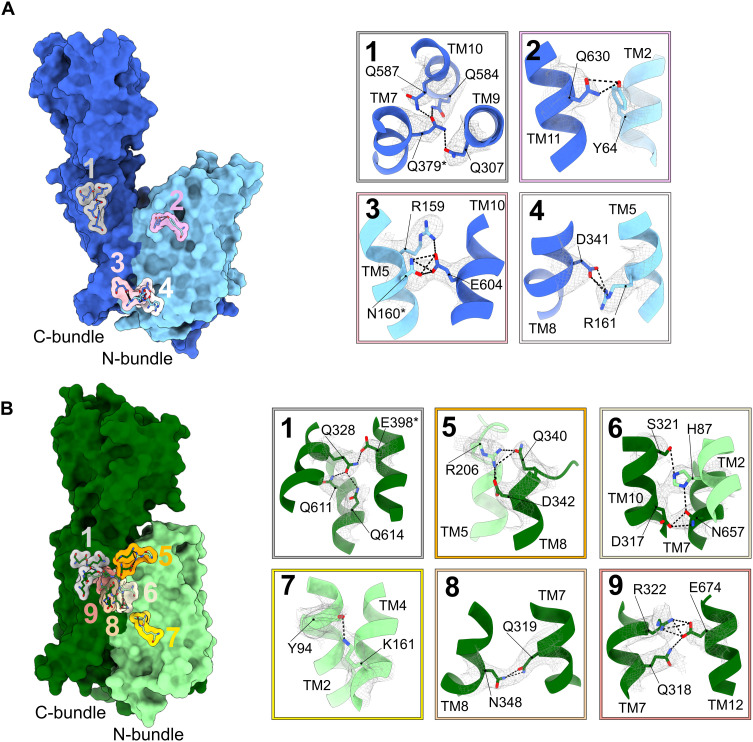
Fig. 4.
Interactions stabilizing the outward-facing open state of Hs PepT1 and the inward-facing partially occluded state of Hs PepT2.
The locations of key interactions are shown and labelled on ( A ) Hs PepT1 and ( B ) Hs PepT2. Corresponding close-up views show the cryo-EM densities of the side chains forming the interactions as in...
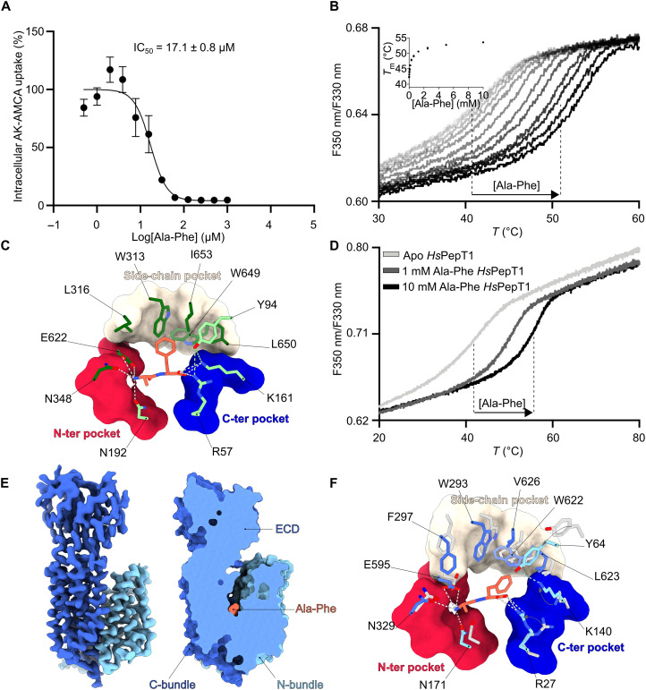
Fig. 5.
Structural basis for substrate recognition in human POTs.
( A ) Concentration-dependent competition assay of the u03b2-Ala-Lys peptide coupled to the fluorescent AMCA moiety (AK-AMCA) in Hs PepT2 with the dipeptide Ala-Phe. The average uptake value for each ...
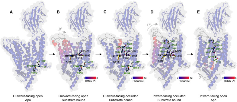
Fig. 6.
Mechanism for substrate recognition and transport in human POTs based on the presented structures.
( A ) In the first step of the transport cycle, the transporter is in an outward-facing open state stabilized by two salt bridges between R159-E604 and R161-D341. ( B ) Upon peptide bindingu2014accomm...
Figure images are served from the NIH/NLM PubMed Central Open Access Subset or Europe PMC; copyright remains with the publishers and authors.
💬 Discussion
0 commentsNo comments yet. Be the first to start a discussion!
Leave a Comment