Abstract
RNA polymerase II (Pol II) transcribes all protein-coding genes and many noncoding RNAs in eukaryotic genomes. Although Pol II is a complex, 12-subunit enzyme, it lacks the ability to initiate transcription and cannot consistently transcribe through long DNA sequences. To execute these essential functions, an array of proteins and protein complexes interact with Pol II to regulate its activity. In this review, we detail the structure and mechanism of over a dozen factors that govern Pol II initiation (e.g., TFIID, TFIIH, and Mediator), pausing, and elongation (e.g., DSIF, NELF, PAF, and P-TEFb). The structural basis for Pol II transcription regulation has advanced rapidly in the past decade, largely due to technological innovations in cryoelectron microscopy. Here, we summarize a wealth of structural and functional data that have enabled a deeper understanding of Pol II transcription mechanisms; we also highlight mechanistic questions that remain unanswered or controversial.
🔬 Techniques
🧬 Organisms
💻 Software
✨ Fluorophores
🧪 Sample Preparation
🔬 Cell Lines
🏭 Microscope Brands
🧪 Reagent Suppliers
📷 Detectors
💻 Software Details
💾 Data Repositories
🏛️ Research Organizations (ROR)
Affiliated research institutions:
📊 Figures
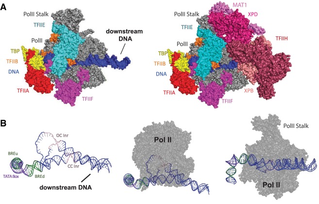
Figure 1.
Overview of the PIC and DNA path in closed and open complex. ( A ) The preinitiation complex (PIC) consists of TFIIA (red), TFIIB (orange), TFIID (not pictured), TFIIE (cyan), TFIIF (magenta), TFIIH (...
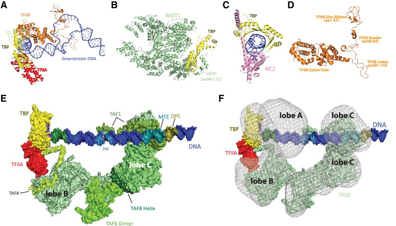
Figure 2.
TBP is a regulatory hub within the larger TFIID complex. ( A ) Structure of TBP (yellow), TFIIA (red), and TFIIB (orange) bound to promoter DNA in an open PIC complex. The TFIIB zinc ribbon (amino aci...
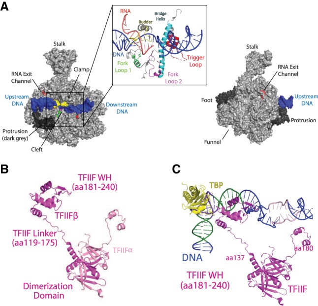
Figure 3.
Pol II and TFIIF. ( A ) Bovine Pol II (gray) shown in two orientations (rotated 180u00b0). DNA is colored in blue, and the Pol II stalk, clamp, foot, funnel, and RNA exit channel are marked; the protr...
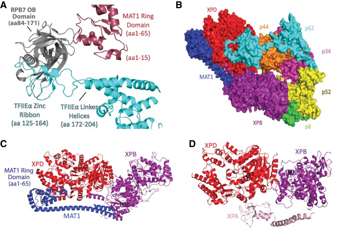
Figure 4.
Structural details for TFIIE and TFIIH. ( A ) TFIIE and TFIIH converge at the Pol II stalk. The Ring domain in the TFIIH subunit MAT1 interacts with the OB domain of RPB7 and the TFIIE linker helices....
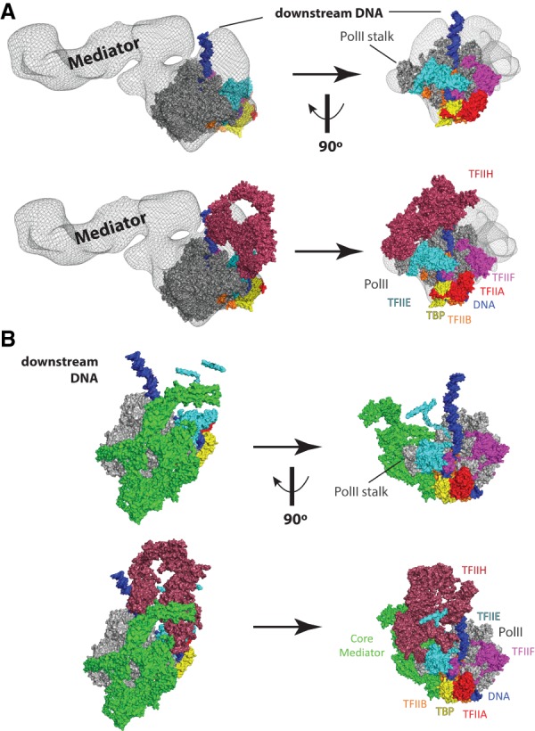
Figure 5.
PIC structural models that include the Mediator complex. ( A ) Model of a partial human PIC that includes Mediator. Figure was prepared by rendering a Mediatoru2013Pol IIu2013TFIIF cryoEM density ( Be...
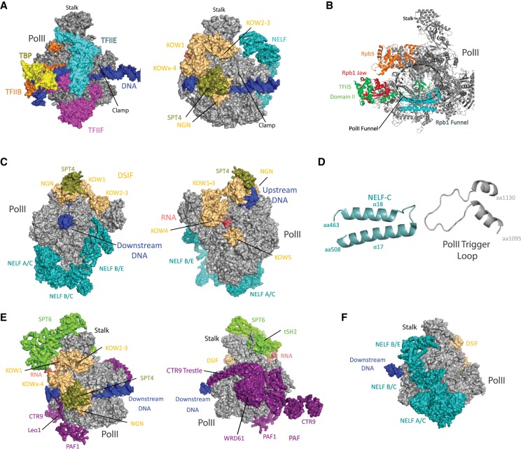
Figure 6.
Pol II elongation complexes bound to pausing (NELF) or elongation factors (DSIF, PAF, SPT6). ( A ) Structures of partial PICs that emphasize how DSIF binds Pol II surfaces occupied by TFIIB, TFIIE, an...
Figure images are served from the NIH/NLM PubMed Central Open Access Subset or Europe PMC; copyright remains with the publishers and authors.
💬 Discussion
0 commentsNo comments yet. Be the first to start a discussion!
Leave a Comment