Abstract
Transient receptor potential melastatin 2 (TRPM2) is a Ca2+-permeable cation channel required for immune cell activation, insulin secretion, and body heat control. TRPM2 is activated by cytosolic Ca2+, phosphatidyl-inositol-4,5-bisphosphate and ADP ribose. Here, we present the ~3 Å resolution electron cryo-microscopic structure of TRPM2 from Nematostella vectensis, 63% similar in sequence to human TRPM2, in the Ca2+-bound closed state. Compared to other TRPM channels, TRPM2 exhibits unique structural features that correlate with its function. The pore is larger and more negatively charged, consistent with its high Ca2+ selectivity and larger conductance. The intracellular Ca2+ binding sites are connected to the pore and cytosol, explaining the unusual dependence of TRPM2 activity on intra- and extracellular Ca2+. In addition, the absence of a post-filter motif is likely the cause of the rapid inactivation of human TRPM2. Together, our cryo-EM and electrophysiology studies provide a molecular understanding of the unique gating mechanism of TRPM2.
🔬 Techniques
🔭 Microscopes
🧬 Organisms
💻 Software
✨ Fluorophores
🧪 Sample Preparation
🔬 Cell Lines
🏭 Microscope Brands
🧪 Reagent Suppliers
💻 Software Details
💾 Data Repositories
🏷️ Research Resource Identifiers (RRIDs)
Verified research resources used in this paper:
🏛️ Research Organizations (ROR)
Affiliated research institutions:
📋 Methods
Key resources table
Reagent type (species) or resource Designation Source or reference Identifiers Additional information cell line ( Spodoptera frugiperda ) Sf9 ATCC CRL-1711 cell line ( Homo sapiens ) HEK293S GnTI- ATCC CRL-3022 biological sample ( Xenopus laevis ) Xenopus laevis oocytes African Reptile Park < mandyvorster@xsinet.co.za> RRID: NXR_0.0080 commercial assay or kit CNBR-activated sepharose beads GE Healthcare 17043001 commercial assay or kit Superose 6, 10/300 GL GE Healthcare 17517201 commercial assay or kit HiSpeed Plasmid Midi Kit Qiagen Catalog #12643 commercial assay or kit QuikChange XL Site- Directed Mutagenesis Kit Agilent Technologies Catalog #200521 commercial assay or kit mMESSAGE mMACHINE T7 Transcription Kit ThermoFisher Catalog #AM1344 chemical compound, drug 2,2-didecylpropane-1,3- bis-β-D-maltopyranoside (LMNG) Anatrace NG310 chemical compound, drug Cholesteryl hemisuccinate (CHS) Anatrace CH210 chemical compound, drug Digitonin Sigma-Aldrich D141 chemical compound, drug sf-900 II SFM medium Gibco Cat#10902088 chemical compound, drug Cellfectin II reagents Invitrogen Cat#10362100 chemical compound, drug Freestyle 293 medium Gibco Cat#12338018 chemical compound, drug HI FBS Gibco Cat#16140071 chemical compound, drug Antibiotic-Antimycotic (100X) Gibco Cat#15240062 chemical compound, drug Gentamicin sulphate Sigma-Aldrich G1397-10mL chemical compound, drug Collegenase type II Gibco by life technologies 17107–0125 chemical compound, drug Adenosine 5′- diphosphoribose sodium salt Sigma-Aldrich A0752 chemical compound, drug PtdIns-(4,5)-P2 (1,2- dioctanoyl) (sodium salt) Cayman Chemical 64910 software, algorithm Seriel EM DOI: 10.1016/j.jsb.2005.07.007 http://bio3d.colorado.edu/SerialEM software, algorithm MotionCor2 DOI: 10.1038/nmeth.4193 http://msg.ucsf.edu/em/software/motioncor2.html software, algorithm Gctf DOI: 10.1016/j.jsb.2015.11.003 https://www.mrc-lmb.cam.ac.uk /kzhang/ software, algorithm Gautomatch other https://www.mrc-lmb.cam.ac.uk /kzhang/ Downloaded from a personal URL software, algorithm RELION 2.1 DOI: 10.7554/eLife.18722 http://www2.mrc-lmb.cam.ac.uk /relion software, algorithm SWISS-MODEL DOI: 10.1093/nar/gku340 https://swissmodel.expasy.org software, algorithm COOT DOI: 10.1107/S0907444910007493 https://www2.mrc- lmb.cam.ac.uk/personal/ pemsley/coot software, algorithm PHENIX DOI: DOI: 10.1107/S0907444909052925 https://www.phenix-online.org software, algorithm Blocres DOI: 10.1016/j.jsb.2006.06.006 https://lsbr.niams.nih.gov/bsoft /programs/blocres.html software, algorithm MolProbity DOI: 10.1107/S0907444909042073; 10.1093/nar/gkm216 http://molprobity.biochem.duke .edu software, algorithm Chimera DOI: 10.1002/jcc.20084 https://www.cgl.ucsf.edu/chimera software, algorithm Pymol PyMOL http://www.pymol.org software, algorithm HOLE PMID: 9195488 http://www.holeprogram.org software, algorithm APBS DOI: 10.1093/nar/gkm276; 10.1073/pnas.181342398 http://www.poissonboltzmann.org software, algorithm Pclamp9 Molecular Devices RRID: SCR_011323 other R1.2/1.3 400 mesh Au holey carbon grids Quantifoil 1210627 Cell culture Insect cells were cultured at 28°C in sf-900 II SFM medium (GIBCO) supplemented with 5% FBS and 1% Antibiotic-Antimycotic. Mammalian cells were grown at 37°C in Freestyle 293 (GIBCO) supplemented with 2% FBS and 1% Antibiotic-Antimycotic. All cells were maintained with 8% CO 2 and 80% humidity. Protein expression and purification The Nematostella vectensis (nv) TRPM2 gene was synthesized into the pRML-13 BacMam expression vector (generous gift from Eric Gouaux) with a C-terminal GFP tag attached (General Biosystems). The plasmid was transformed into DH10 Bac cells (Invitrogen, Waltham, MA USA) to produce bacmid DNA, which was transfected into Sf9 cells (ATCC, Catalog#: ATCC CRL-1711) to generate recombinant baculoviruses. 10% (v/v) P3 virus was added to HEK 293S GnTI - cells (ATCC, Catalog#: ATCC CRL-3022) at 3 × 10 6 cells/ml. After 12 hr incubation at 37°C, protein expression was induced by 10 mM sodium butyrate at 30°C for 48 hr ( Goehring et al., 2014 ). Cells were harvested by centrifugation at 4,000 rpm for 20 min. For protein purification, the cells were resuspended and homogenized in lysis buffer (50 mM Tris-HCl pH 8.0, 2 mM MgCl 2 , 200 mM NaCl, 20% Glycerol, and 1 mM DTT) supplemented with protease inhibitors (1 mM phenylmethanesulfonyl fluoride (PMSF), 1 mM benzamidine, 1 μg/ml aprotinin, 100 μg/ml trypsin inhibitor, 1 μg/ml leupeptin, and 1 μg/ml pepstatin) and DNase (2 μg/ml). Membranes were solubilized with 1% 2,2-didecylpropane-1,3-bis-β-D-maltopyranoside (LMNG) and 0.1% cholesteryl hemisuccinate (CHS) at 4°C for 2 hr. After centrifugation at 75,000 g for 1 hr, the supernatant was mixed with GFP nanobody-coupled resin at 4°C for 2 hr. The resin was washed with 20 column volumes of Buffer A (20 mM Tris-HCl pH 8.0, 150 mM NaCl, 0.06% digitonin, and 1 mM DTT) to exchange LMNG and CHS with digitonin, and then incubated with PreScission protease (~10:1 w/w protein-to-enzyme ratio) at 4°C overnight to remove the C-terminal GFP tag. The GST-tagged protease was removed by binding to a glutathione-sepharose resin (GE HEalthcare). The concentrated nvTRPM2 protein was further purified by gel filtration in Buffer A on a Superose 6 10/300 column (GE Healthcare). EM sample preparation, data collection, and processing Gel filtration peak fractions in Buffer A were concentrated to 5 mg/ml protein, 3 μl of fresh protein was placed onto Quantifoil R1.2/1.3 400 Au holey carbon grids (Quantifoil) and blotted using Vitrobot (FEI). Humidity was set to 100%, blotting time to 3 s and force to 0. After flash freezing in liquid ethane, the grids were stored in liquid nitrogen until screening and data collection. The grids were initially screened on a 200 kV Talos Arctica (FEI) microscope, selected grids were then loaded into a 300 kV Titan Krios (FEI) microscope with a K2 summit detector (Gatan). Data were collected in super-resolution mode using Serial EM ( Mastronarde, 2005 ). Physical pixel size was 1.03 Å and dose rate was 8 electrons/pixel/second. Images were exposed for 10 s, subdivided into 50 frames, amounting to a total dose of ~75 electrons/Å 2 . A total of 1619 images were collected. After manual inspection to remove poor quality images, 1550 images were used for further processing. Beam-induced sample motion was corrected for using MotionCor2 ( Zheng et al., 2017 ). Contrast transfer function (CTF) estimation was performed using Gctf ( Zhang, 2016 ). Particle auto-picking was done by Gautomatch ( http://www.mrc-lmb.cam.ac.uk/kzhang ), selected particles were manually inspected to remove false-positives and supplement false-negatives. Finally, a total number of 196,198 particles were input to RELION2 ( Kimanius et al., 2016 ) for further classification and refinement. 2D classification sorted out 144,717 good particles which were used for subsequent 3D classification. The initial reference map for 3D classification was generated by ab-initio reconstruction in cryoSPARC ( Punjani et al., 2017 ). After 3D classification, the two good classes out of three, containing 104,268 particles, were combined for 3D auto-refine. The refinement, first carried out by using a loose mask which included the entire micelle densities, yielded a 3.22 Å map. Subsequent application of a tighter mask just around the protein density for the final local searches of 3D auto-refine increased the resolution to 3.11 Å. After post-processing, the final resolution was further improved to 3.07 Å using the 0.143 cutoff criterion. The map shown in this paper was sharpened with a B-factor of −101 Å 2 and low-pass filtered to 3.07 Å during post-processing.
Show full methods section
Key resources table
Reagent type (species) or resource Designation Source or reference Identifiers Additional information cell line ( Spodoptera frugiperda ) Sf9 ATCC CRL-1711 cell line ( Homo sapiens ) HEK293S GnTI- ATCC CRL-3022 biological sample ( Xenopus laevis ) Xenopus laevis oocytes African Reptile Park < mandyvorster@xsinet.co.za> RRID: NXR_0.0080 commercial assay or kit CNBR-activated sepharose beads GE Healthcare 17043001 commercial assay or kit Superose 6, 10/300 GL GE Healthcare 17517201 commercial assay or kit HiSpeed Plasmid Midi Kit Qiagen Catalog #12643 commercial assay or kit QuikChange XL Site- Directed Mutagenesis Kit Agilent Technologies Catalog #200521 commercial assay or kit mMESSAGE mMACHINE T7 Transcription Kit ThermoFisher Catalog #AM1344 chemical compound, drug 2,2-didecylpropane-1,3- bis-β-D-maltopyranoside (LMNG) Anatrace NG310 chemical compound, drug Cholesteryl hemisuccinate (CHS) Anatrace CH210 chemical compound, drug Digitonin Sigma-Aldrich D141 chemical compound, drug sf-900 II SFM medium Gibco Cat#10902088 chemical compound, drug Cellfectin II reagents Invitrogen Cat#10362100 chemical compound, drug Freestyle 293 medium Gibco Cat#12338018 chemical compound, drug HI FBS Gibco Cat#16140071 chemical compound, drug Antibiotic-Antimycotic (100X) Gibco Cat#15240062 chemical compound, drug Gentamicin sulphate Sigma-Aldrich G1397-10mL chemical compound, drug Collegenase type II Gibco by life technologies 17107–0125 chemical compound, drug Adenosine 5′- diphosphoribose sodium salt Sigma-Aldrich A0752 chemical compound, drug PtdIns-(4,5)-P2 (1,2- dioctanoyl) (sodium salt) Cayman Chemical 64910 software, algorithm Seriel EM DOI: 10.1016/j.jsb.2005.07.007 http://bio3d.colorado.edu/SerialEM software, algorithm MotionCor2 DOI: 10.1038/nmeth.4193 http://msg.ucsf.edu/em/software/motioncor2.html software, algorithm Gctf DOI: 10.1016/j.jsb.2015.11.003 https://www.mrc-lmb.cam.ac.uk /kzhang/ software, algorithm Gautomatch other https://www.mrc-lmb.cam.ac.uk /kzhang/ Downloaded from a personal URL software, algorithm RELION 2.1 DOI: 10.7554/eLife.18722 http://www2.mrc-lmb.cam.ac.uk /relion software, algorithm SWISS-MODEL DOI: 10.1093/nar/gku340 https://swissmodel.expasy.org software, algorithm COOT DOI: 10.1107/S0907444910007493 https://www2.mrc- lmb.cam.ac.uk/personal/ pemsley/coot software, algorithm PHENIX DOI: DOI: 10.1107/S0907444909052925 https://www.phenix-online.org software, algorithm Blocres DOI: 10.1016/j.jsb.2006.06.006 https://lsbr.niams.nih.gov/bsoft /programs/blocres.html software, algorithm MolProbity DOI: 10.1107/S0907444909042073; 10.1093/nar/gkm216 http://molprobity.biochem.duke .edu software, algorithm Chimera DOI: 10.1002/jcc.20084 https://www.cgl.ucsf.edu/chimera software, algorithm Pymol PyMOL http://www.pymol.org software, algorithm HOLE PMID: 9195488 http://www.holeprogram.org software, algorithm APBS DOI: 10.1093/nar/gkm276; 10.1073/pnas.181342398 http://www.poissonboltzmann.org software, algorithm Pclamp9 Molecular Devices RRID: SCR_011323 other R1.2/1.3 400 mesh Au holey carbon grids Quantifoil 1210627 Cell culture Insect cells were cultured at 28°C in sf-900 II SFM medium (GIBCO) supplemented with 5% FBS and 1% Antibiotic-Antimycotic. Mammalian cells were grown at 37°C in Freestyle 293 (GIBCO) supplemented with 2% FBS and 1% Antibiotic-Antimycotic. All cells were maintained with 8% CO 2 and 80% humidity. Protein expression and purification The Nematostella vectensis (nv) TRPM2 gene was synthesized into the pRML-13 BacMam expression vector (generous gift from Eric Gouaux) with a C-terminal GFP tag attached (General Biosystems). The plasmid was transformed into DH10 Bac cells (Invitrogen, Waltham, MA USA) to produce bacmid DNA, which was transfected into Sf9 cells (ATCC, Catalog#: ATCC CRL-1711) to generate recombinant baculoviruses. 10% (v/v) P3 virus was added to HEK 293S GnTI - cells (ATCC, Catalog#: ATCC CRL-3022) at 3 × 10 6 cells/ml. After 12 hr incubation at 37°C, protein expression was induced by 10 mM sodium butyrate at 30°C for 48 hr ( Goehring et al., 2014 ). Cells were harvested by centrifugation at 4,000 rpm for 20 min. For protein purification, the cells were resuspended and homogenized in lysis buffer (50 mM Tris-HCl pH 8.0, 2 mM MgCl 2 , 200 mM NaCl, 20% Glycerol, and 1 mM DTT) supplemented with protease inhibitors (1 mM phenylmethanesulfonyl fluoride (PMSF), 1 mM benzamidine, 1 μg/ml aprotinin, 100 μg/ml trypsin inhibitor, 1 μg/ml leupeptin, and 1 μg/ml pepstatin) and DNase (2 μg/ml). Membranes were solubilized with 1% 2,2-didecylpropane-1,3-bis-β-D-maltopyranoside (LMNG) and 0.1% cholesteryl hemisuccinate (CHS) at 4°C for 2 hr. After centrifugation at 75,000 g for 1 hr, the supernatant was mixed with GFP nanobody-coupled resin at 4°C for 2 hr. The resin was washed with 20 column volumes of Buffer A (20 mM Tris-HCl pH 8.0, 150 mM NaCl, 0.06% digitonin, and 1 mM DTT) to exchange LMNG and CHS with digitonin, and then incubated with PreScission protease (~10:1 w/w protein-to-enzyme ratio) at 4°C overnight to remove the C-terminal GFP tag. The GST-tagged protease was removed by binding to a glutathione-sepharose resin (GE HEalthcare). The concentrated nvTRPM2 protein was further purified by gel filtration in Buffer A on a Superose 6 10/300 column (GE Healthcare). EM sample preparation, data collection, and processing Gel filtration peak fractions in Buffer A were concentrated to 5 mg/ml protein, 3 μl of fresh protein was placed onto Quantifoil R1.2/1.3 400 Au holey carbon grids (Quantifoil) and blotted using Vitrobot (FEI). Humidity was set to 100%, blotting time to 3 s and force to 0. After flash freezing in liquid ethane, the grids were stored in liquid nitrogen until screening and data collection. The grids were initially screened on a 200 kV Talos Arctica (FEI) microscope, selected grids were then loaded into a 300 kV Titan Krios (FEI) microscope with a K2 summit detector (Gatan). Data were collected in super-resolution mode using Serial EM ( Mastronarde, 2005 ). Physical pixel size was 1.03 Å and dose rate was 8 electrons/pixel/second. Images were exposed for 10 s, subdivided into 50 frames, amounting to a total dose of ~75 electrons/Å 2 . A total of 1619 images were collected. After manual inspection to remove poor quality images, 1550 images were used for further processing. Beam-induced sample motion was corrected for using MotionCor2 ( Zheng et al., 2017 ). Contrast transfer function (CTF) estimation was performed using Gctf ( Zhang, 2016 ). Particle auto-picking was done by Gautomatch ( http://www.mrc-lmb.cam.ac.uk/kzhang ), selected particles were manually inspected to remove false-positives and supplement false-negatives. Finally, a total number of 196,198 particles were input to RELION2 ( Kimanius et al., 2016 ) for further classification and refinement. 2D classification sorted out 144,717 good particles which were used for subsequent 3D classification. The initial reference map for 3D classification was generated by ab-initio reconstruction in cryoSPARC ( Punjani et al., 2017 ). After 3D classification, the two good classes out of three, containing 104,268 particles, were combined for 3D auto-refine. The refinement, first carried out by using a loose mask which included the entire micelle densities, yielded a 3.22 Å map. Subsequent application of a tighter mask just around the protein density for the final local searches of 3D auto-refine increased the resolution to 3.11 Å. After post-processing, the final resolution was further improved to 3.07 Å using the 0.143 cutoff criterion. The map shown in this paper was sharpened with a B-factor of −101 Å 2 and low-pass filtered to 3.07 Å during post-processing.
Model building and refinement
The transmembrane domain (TMD) was modeled using SWISS-MODEL ( Biasini et al., 2014 ) based on the TRPV1 structure (PDB: 3J5P) ( Liao et al., 2013 ), roughly fitted into the cryo-EM map in Chimera ( Pettersen et al., 2004 ), and then manually adjusted to the density map in Coot ( Emsley et al., 2010 ). All other regions of the protein were built de novo in Coot, since the densities for most of the side chains were quite distinct. The N-Acetylglucosamine modifications of residue N1017 were very clear, and further confirmed the validity of model building around this region. Some obvious phospholipid-like densities were seen around the TMD. As these accounted only for parts of phospholipids, and the densities for the head groups were particularly poorly resolved, they were filled with various truncated versions of 1-palmitoyl-2-oleoyl-sn-glycero-3-phosphocholine (POPC) to maximally fit the densities. In addition, a few flatter densities that could not be fitted with phospholipids were filled with cholesterol. The final structure model (tetramer) contained 4244 protein residues (6–23, 41–51, 86–546, 573–755, 812–1136, and 1148–1210), 4 Ca 2+ ions, 3 Na + ions, 60 lipid molecules, and 8 N-Acetylglucosamine molecules. The EM density for the C-terminal NUDT9H domain (residues 1271–1551) was not observed. Because the very N-terminal segments (6–23 and 41–51) had poor density and were disconnected from the rest of the protein, their registers are somewhat uncertain and they were built as poly-alanines. The refinement was performed in Phenix ( Adams et al., 2010 ) with secondary structure and C4 non-crystallographic symmetry (NCS) restraints. For cross-validation ( Brown et al., 2015 ), the two half maps generated by 3D auto-refine were sharpened with the same B-factor and low-pass filter as used for post-processing. The model was randomly displaced by 0.5 Å and then refined against half map1 using Phenix. The Fourier shell correlation (FSC) curves were calculated between the refined model and different maps (full map, half map1, and half map2), respectively. The small differences between the FSC curves derived from half map1 and half map2 indicated that the model was not over-fitted during refinement. Local resolution estimation was carried out by Blocres ( Heymann and Belnap, 2007 ). Validation of geometries was performed using MolProbity ( Chen et al., 2010 ; Davis et al., 2007 ). All the structure figures were generated using Pymol ( http://www.pymol.org ), Chimera, HOLE ( Smart et al., 1996 ), and APBS ( Baker et al., 2001 ; Dolinsky et al., 2007 ).
Isolation and maintenance of Xenopus laevis oocytes
Ovarian lobes were removed from anaesthetized Xenopus laevis [RRID: NXR_0.0080 ] following a IACUC-approved protocol. Oocytes were defolliculated by treatment with Type II collagenase (GIBCO) and stored at 18°C in a frog Ringer's solution supplemented with 1.8 mM CaCl 2 plus 50 μg/ml gentamycin sulfate (Sigma). Functional expression of nvTRPM2 in Xenopus laevis oocytes The nv TRPM2 gene was synthesized into the pGEMHE expression vector (General Biosystems). Mutations were introduced using Stratagene QuikChange. cDNA was transcribed in vitro using T7 polymerase (mMESSAGE mMACHINE T7 kit Thermo Scientific), and cRNA stored at −80°C. Xenopus laevis oocytes were injected (Drummond Nanoject) with 0.1–10 ng of WT or mutant nvTRPM2 cRNA, and recordings were done 1–3 days after injection.
Excised inside-out patch-clamp recording
Macroscopic and unitary nvTRPM2 currents were recorded in excised inside-out patches at 25°C in symmetrical 140 mM Na-gluconate based solutions to avoid activation of endogenous Ca 2+ -activated chloride currents, as described earlier for hTRPM2 ( Csanády and Töröcsik, 2009 ). The tip of the patch pipette was filled to ~1 cm height with 140 mM Na-gluconate, 2 mM Mg-gluconate 2 , 10 mM HEPES (pH = 7.4 with NaOH; free [Ca 2+ ]~0.5 μM); 1 mM Na-EGTA or 8 mM Ca(gluconate) 2 was added to obtain free [Ca 2+ ] of ~1 nM ( Figures 1 , 4 and 5D ) or ~1 mM ( Figures 5E and 6 ). The pipette electrode was placed into a 140 mM NaCl-based solution carefully layered on top. Bath solution contained 140 mM Na-gluconate, 2 mM Mg-gluconate 2 , 10 mM HEPES (pH 7.1 with NaOH), and either 1 mM EGTA (to obtain ‘zero’ (~1 nM) Ca 2+ ), or 20 μM to 10 mM Ca-gluconate 2 (to obtain 3 to 1250 μM free [Ca 2+ ]). For unitary current measurements under biionic conditions ( Figure 3G ) the pipette solution contained 10 mM CaCl 2 , 0.5 mM MgCl 2 , and 10 mM HEPES (pH = 7.4 with Ca(OH) 2 ). Na 2 -ADPR (Sigma), Dioctanoyl-PI(4,5)P 2 (Cayman Chemical), and poly-L-lysine (Sigma) were added to the bath solution from 200 mM, 2.5 mM, and 15 mg/ml aqueous stock solutions, respectively. The continuously flowing bath solution was exchanged using computer-driven electronic valves; solution exchange time constant was + 4 mV, the extrapolated reversal potential (~+28 mV) was also verified in, and found roughly consistent with, macroscopic recordings. Fractional current activation in 100 μM ADPR by test concentrations of cytosolic Ca 2+ , or by 125 μM Ca 2+ + 25 μM dioctanoyl-PIP 2 , were calculated by dividing steady current in the test segment ( I ) with that in 125 μM Ca 2+ ( I 125 ) in the same patch. Open probabilities (P o ) normalized to that in 125 μM Ca 2+ (P o;125 ), were calculated as P o /P o;125 =( I / I 125 )/( i / i 125 ) ( i , unitary current; i 125 , unitary current in 125 μM cytosolic Ca 2+ [see Figure 4E ]). To emphasize several orders-of-magnitude reductions in maximal open probability for the Ca 2+ site mutants, normalized currents and open probabilities hence obtained are shown in Figure 4K rescaled by their values measured in 125 μM Ca 2+ + 25 μM PIP 2 , i.e., I /I 125+PIP2 =( I /I 125 )/( I 125+PIP2 /I 125 ) and P o /P o;125+PIP2 =(P o /P o;125 )/(P o;125+PIP2 /P o;125 ). For D921A nvTRPM2 channel currents in the absence of PIP 2 were too small for reliable cursor measurement. Thus, for this mutant i and N ⋅P o in 13, 125, or 1250 μM cytosolic Ca 2+ was estimated using dwell-time analysis, and fractional P o under such conditions ( Figure 4K , right , purple bars ) calculated as P o /P o;125+PIP2 =( i ⋅ N ⋅P o )/(I 125+PIP2 ). Macroscopic current relaxations ( Figure 5D–E ) were fitted by single-exponential functions using least-squares. At negative membrane potentials, in the presence of external Ca 2+ , current decay time courses upon cytosolic Ca 2+ removal ( Figure 5E ) do not reflect channel closing rate, as opening rate remains non-zero under such conditions: thus, these current segments were not fitted.
Statistics
The quantification and statistical analyses for the structural parts are integral outputs of the software and algorithms used. Electrophysiological data are given as mean ± SEM of measurements from ≥5 (typically ~20) segments of recording, from ≥3 (typically ~10) patches.
Data and software availability
Cryo-EM density map of nvTRPM2 has been deposited in the electron microscopy data bank (EMDB) under accession code EMD-7542. Atomic coordinates of nvTRPM2 have been deposited in the protein data bank (PDB) under accession code: 6CO7.
Additional files 10.7554/eLife.36409.018 Transparent reporting form Data availability Cryo-EM density map of nvTRPM2 has been deposited in the electron microscopy data bank (EMDB) under accession code EMD-7542. Atomic coordinates of nvTRPM2 have been deposited in the protein data bank (PDB) under accession code: 6CO7. The following datasets were generated: Zhe Zhang Balázs Tóth Andras Szollosi Jue Chen László Csanády 2018 Structure of the nvTRPM2 channel in complex with Ca2+ http://www.rcsb.org/pdb/search/structidSearch.do?structureId=6CO7 Publicly available at the RCSB Protein Data Bank (accession no. 6CO7) Zhe Zhang Balázs Tóth Andras Szollosi Jue Chen László Csanády 2018 Structure of the nvTRPM2 channel in complex with Ca2+ www.ebi.ac.uk/pdbe/entry/emdb/EMD-7542 Publicly available at the Electron Microscopy Data Bank (accession no. EMD-7542)
📊 Figures
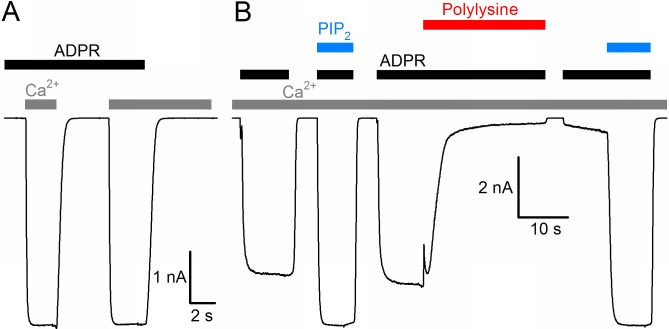
Figure 1.
Basic functional properties of the Nematostella vectensis (nv) TRPM2 channel.
( A ) Macroscopic inward Na + currents in an inside-out patch excised from a Xenopus laevis oocyte overexpressing nvTRPM2, evoked by superfusion of the cytoplasmic patch surface with 100 u03bcM ADPR (...
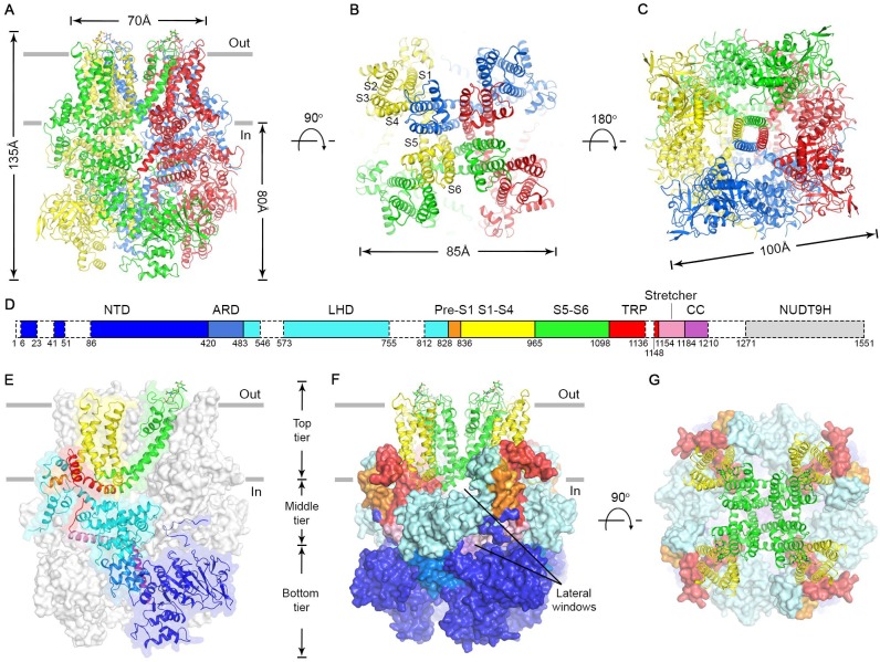
Figure 2.
Cryo-EM structure of the nvTRPM2 channel.
( Au2013C ) Different views of the overall structure of the nvTRPM2 tetramer. Protomers are color coded. Gray bars in ( A ) represent approximate membrane boundaries. Transmembrane helices S1-S6 of on...
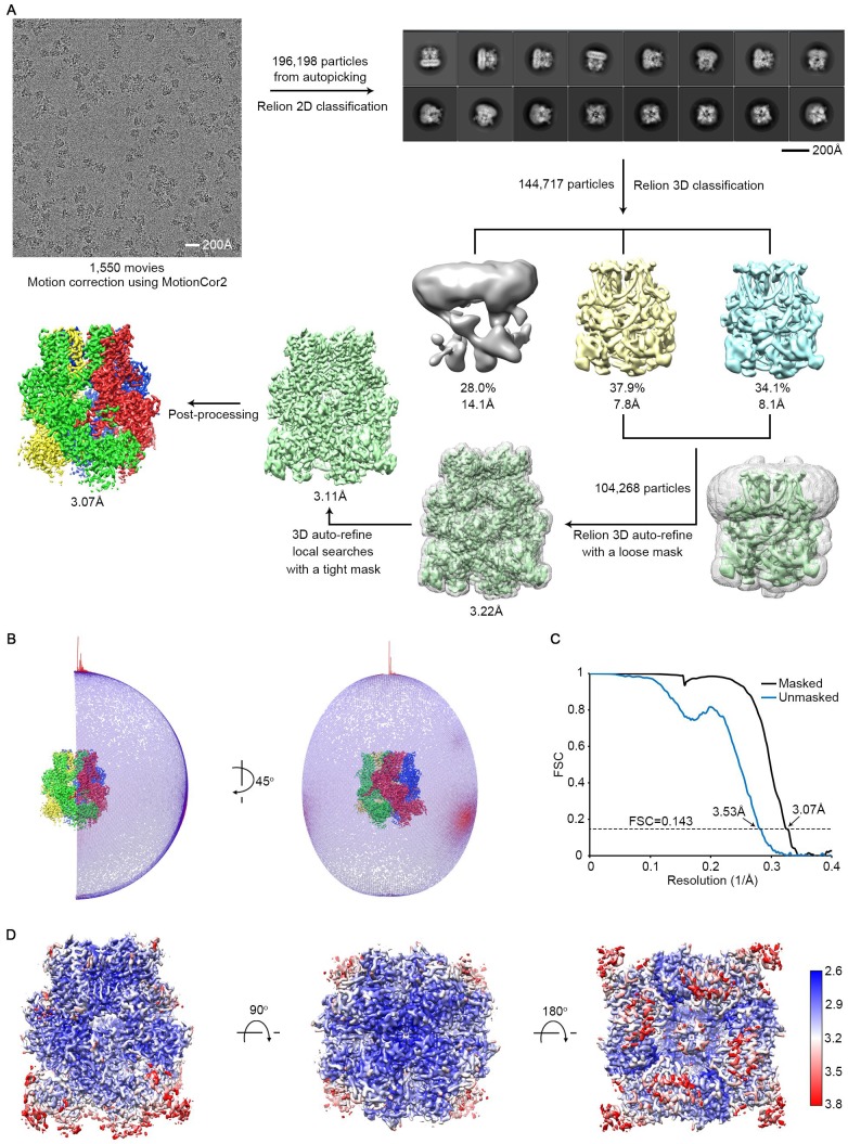
Figure 2u2014figure supplement 1.
Cryo-EM structure determination and evaluation.
( A ) Flow chart of data processing. ( B ) Angular distributions for all the particles used in the final reconstruction. Red and higher cylinders represent more particles, blue and shorter cylinders l...
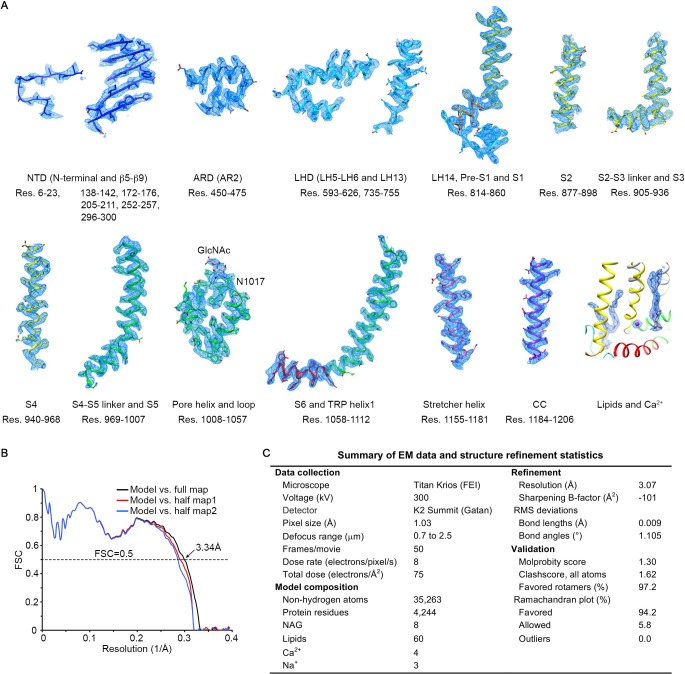
Figure 2u2014figure supplement 2.
Validation of the atomic model.
( A ) Local EM density maps and fitted atomic models for representative regions of the structure. ( B ) Fourier shell correlation (FSC) curves between the model and different maps. The model was refin...
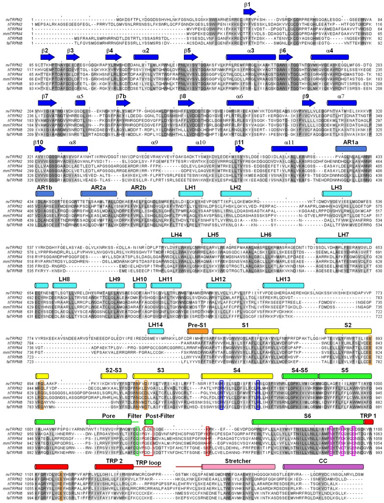
Figure 2u2014figure supplement 3.
Multiple sequence alignment of TRPM proteins.
Sequence alignment of TRPM2, TRPM4, TRPM5, and TRPM8 channel orthologs (h: human, nv: Nematostella vectensis , mm: Mus musculus , fa: Ficedula albicollis ) predicted by Clustal Omega ( Li et al., 2015...
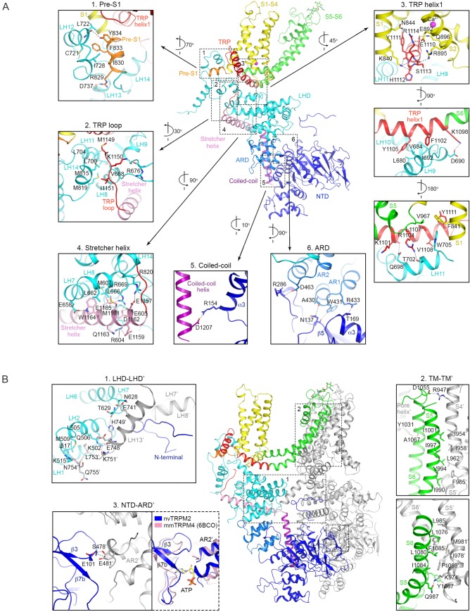
Figure 2u2014figure supplement 4.
Intra- and inter-subunit interactions.
( A ) Intra-subunit interactions. Interactions between different domains within one subunit shown in cartoon representation and color-coded by domains (see Figure 2D ). ( B ) Inter-subunit interaction...
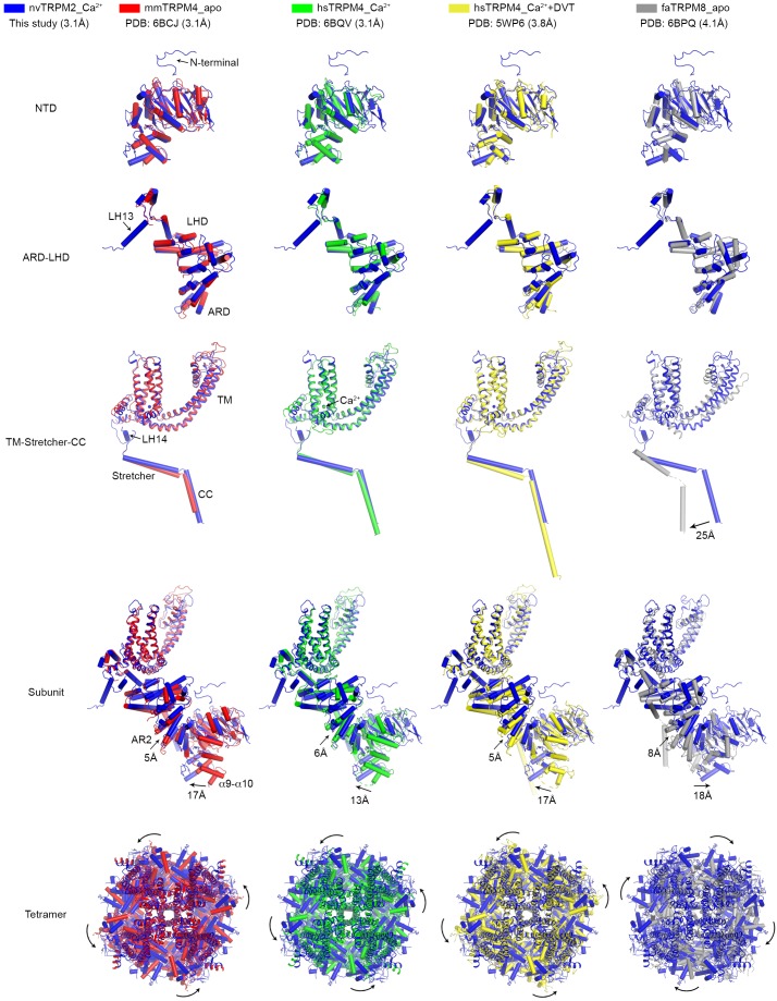
Figure 2u2014figure supplement 5.
Structural comparisons between individual domains, one subunit, or the entire tetramer of nvTRPM2 and other TRPM family channels.
The superposition was performed for individual domains (NTD, ARD-LHD, and TM-Stretcher-CC), the integral subunit, as well as the entire tetramer. The root-mean-square deviations (RMSDs) between nvTRPM...
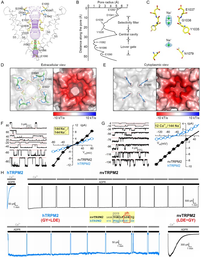
Figure 3.
Pore of the nvTRPM2 channel.
( A ) Ribbon representation of the ion pore, front and rear subunits are removed for clarity. The dotted mesh distinguishes regions that are too tight ( red , radiusu00a0<1.15 u00c5) or just spatio...
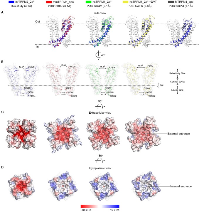
Figure 3u2014figure supplement 1.
Structural comparisons between the ion channel pore of nvTRPM2 and of other TRPM family channels.
( A ) Superposition of the pore region (S5u2013S6) between nvTRPM2 and other TRPM family structures. The same structures as those in Figure 2u2014figure supplement 5 were used for comparison. Only one...
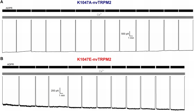
Figure 3u2014figure supplement 2.
Disruption of post-filter salt bridge does not cause inactivation.
( Au2013B ) Lack of inactivation of nvTRPM2 channels in which the E1042-K1047 salt bridge has been disrupted by mutation K1047A ( A ) or K1047E ( B ). Membrane potential was u221220 mV, currents were ...
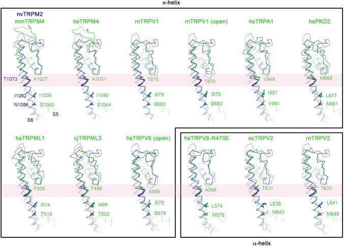
Figure 3u2014figure supplement 3.
Alpha-pi-alpha helical transition in S6 of TRP family channels.
Pairwise structural superposition of the pore domain of one nvTRPM2 subunit ( blue ) with those of other TRP channels ( green ), including mm ( Mus musculus ) and hs ( Homo sapiens ) TRPM4 (PDB: 6BCJ ...
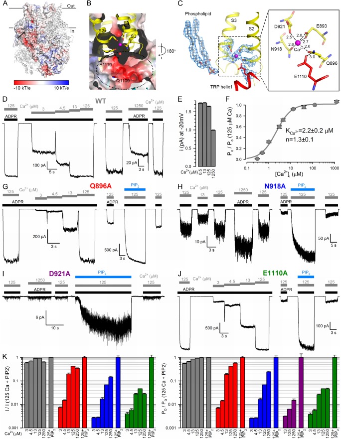
Figure 4.
The nvTRPM2 Ca 2+ binding site.
( A ) The Ca 2+ binding site is located close to the inner leaflet of the membrane. One subunit is represented as electrostatic surface, the remaining three are shown as ribbon. The position of the Ca...
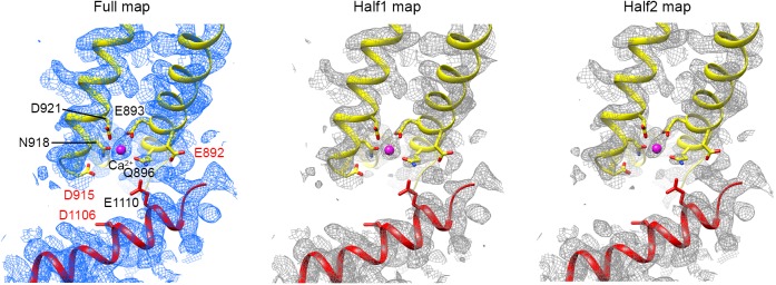
Figure 4u2014figure supplement 1.
Density maps around the Ca 2+ binding site.
The full map and both half maps are presented. Residues that participate in Ca 2+ binding are labeled with black font . Some adjacent acid amino acids not involved in hydrogen bonds or ion bindings ar...
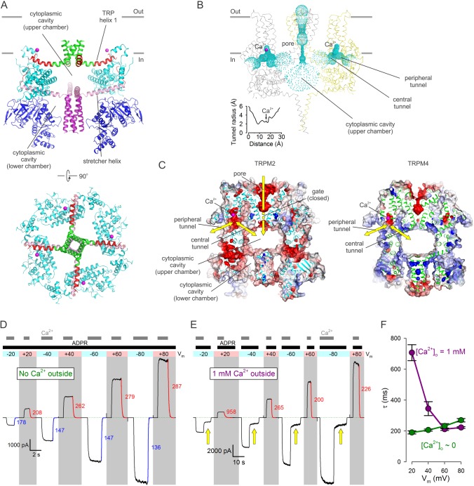
Figure 5.
Cytoplasmic cavities and tunnels.
( A ) Architecture of the cytoplasmic cavity viewed from an angle parallel ( top ) or perpendicular ( bottom ) to the membrane plane. The NTD ( blue ), the LHD ( cyan ), the cytoplasmic ends of transm...
Video 1.
Cytosolic chambers and tunnels in nvTRPM2.
( Right ) Vertical cross section along nvTRPM2 pore axis: 4 u00c5 slab represented in spacefill with surface rendering. Domain color coding as in Figure 1E . Sodium ions in the pore and bound Ca 2+ io...
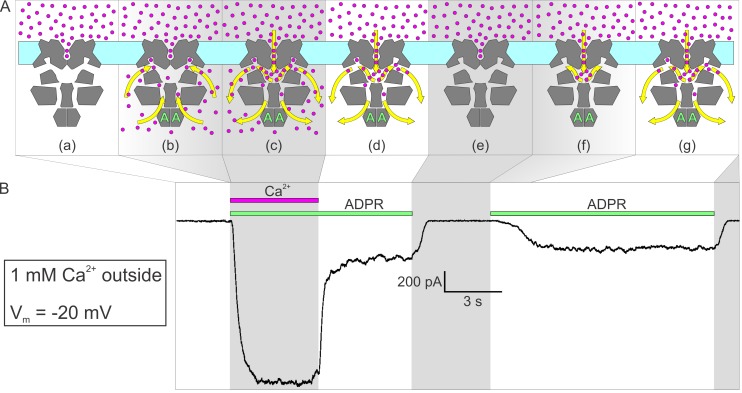
Figure 6.
Activation by ADPR of nvTRPM2 current in the presence of external but absence of cytosolic Ca 2+ .
( A ) Cartoon interpretation of the molecular events that occur during consecutive time intervals ( sections a-g , also identified by intermittent gray shading in ( A )-( B )) of the current recording...
Figure images are served from the NIH/NLM PubMed Central Open Access Subset or Europe PMC; copyright remains with the publishers and authors.
💬 Discussion
0 commentsNo comments yet. Be the first to start a discussion!
Leave a Comment