Abstract
The current Zika virus (ZIKV) epidemic is characterized by severe pathogenicity in both children and adults. Sequence changes in ZIKV since its first isolation are apparent when pre-epidemic strains are compared with those causing the current epidemic. However, the residues that are responsible for ZIKV pathogenicity are largely unknown. Here we report the cryo-electron microscopy (cryo-EM) structure of the immature ZIKV at 9-Å resolution. The cryo-EM map was fitted with the crystal structures of the precursor membrane and envelope glycoproteins and was shown to be similar to the structures of other known immature flaviviruses. However, the immature ZIKV contains a partially ordered capsid protein shell that is less prominent in other immature flaviviruses. Furthermore, six amino acids near the interface between pr domains at the top of the spikes were found to be different between the pre-epidemic and epidemic ZIKV, possibly influencing the composition and structure of the resulting viruses.
🔬 Techniques
🔭 Microscopes
💻 Software
🧪 Sample Preparation
🏭 Microscope Brands
📷 Detectors
💻 Software Details
💾 Data Repositories
🏛️ Research Organizations (ROR)
Affiliated research institutions:
📋 Methods
Virus preparation and purification Approximately 1 × 10 9 C6/36 cells (ATCC CRL-1660) were infected with Zika virus (strain H/PF/2013) at a multiplicity of infection of 4 for 16hrs at 30°C. The inoculum was removed and cells were rinsed three times with phosphate buffered saline (pH 8.0) and incubated for two hours at 30°C in minimal essential media containing 2% fetal bovine serum, 25mM HEPES pH 7.3, and 30mM NH 4 Cl. The process was repeated two more times for a total of three incubations at 30°C in cell culture media containing 30mM NH 4 Cl. Media was collected 72 hours post infection and immature virus particles were purified according to previously described methods 12 . Briefly, virus particles were precipitated from the media by overnight mixing with 8% polyethylene glycol 8000 at 4°C. This mixture was then pelleted at 8900× g for 50 minutes at 4°C. Resuspended particles were pelleted through a 24% sucrose cushion, resuspended in 0.5mL NTE buffer (20mM Tris pH 8.0, 120mM NaCl, 1mM EDTA) and purified with a discontinuous gradient in 5% intervals from 35% to 10% K-tartrate, 20mM Tris pH 8.0 and 1mM EDTA. Immature virus was extracted from the gradient, concentrated and buffer exchanged into NTE buffer.
Cryo-electron microscopy data collection
The purified immature ZIKV sample was plunge frozen on ultrathin lacey carbon EM grids (Electron Microscopy Sciences). The grids were examined using an FEI Titan Krios electron microscope with a Gatan K2 Summit detector. Cryo-EM images were collected at a magnification of 22,500× in the “super-resolution” data collection mode. The pixel size of the collected images was 0.65 Å. The total exposure time for producing one image composed of 38 frames was 7.6 s and required a dose rate of 4.7 electrons Å −2 s −1 . A total of 3341 images were collected and 14351 particles were boxed manually using the e2boxer program from the EMAN2 software 26 .
Show full methods section
Virus preparation and purification Approximately 1 × 10 9 C6/36 cells (ATCC CRL-1660) were infected with Zika virus (strain H/PF/2013) at a multiplicity of infection of 4 for 16hrs at 30°C. The inoculum was removed and cells were rinsed three times with phosphate buffered saline (pH 8.0) and incubated for two hours at 30°C in minimal essential media containing 2% fetal bovine serum, 25mM HEPES pH 7.3, and 30mM NH 4 Cl. The process was repeated two more times for a total of three incubations at 30°C in cell culture media containing 30mM NH 4 Cl. Media was collected 72 hours post infection and immature virus particles were purified according to previously described methods 12 . Briefly, virus particles were precipitated from the media by overnight mixing with 8% polyethylene glycol 8000 at 4°C. This mixture was then pelleted at 8900× g for 50 minutes at 4°C. Resuspended particles were pelleted through a 24% sucrose cushion, resuspended in 0.5mL NTE buffer (20mM Tris pH 8.0, 120mM NaCl, 1mM EDTA) and purified with a discontinuous gradient in 5% intervals from 35% to 10% K-tartrate, 20mM Tris pH 8.0 and 1mM EDTA. Immature virus was extracted from the gradient, concentrated and buffer exchanged into NTE buffer.
Cryo-electron microscopy data collection
The purified immature ZIKV sample was plunge frozen on ultrathin lacey carbon EM grids (Electron Microscopy Sciences). The grids were examined using an FEI Titan Krios electron microscope with a Gatan K2 Summit detector. Cryo-EM images were collected at a magnification of 22,500× in the “super-resolution” data collection mode. The pixel size of the collected images was 0.65 Å. The total exposure time for producing one image composed of 38 frames was 7.6 s and required a dose rate of 4.7 electrons Å −2 s −1 . A total of 3341 images were collected and 14351 particles were boxed manually using the e2boxer program from the EMAN2 software 26 .
Three-dimensional reconstruction and data analysis
Non-reference two-dimensional classification was performed using the Relion software package 27 resulting in the selection of 9315 particles. This dataset was split into two equal subsets as required by the “gold standard” for determining the quality of the cryo-EM reconstruction. The jspr program 28 was used for initial model generation and refinement of the orientations of the selected particles. The selected particles were used to generate a cryo-EM map at an average resolution of 9.34 Å based on the 0.143 Fourier shell correlation criterion. After the application of soft spherical masks, the resolution improved to 9.14 Å. Fitting of crystal structures into the cryo-EM map The crystal structure of immature dengue virus-2 prM-E heterodimer (PDB ID: 3C6E) was used for fitting into a trimeric spike in the immature ZIKV map. Sequential fitting of the three prM-E molecules into the trimeric spike density was carried out using the UCSF Chimera software 29 . The Cα backbone of the transmembrane helices of M and E proteins from the mature ZIKV structure (PDB ID: 5IRE) were also fitted into the transmembrane regions of the three prM-E heterodimers. The solution structure of DENV-2 capsid protein (PDB ID: 1R6R) was placed into the density between the inner layer of the viral membrane and the RNA core of immature ZIKV, according to previously suggested orientation of the capsid protein in the virion 17 . The mature ZIKV E protein’s ectodomain structure (PDB ID: 5IRE) 8 was split into two parts, with one molecule containing the domain I plus III and the second molecule containing domain II. These different parts were superposed independently on the immature dengue prM-E molecule for determining the interaction regions between E proteins from adjacent spikes. Interface regions between different protein partners were identified as residues with less than 6 Å distance between their corresponding Cα backbones.
Multiple sequence alignment
Sequence alignments of the pr domain and E glycoprotein amongst flaviviruses were carried out using the MAFFT program 30 and rendered using the ESPript software 31 . The sequences for comparison were obtained from the ViPR resource database 32 . The representative flavivirus strains used in the comparison were Zika-Asian (H/PF/2013 strain), Zika-African (1968-Nigeria strain), West Nile virus (NY99 strain), Japanese encephalitis virus (SA14), yellow fever virus (Asibi strain) and the four dengue virus serotypes (Western Pacific, S16803 , CH53489 and IND1979 strains). In the manuscript, the residue numbers for pr and E are used based on the ZIKV sequence and structure, though the numbers assigned to the residues during multiple sequence alignment may have slightly shifted in Supplementary Fig. 4 .
Supplementary Material 1 2
📊 Figures
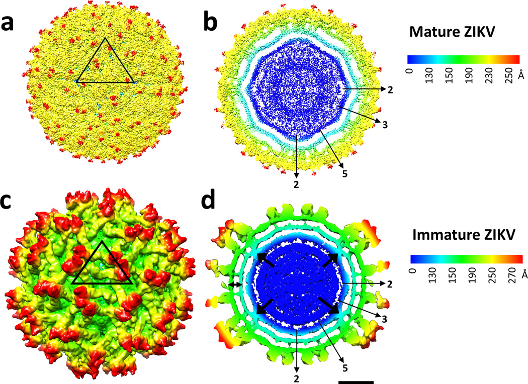
Figure 1
Cryo-EM structure of immature Zika virus. Surface view ( a and c ) and cross-section ( b and d ) of mature 8 and immature ZIKV colored radially. Black arrows indicate the density between the inner RNA...
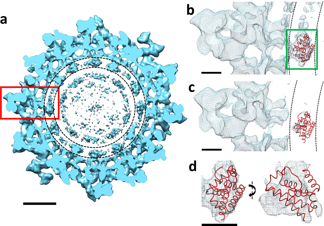
Figure 2
Cross-section of the immature ZIKV contoured to show the inner capsid shell. ( a ) Cross-section of immature ZIKV projected down an icosahedral 2-fold axis. The putative location of the inner capsid s...
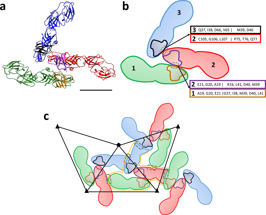
Figure 3
Interface between prM-E heterodimers. ( a ) Ribbon diagram of prM-E heterodimers in a trimeric spike of immature ZIKV. The prM-E heterodimers are colored in green, red and blue for the E proteins wher...
Figure images are served from the NIH/NLM PubMed Central Open Access Subset or Europe PMC; copyright remains with the publishers and authors.
💬 Discussion
0 commentsNo comments yet. Be the first to start a discussion!
Leave a Comment