Abstract
Ca2+/calmodulin-dependent protein kinase II (CaMKII) and long-term potentiation (LTP) were discovered within a decade of each other and have been inextricably intertwined ever since. However, like many marriages, it has had its up and downs. Based on the unique biochemical properties of CaMKII, it was proposed as a memory molecule before any physiological linkage was made to LTP. However, as reviewed here, the convincing linkage of CaMKII to synaptic physiology and behavior took many decades. New technologies were critical in this journey, including in vitro brain slices, mouse genetics, single-cell molecular genetics, pharmacological reagents, protein structure, and two-photon microscopy, as were new investigators attracted by the exciting challenge. This review tracks this journey and assesses the state of this marriage 40 years on. The collective literature impels us to propose a relatively simple model for synaptic memory involving the following steps that drive the process: 1) Ca2+ entry through N-methyl-d-aspartate (NMDA) receptors activates CaMKII. 2) CaMKII undergoes autophosphorylation resulting in constitutive, Ca2+-independent activity and exposure of a binding site for the NMDA receptor subunit GluN2B. 3) Active CaMKII translocates to the postsynaptic density (PSD) and binds to the cytoplasmic C-tail of GluN2B. 4) The CaMKII-GluN2B complex initiates a structural rearrangement of the PSD that may involve liquid-liquid phase separation. 5) This rearrangement involves the PSD-95 scaffolding protein, α-amino-3-hydroxy-5-methyl-4-isoxazolepropionic acid receptors (AMPARs), and their transmembrane AMPAR-regulatory protein (TARP) auxiliary subunits, resulting in an accumulation of AMPARs in the PSD that underlies synaptic potentiation. 6) The stability of the modified PSD is maintained by the stability of the CaMKII-GluN2B complex. 7) By a process of subunit exchange or interholoenzyme phosphorylation CaMKII maintains synaptic potentiation in the face of CaMKII protein turnover. There are many other important proteins that participate in enlargement of the synaptic spine or modulation of the steps that drive and maintain the potentiation. In this review we critically discuss the data underlying each of the steps. As will become clear, some of these steps are more firmly grounded than others, and we provide suggestions as to how the evidence supporting these steps can be strengthened or, based on the new data, be replaced. Although the journey has been a long one, the prospect of having a detailed cellular and molecular understanding of learning and memory is at hand.
🔬 Techniques
🧪 Sample Preparation
💾 Data Repositories
🏛️ Research Organizations (ROR)
Affiliated research institutions:
📊 Figures
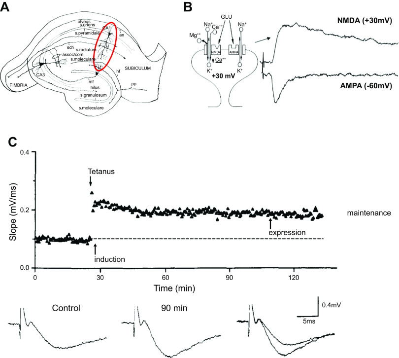
Figure 1.
A diagram of the hippocampal slice and example of long-term potentiation (LTP). A : diagram of the hippocampal slice preparation. The red circle highlights the CA1 region where most studies on LTP hav...
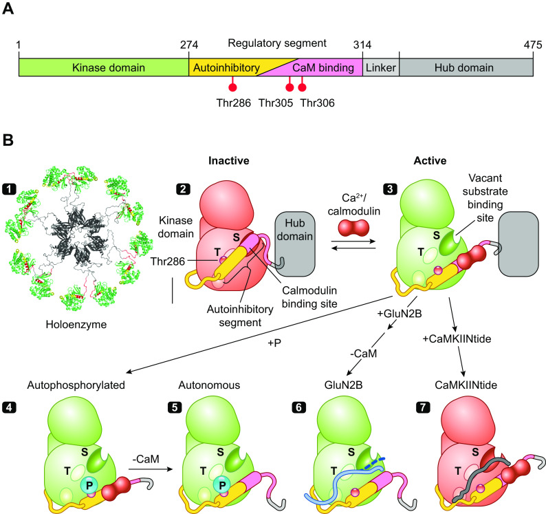
Figure 2.
The structure and regulation of Ca 2+ /calmodulin (CaM)-dependent protein kinase IIu03b1 (CaMKIIu03b1). A : the domain structure of CaMKII with kinase domain followed by regulatory segment, linker, an...
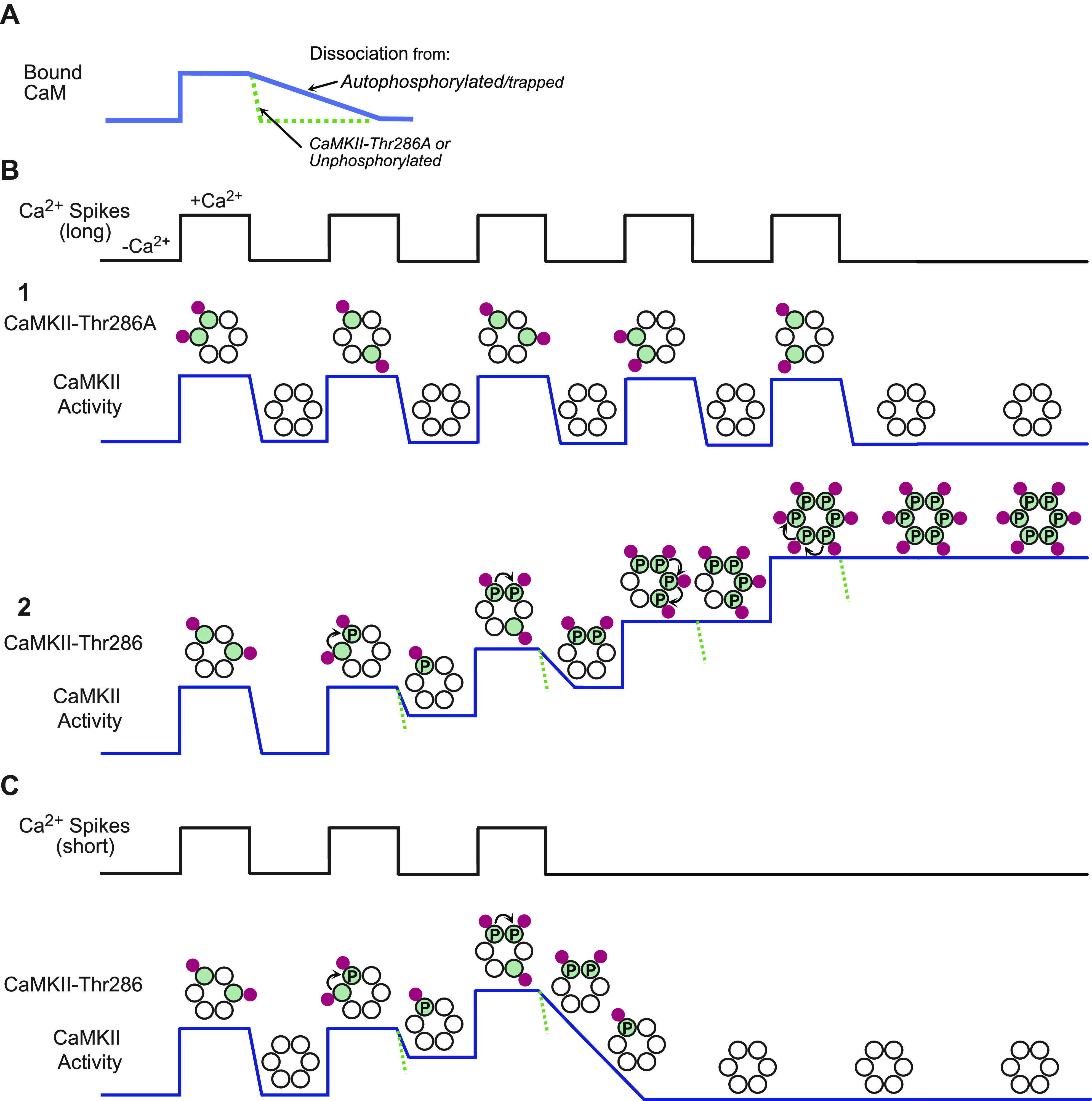
Figure 3.
Ca 2+ /calmodulin (CaM)-dependent protein kinase II (CaMKII) and integration of Ca 2+ stimuli. A : graph illustrating that CaM binds CaMKII in response to a rise in Ca 2+ and quickly dissociates as th...
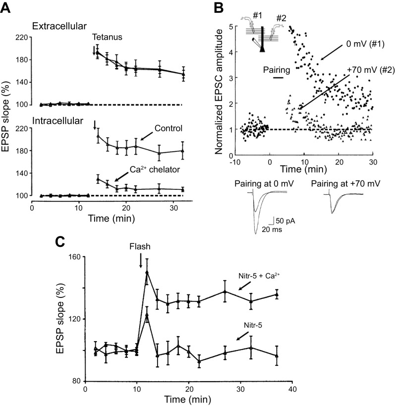
Figure 4.
Ca 2+ is necessary and sufficient for long-term potentiation (LTP). A : in this series of experiments an extracellular electrode records the field potential LTP from a population of neurons ( top ). S...
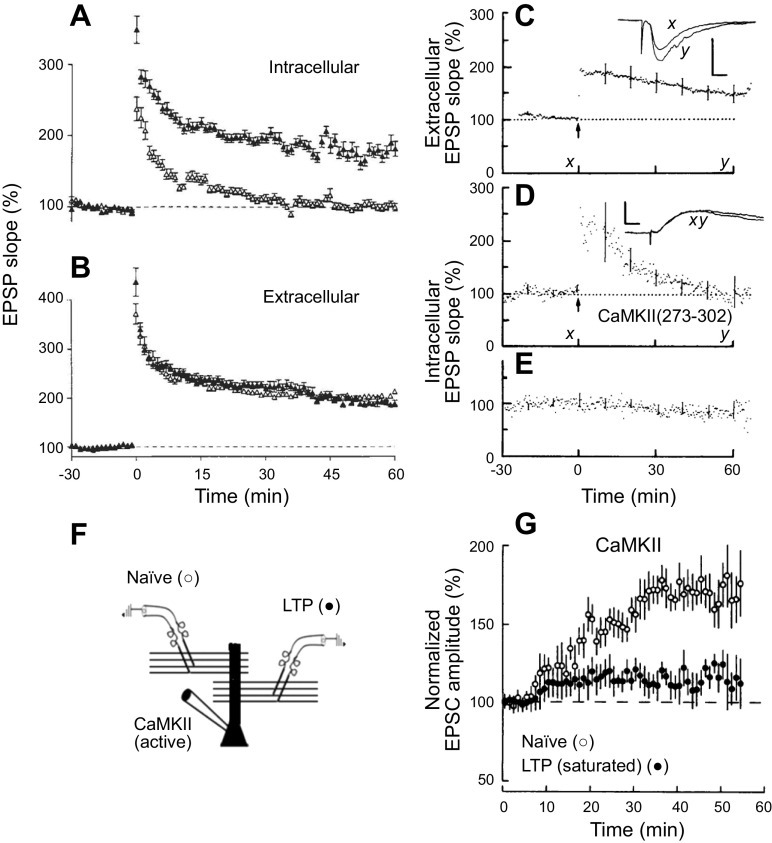
Figure 5.
Ca 2+ /calmodulin (CaM)-dependent protein kinase II (CaMKII) is required and sufficient for long-term potentiation (LTP). A and B : inhibition of CaMKII blocks LTP. Effect of intracellular application...
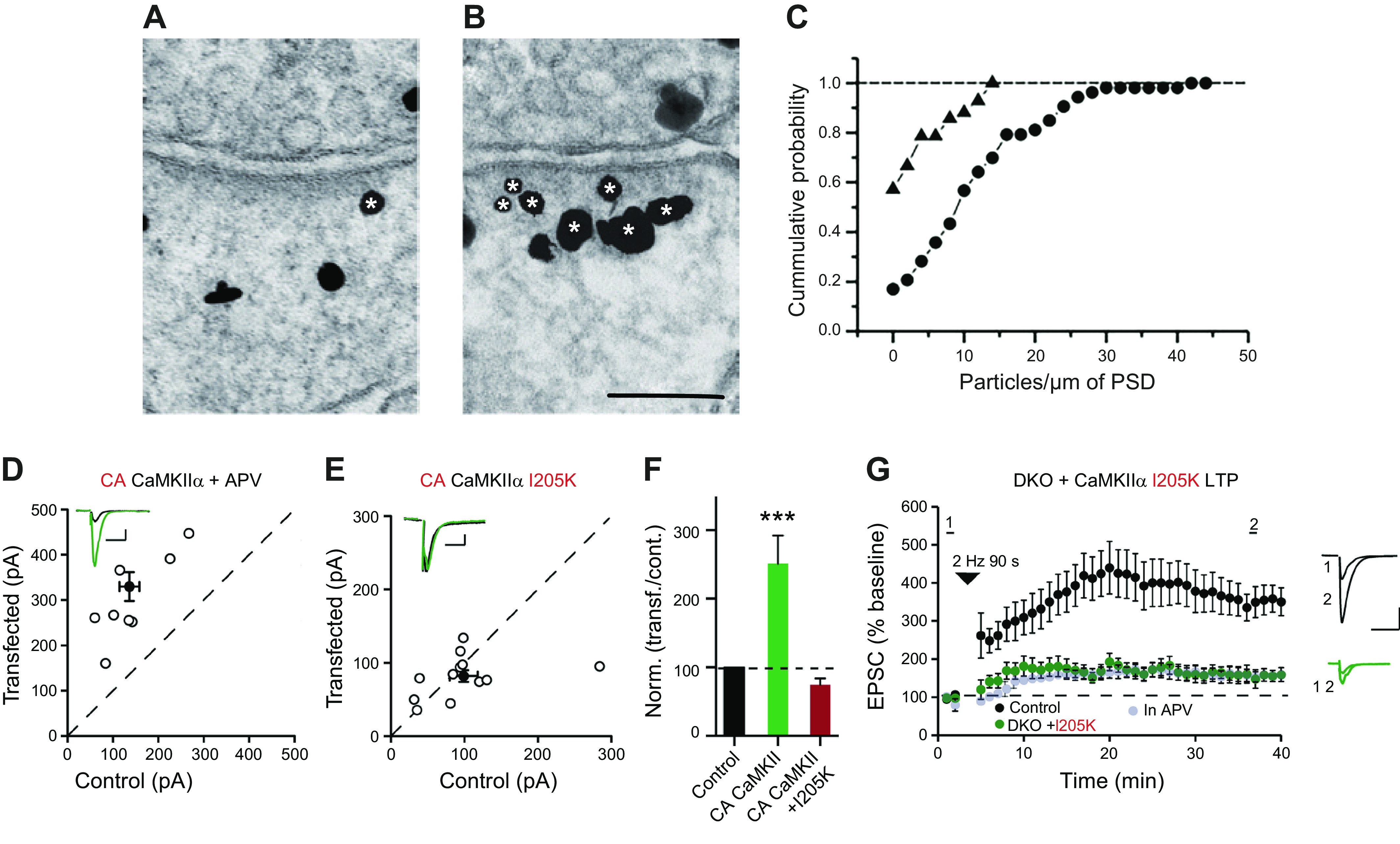
Figure 6.
Activity-dependent Ca 2+ /calmodulin (CaM)-dependent protein kinase II (CaMKII) accumulation at the postsynaptic density (PSD) and its actions require binding of CaMKII to N -methyl- d -aspartate rece...
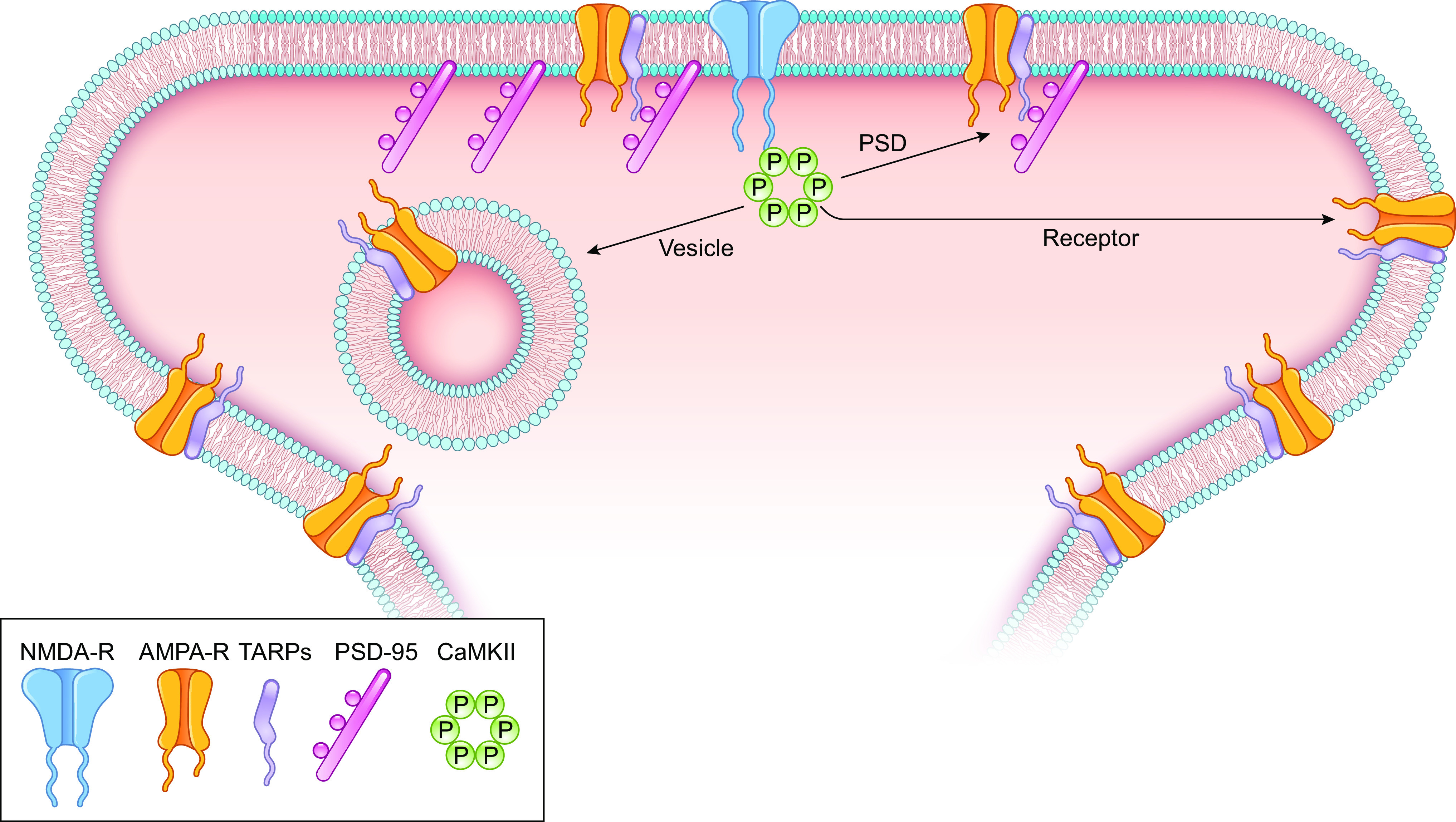
Figure 7.
Potential downstream targets of Ca 2+ /calmodulin-dependent protein kinase II (CaMKII) underlying long-term potentiation (LTP). 1 ) CaMKII modifies u03b1-amino-3-hydroxy-5-methyl-4-isoxazolepropionic ...
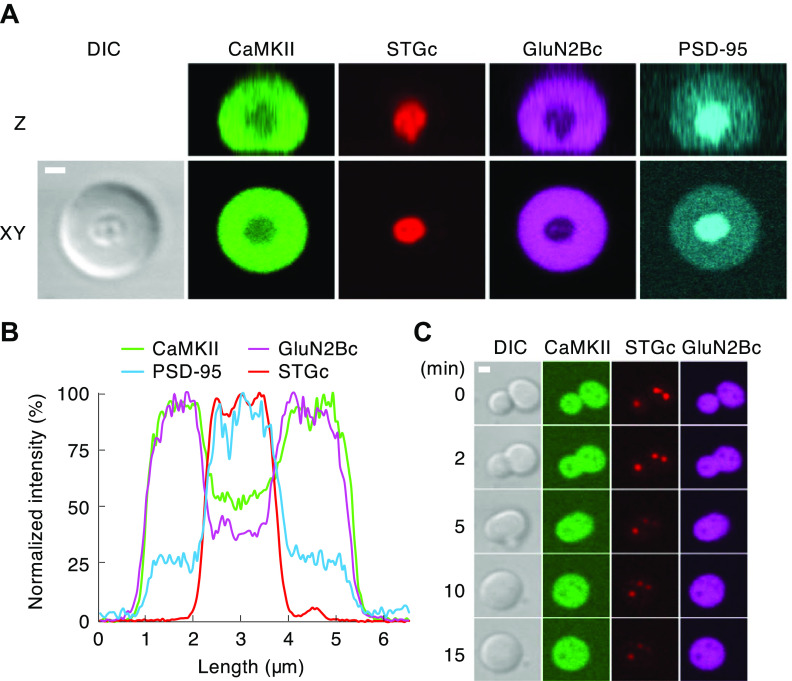
Figure 8.
Segregation of u03b1-amino-3-hydroxy-5-methyl-4-isoxazolepropionic acid receptor (AMPAR) and N -methyl- d -aspartate receptor (NMDAR) in protein condensate by active Ca 2+ /calmodulin (CaM)-dependent ...
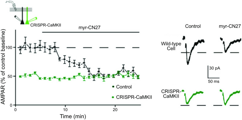
Figure 9.
Blocking Ca 2+ /calmodulin-dependent protein kinase II (CaMKII) selectively depresses u03b1-amino-3-hydroxy-5-methyl-4-isoxazolepropionic acid receptor (AMPAR) excitatory postsynaptic currents (EPSCs)...
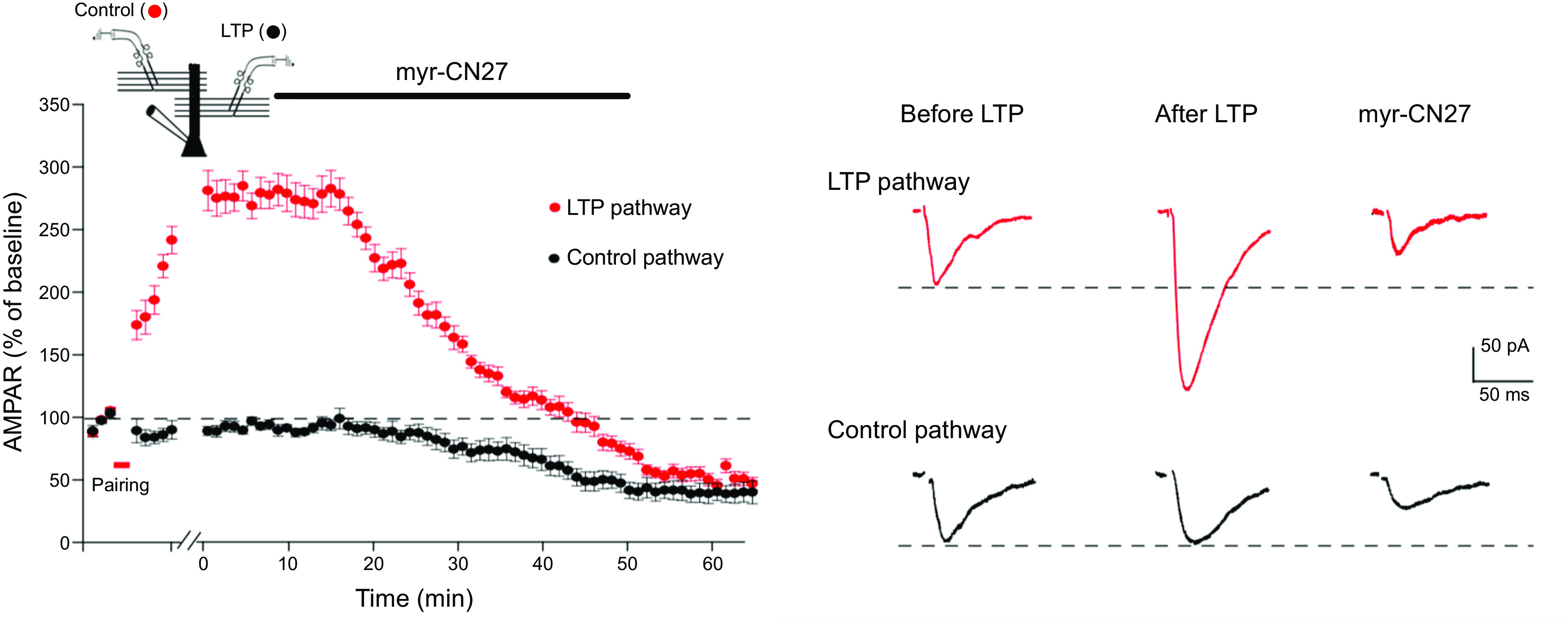
Figure 10.
Blocking Ca 2+ /calmodulin-dependent protein kinase II (CaMKII) fully reverses established long-term potentiation (LTP). Diagram shows the two-pathway experimental setup. To record the response from t...
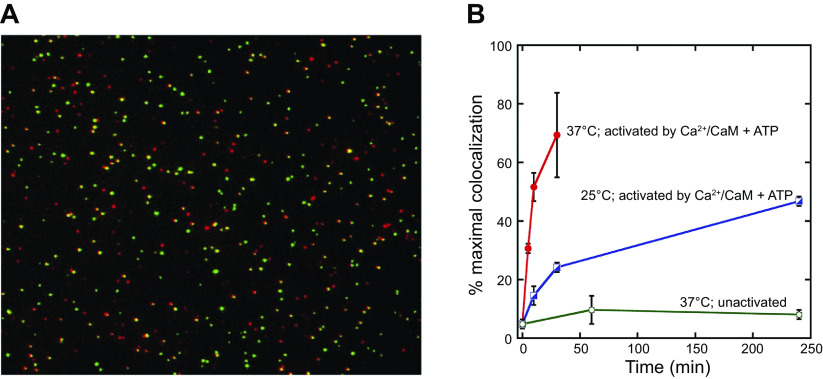
Figure 11.
Single-molecule assay for subunit exchange reveals activation-dependent subunit exchange. A : a representative single-molecule total internal reflection fluorescence (TIRF) image, with red and green c...
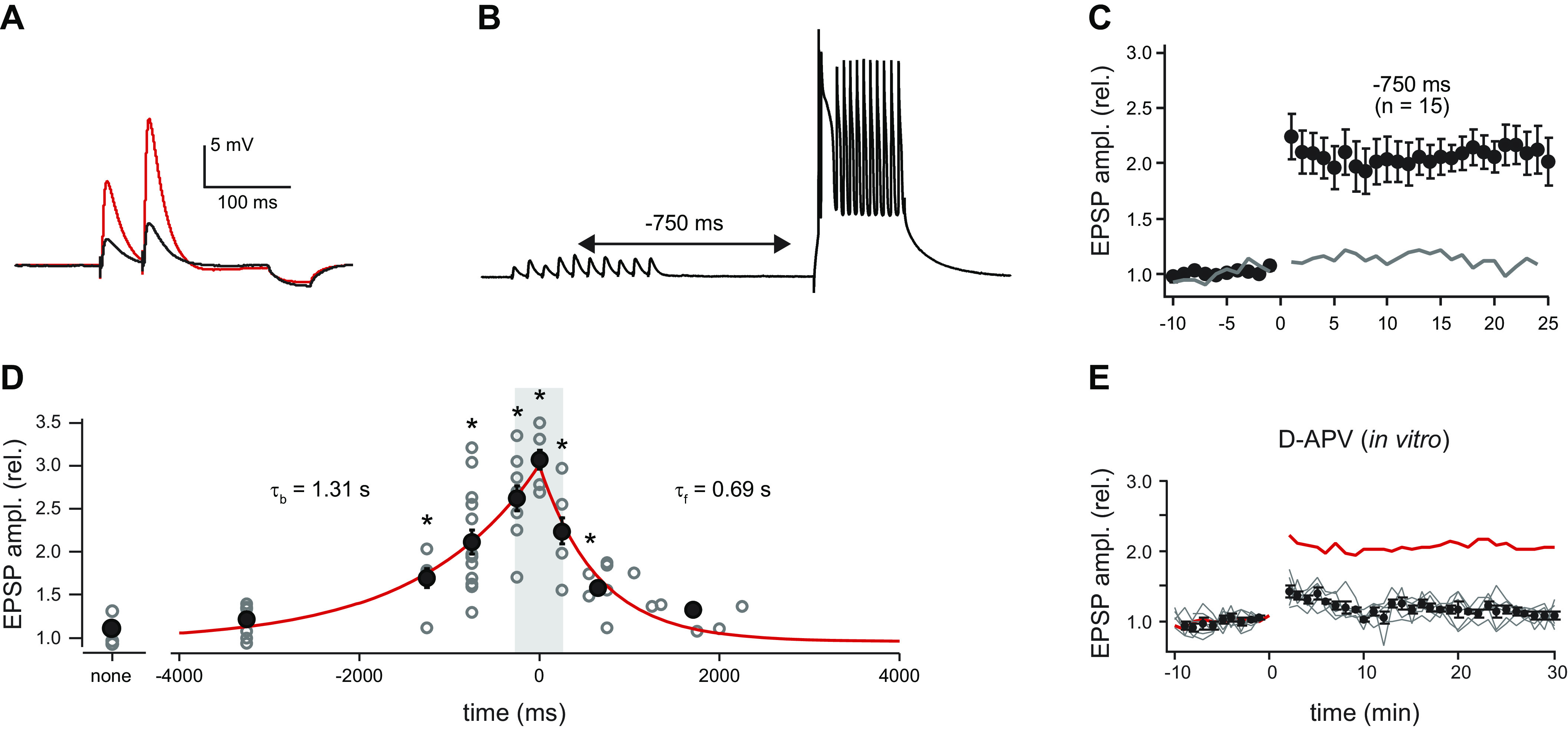
Figure 12.
Behavioral timescale synaptic plasticity. A : excitatory postsynaptic potentials (EPSPs) used to determine synaptic strength (50-ms interval). Black trace is average baseline EPSP; red trace is averag...
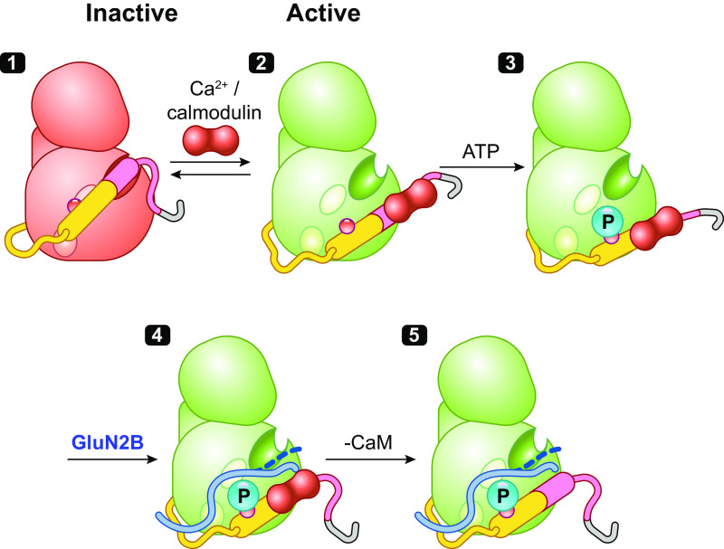
Figure 13.
Proposed sequence of events underlying activation of Ca 2+ /calmodulin-dependent protein kinase II (CaMKII). We next turn to how the CaMKII-GluN2B complex enhances synaptic transmission. Three models ...
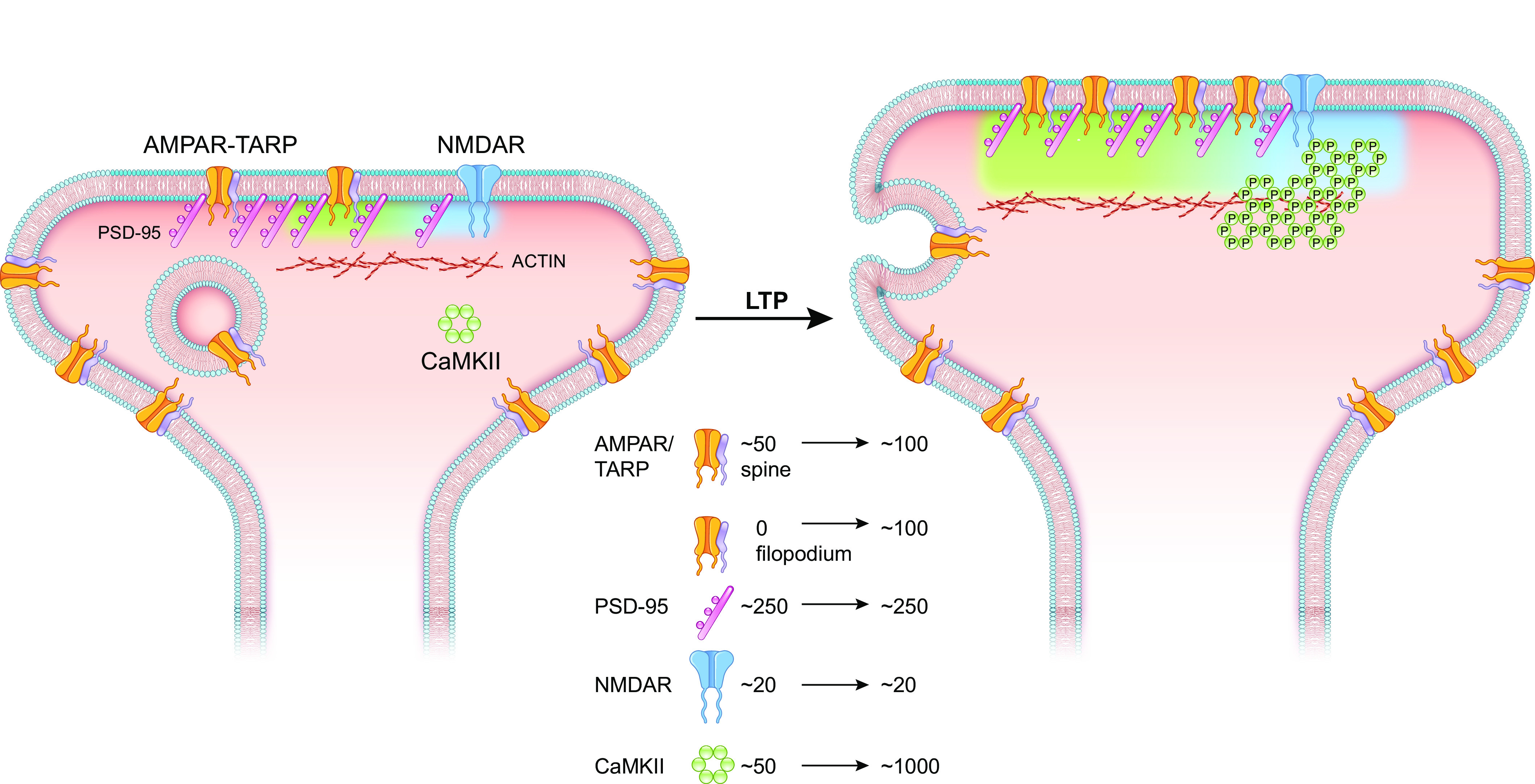
Figure 14.
The proposed sequence of events underlying long-term potentiation (LTP). This figure focuses on spine synapses, but a substantial number of synapses are formed on filopodia and assumed to be like spin...
Figure images are served from the NIH/NLM PubMed Central Open Access Subset or Europe PMC; copyright remains with the publishers and authors.
💬 Discussion
0 commentsNo comments yet. Be the first to start a discussion!
Leave a Comment