Abstract
Cold allodynia is a common feature of neuropathic pain however the underlying mechanisms of this enhanced sensitivity to cold are not known. Recently the transient receptor potential (TRP) channels TRPM8 and TRPA1 have been identified and proposed to be molecular sensors for cold. Here we have investigated the expression of TRPM8 and TRPA1 mRNA in the dorsal root ganglia (DRG) and examined the cold sensitivity of peripheral sensory neurons in the chronic construction injury (CCI) model of neuropathic pain in mice.In behavioral experiments, chronic constriction injury (CCI) of the sciatic nerve induced a hypersensitivity to both cold and the TRPM8 agonist menthol that developed 2 days post injury and remained stable for at least 2 weeks. Using quantitative RT-PCR and in situ hybridization we examined the expression of TRPM8 and TRPA1 in DRG. Both channels displayed significantly reduced expression levels after injury with no change in their distribution pattern in identified neuronal subpopulations. Furthermore, in calcium imaging experiments, we detected no alterations in the number of cold or menthol responsive neurons in the DRG, or in the functional properties of cold transduction following injury. Intriguingly however, responses to the TRPA1 agonist mustard oil were strongly reduced.Our results indicate that injured sensory neurons do not develop abnormal cold sensitivity after chronic constriction injury and that alterations in the expression of TRPM8 and TRPA1 are unlikely to contribute directly to the pathogenesis of cold allodynia in this neuropathic pain model.
🔬 Techniques
✨ Fluorophores
🧪 Sample Preparation
🔬 Cell Lines
🏭 Microscope Brands
🧪 Reagent Suppliers
📷 Detectors
🏛️ Research Organizations (ROR)
Affiliated research institutions:
📋 Methods
Surgical procedures
All experiments were conducted on C57/B6 mice or Wistar rats with the approval of the state animal care and use committee (Landesamt für Arbeitsschutz, Gesundheit und Technische Sicherheit Berlin). A chronic constriction injury (CCI) of the sciatic nerve was used to model neuropathic pain. Briefly, the right sciatic nerve was exposed at mid-thigh level under isoflurane anesthesia. 3 loose silk ligatures were tied around the nerve and the incision was closed. For sham controls, the sciatic nerve was exposed but not ligated. Intraplantar injection of Complete Freunds Adjuvant (CFA) was used to model inflammatory pain. 20 µl CFA (50 µg of desiccated M. Butyricum diluted into 20 µl Incomplete Freunds Adjuvant from Difco Laboratories Detroit, USA) was injected into the right hind paw under brief isoflurane (Rhodia Organic Fine Ltd., Bristol, UK) anesthesia. The inflammation was confined to the right paw throughout the observation period. Animals were killed after 48 hours and tissue was prepared as described below. Behavioral experiments For all behavioral experiments, mice were habituated to the test procedure for 7 days before surgery. Responses were taken 1 day prior to surgery and at 2, 4, 7 and 14 days postoperative. The acetone test was used to assess cold allodynia in mice. In this test, evaporative cooling of locally applied acetone is used to evoke nociceptive behavior in CCI mice [57] , [58] . 40 µl of acetone was applied to the dorsal hind paw ipsilateral to the injury and the behavior was assigned an arbitrary score. A score of 0 indicated no response, 0.5, a licking response, 1, flinching and brushing of the paw, 2, strong flinching, and 3, strong flinching and licking. Behavior was observed during the first 30 seconds after acetone application and measurements were repeated 2 times with a 1 minute interval to obtain a mean value. Dorsal root ganglia from animals displaying cold allodynia were used for qRT-PCR, in situ hybridization and calcium imaging assays. To assess the effects of menthol on nociceptive behavior in CCI mice, 40 µl of 250 mM (-)-menthol (Sigma, Germany) dissolved in 90%DMSO and 10%PBS or vehicle alone was applied to the dorsal surface of the ipsilateral hind paw. Mice were observed for 5 minutes and the time spent licking the hind paw was measured. Experiments were repeated in control mice.
Show full methods section
Surgical procedures
All experiments were conducted on C57/B6 mice or Wistar rats with the approval of the state animal care and use committee (Landesamt für Arbeitsschutz, Gesundheit und Technische Sicherheit Berlin). A chronic constriction injury (CCI) of the sciatic nerve was used to model neuropathic pain. Briefly, the right sciatic nerve was exposed at mid-thigh level under isoflurane anesthesia. 3 loose silk ligatures were tied around the nerve and the incision was closed. For sham controls, the sciatic nerve was exposed but not ligated. Intraplantar injection of Complete Freunds Adjuvant (CFA) was used to model inflammatory pain. 20 µl CFA (50 µg of desiccated M. Butyricum diluted into 20 µl Incomplete Freunds Adjuvant from Difco Laboratories Detroit, USA) was injected into the right hind paw under brief isoflurane (Rhodia Organic Fine Ltd., Bristol, UK) anesthesia. The inflammation was confined to the right paw throughout the observation period. Animals were killed after 48 hours and tissue was prepared as described below. Behavioral experiments For all behavioral experiments, mice were habituated to the test procedure for 7 days before surgery. Responses were taken 1 day prior to surgery and at 2, 4, 7 and 14 days postoperative. The acetone test was used to assess cold allodynia in mice. In this test, evaporative cooling of locally applied acetone is used to evoke nociceptive behavior in CCI mice [57] , [58] . 40 µl of acetone was applied to the dorsal hind paw ipsilateral to the injury and the behavior was assigned an arbitrary score. A score of 0 indicated no response, 0.5, a licking response, 1, flinching and brushing of the paw, 2, strong flinching, and 3, strong flinching and licking. Behavior was observed during the first 30 seconds after acetone application and measurements were repeated 2 times with a 1 minute interval to obtain a mean value. Dorsal root ganglia from animals displaying cold allodynia were used for qRT-PCR, in situ hybridization and calcium imaging assays. To assess the effects of menthol on nociceptive behavior in CCI mice, 40 µl of 250 mM (-)-menthol (Sigma, Germany) dissolved in 90%DMSO and 10%PBS or vehicle alone was applied to the dorsal surface of the ipsilateral hind paw. Mice were observed for 5 minutes and the time spent licking the hind paw was measured. Experiments were repeated in control mice.
Quantitative reverse transcription-PCR Lumbar L3-L6 dorsal root ganglia
(DRG) were dissected from CCI and control mice, pooled and total RNA extracted using the RNeasy mini kit (Qiagen, Germany). Samples were quantified using a spectrophotometer (Gene Quant II, Pharmacia Biotech, UK) and reverse transcribed with AMV-reverse transcriptase (Roche, Germany). Quantitative PCR was performed using the Lightcycler system (Roche, Germany) utilizing SYBR green to detect amplification. PCR primers were designed to amplify 400 bp regions from TRPM8, TRPA1, TREK-1, galanin and the housekeeping gene GAPDH as a reference. These primers were tested at a range of annealing temperatures and were found to amplify 1 PCR product as determined by melting curve analysis and agarose gel electrophoresis. A standard concentration curve for each cDNA was constructed from serial dilutions of linearized plasmid DNA. Experiments were performed in triplicate and data was normalized to GAPDH levels. In further experiments, individual L4 and L5 ganglia were dissected from control and CCI rats and processed as above for qRT-PCR of TRPM8, TRPA1 and GAPDH mRNA. In situ hybridization and immunohistochemistry Mouse tissue was fixed by transcardial perfusion of 4% paraformaldehyde (PFA) in PBS. Lumbar L3-L6 DRG were dissected, post-fixed for a further 2 hours in PFA and cryoprotected overnight in 30% sucrose. Non-radioactive in situ hybridization was performed using digoxygenin (DIG) (Roche, Germany) labeled RNA probes on 10 µm frozen sections. Antisense probes corresponded to nucleotides 562–1465, 262–808, 673–1087 and full length for TRPA1 and 963–1533, 242–870 and full length for TRPM8. Equivelant sense probes displayed no signal. Sections were treated with 1 µg/ml proteinase K (Sigma, Germany) for 5 minutes, acetylated for 10 minutes with 0.25% (v/v) acetic anhydride in 0.1 M triethanolamine, prehybridized for 4 hours at 56°C and hybridized with RNA probes overnight at 56°C. Following post hybridization washes and blocking, sections were incubated for 30 minutes in 1∶100 anti-DIG antibody conjugated with horseradish peroxidase (Roche, Germany) and signal was visualized using tyramide signal amplification (PerkinElmer, Germany).
Immunohistochemistry and isolectin B4
(IB4) staining followed in situ hybridization. FITC-labeled IB4 (Sigma, Germany) was used at a concentration of 10 µg/ml, anti-CGRP (polyclonal, Sigma, Germany) at a 1∶2000 dilution, and anti-NF200 (clone N52, Sigma, Germany) at 1∶4000 dilution. All cell counts and quantification were conducted by an observer blinded to the experimental condition.
Calcium imaging Mouse lumbar L3-L6
DRG were dissected from CCI and control mice and incubated with 1 mg/ml collagenase IV (Sigma, Germany) and 0.05% trypsin (Biochrom, Berlin, Germany) for 30 minutes each at 37°C. The DRG were suspended in DMEM/Hams-F12 medium (Invitrogen, Germany) containing 10% heat-inactivated horse serum (Biochrom, Berlin, Germany), 1 mM glutamine (Invitrogen, Germany) 0.8% glucose (Sigma, Germany), 100U penicillin, and 100 µg/ml streptomycin (Biochrom, Berlin, Germany). DRG were dissociated using 18G, 22G, 25G needles, and debris was removed with a 40 µm cell strainer (BD Biosciences Europe, Belgium). Cells were plated in a droplet of medium on poly-L-lysine (100 µg/ml, Sigma, Germany) coated coverslips and left to adhere for 30 minutes before the coverslip was flooded. Experiments were conducted 1–3 hours after plating of cells. Ratiometric calcium imaging was performed with FURA-2/AM dye (Invitrogen, Germany) and analyzed using Tillvision software (Till Photonics, Germany). Cells were loaded with 3 µM Fura-2/AM and placed in a recording chamber containing calcium imaging buffer (CIB: 140 mM NaCl, 4 mM KCl, 2 mM CaCl 2 , 1 mM MgCl 2 , 5 mM Glucose, 10 mM HEPES, pH 7.4). Pairs of images were collected every 2 seconds at alternating exposures of 340 nM and 380 nM (exposure time 70 ms) using a Polychrome V monochromator and CCD Imago camera (Till Photonics, Germany). Following subtraction of background fluorescence the ratio of fluorescence at 340 nm and 380 nm was calculated. Coverslips were superfused with CIB buffer at approximately 2 ml/min. Drugs were applied via a gravity driven perfusion system that allowed rapid exchange of solutions. Cold stimuli were applied using a peltier device (ESF electronic, Germany) and temperature changes were monitored with a thermocouple placed within the flow of buffer and close to the cells. Following selection of a suitable field of view, coverslips were maintained at 31°C for 5 minutes. A cold stimulus was then applied which cooled the cells from 31°C until an endpoint of 8°C had been reached. Cooling occurred at approximately 1°C per second and was reproducible such that in more than 95% of experiments the cold endpoint was reached within 30 seconds. After a recovery period of 5 minutes at 31°C, either (-)-menthol (100 µM unless indicated), or in separate experiments, mustard oil (50 µM based on dose response date (not shown)) was applied for 1 minute. Drugs were washed out for 3–5 minutes before 40 mM KCl was applied for 10 seconds to determine the total number of living cells. A response was designated as a 20% increase in fluorescence ratio from baseline. The number of responders to cold, (-)-menthol or mustard oil was expressed as a percentage of KCl responsive cells. The maximum amplitude of the response, the temperature threshold of cold responses and the cell diameter were also determined. Cold threshold temperatures were calculated as the temperature at which fluorescence ratio increased by 0.5% upon cooling. These relatively stringent selection criteria for cold responsive cells plus the fact that we started our recordings at 31°C (rather than 35–37°C used by some researchers) may account for the low temperature thresholds we observed.
Data analysis
Statistical analysis was performed using Sigmastat software (Systat, San Jose, CA). Significance was tested using a two-way repeated measures ANOVA in behavioral experiments, and a Student's T-test or Mann-Whitney-U-test in expression and calcium imaging experiments.
Surgical procedures
All experiments were conducted on C57/B6 mice or Wistar rats with the approval of the state animal care and use committee (Landesamt für Arbeitsschutz, Gesundheit und Technische Sicherheit Berlin). A chronic constriction injury (CCI) of the sciatic nerve was used to model neuropathic pain. Briefly, the right sciatic nerve was exposed at mid-thigh level under isoflurane anesthesia. 3 loose silk ligatures were tied around the nerve and the incision was closed. For sham controls, the sciatic nerve was exposed but not ligated. Intraplantar injection of Complete Freunds Adjuvant (CFA) was used to model inflammatory pain. 20 µl CFA (50 µg of desiccated M. Butyricum diluted into 20 µl Incomplete Freunds Adjuvant from Difco Laboratories Detroit, USA) was injected into the right hind paw under brief isoflurane (Rhodia Organic Fine Ltd., Bristol, UK) anesthesia. The inflammation was confined to the right paw throughout the observation period. Animals were killed after 48 hours and tissue was prepared as described below.
📊 Figures
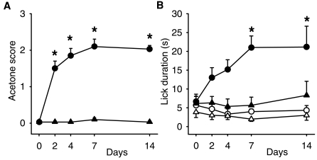
Figure 1
Time course of cold and (-)-menthol sensitivity following sciatic nerve ligation.
(a) Cold sensitivity assessed by acetone response score where 0u200a=u200ano response, 0.5u200a=u200alicking, 1u200a=u200aflinching and brushing of the paw, 2u200a=u200astrong flinching, 3u200a=u200as...
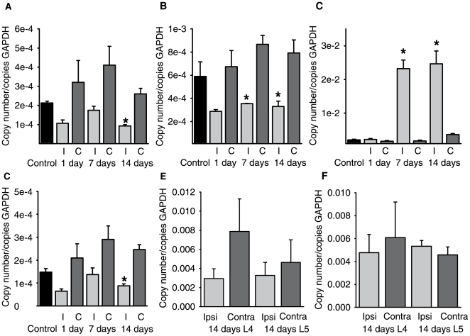
Figure 2
Quantitative reverse-transcription PCR.
mRNA levels for (a), TRPM8 (b), TRPA1 (c), galanin, and (d) TREK-1 in the mouse. Levels are expressed relative to GAPDH in control animals and at 2, 7 and 14 days after surgery. I indicates ipsilatera...
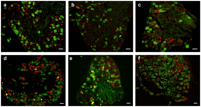
Figure 3
Distribution of TRPM8 and TRPA1 in identified neuronal subpopulations in DRG.
Combined in situ hybridization of TRPM8 (red) with (a), immunohistochemistry (green) for CGRP, (b), IB4 and (c), NF200. TRPA1 mRNA expression (red) with (d), CGRP, (e), IB4 and (f), NF200 (green). Sca...
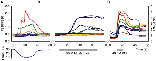
Figure 4
Representative recordings of Ca 2+ transients in DRG neurons from control mice.
Responses to (a), cooling, (b), mustard oil and (c), KCl (note different Y-axis scale for KCl).
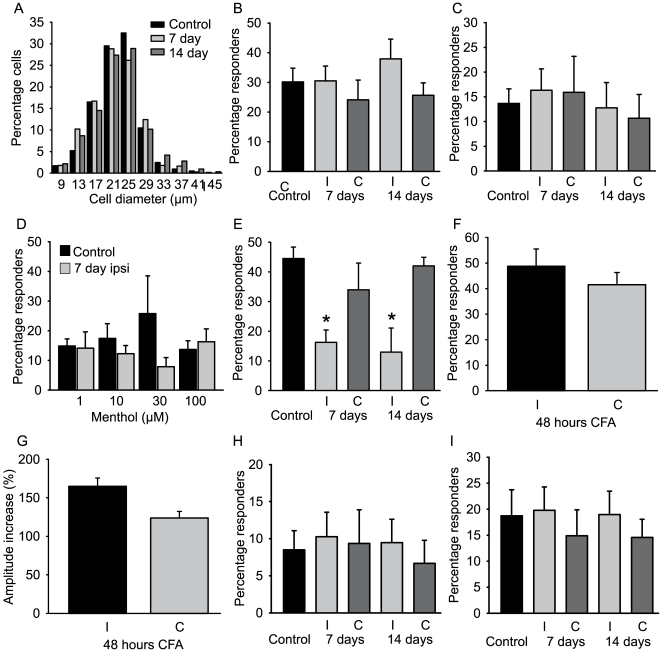
Figure 5
Proportions of cold, (-)-menthol and mustard oil responsive DRG neurons in control animals, at 7 and 14 days post-CCI and at 48 hours post CFA.
(a), Cell-size histogram in control mice and in mice at 7 and 14 days post CCI injury. (b), Percentage of cold-responsive neurons in the DRG (nu200a=u200a6u201315 mice). (c), Percentage of neurons res...
Figure images are served from the NIH/NLM PubMed Central Open Access Subset or Europe PMC; copyright remains with the publishers and authors.
💬 Discussion
0 commentsNo comments yet. Be the first to start a discussion!
Leave a Comment