Abstract
The integration of visual stimuli and motor feedback is critical for successful visually guided navigation. These signals have been shown to shape neuronal activity in the primary visual cortex (V1), in an experience-dependent manner. Here, we examined whether visual, reward, and self-motion-related inputs are integrated in order to encode behaviorally relevant locations in V1 neurons. Using a behavioral task in a virtual environment, we monitored layer 2/3 neuronal activity as mice learned to locate a reward along a linear corridor. With learning, a subset of neurons became responsive to the expected reward location. Without a visual cue to the reward location, both behavioral and neuronal responses relied on self-motion-derived estimations. However, when visual cues were available, both neuronal and behavioral responses were driven by visual information. Therefore, a population of V1 neurons encode behaviorally relevant spatial locations, based on either visual cues or on self-motion feedback when visual cues are absent.
🔬 Techniques
✨ Fluorophores
🧪 Sample Preparation
🏭 Microscope Brands
🔴 Lasers
💻 Software Details
💻 Code & Software
💾 Data Repositories
🏛️ Research Organizations (ROR)
Affiliated research institutions:
📋 Methods
Animals
All animal experiments were approved by the Animal Welfare and Ethical Review Board (AWERB) of the University of Edinburgh and were performed under a project license granted by the UK Home Office, and conformed with the UK Animals (Scientific Procedures) Act 1986 and the European Directive 86/609/EEC on the protection of animals used for experimental purposes. Seven male and female mice with a C57BL/6 background ( Sst tm2.1(cre)Zjh /J [RRID:IMSR_JAX:013044] cross-bred with B6.Cg- Gt(ROSA)26Sor tm14(CAG-tdTomato)Hze /J [RRID:IMSR_JAX:007914]; Jackson Laboratory, ME, USA), aged 6–7 weeks, were used for the experiments. Animals were group housed in a reverse day/night cycle.
Surgical Procedures
For cranial window implantation and virus injection, mice were anaesthetized with isoflurane (4% for induction and 1%–2% maintenance during surgery) and mounted on a stereotaxic frame (David Kopf Instruments, CA, USA). Eye cream was applied to protect the eyes (Bepanthen; Bayer, Germany), and analgesics and anti-inflammatory drugs were injected subcutaneously (Vetergesic, buprenorphine, 0.1 mg/kg of body weight; carprofen, 0.15 mg; and dexamethasone, 2 μg). A section of scalp was removed and the underlying bone cleaned before a craniotomy (around 2 × 2 mm) was made over the left V1 (centered 2.5 mm lateral and 0.5 mm anterior to lambda). Then adeno-associated virus (AAV) (AAV1.Syn.GCaMP6f.WPRE.SV40; University of Pennsylvania Vector Core, PA, USA) was injected, to drive the expression of the fluorescent calcium indicator GCaMP6f in all neurons, using a pipette with 20-μm tip diameter (Nanoject; Drummond Scientific, PA, USA) at a speed of 10 nL min −1 at three different depths (around 250, 400, and 600 μm deep; 50 nL per site). The craniotomy was then sealed with a glass coverslip and fixed with cyanoacrylate glue. A custom-built head post was implanted on the exposed skull with glue and cemented with dental acrylic (Paladur; Heraeus Kulzer, Germany). Animals were returned to their home cage for 2–3 weeks to allow for virus expression and clearing of the imaging window ( Holtmaat et al., 2009 ) before habituation and imaging.
Show full methods section
Animals
All animal experiments were approved by the Animal Welfare and Ethical Review Board (AWERB) of the University of Edinburgh and were performed under a project license granted by the UK Home Office, and conformed with the UK Animals (Scientific Procedures) Act 1986 and the European Directive 86/609/EEC on the protection of animals used for experimental purposes. Seven male and female mice with a C57BL/6 background ( Sst tm2.1(cre)Zjh /J [RRID:IMSR_JAX:013044] cross-bred with B6.Cg- Gt(ROSA)26Sor tm14(CAG-tdTomato)Hze /J [RRID:IMSR_JAX:007914]; Jackson Laboratory, ME, USA), aged 6–7 weeks, were used for the experiments. Animals were group housed in a reverse day/night cycle.
Surgical Procedures
For cranial window implantation and virus injection, mice were anaesthetized with isoflurane (4% for induction and 1%–2% maintenance during surgery) and mounted on a stereotaxic frame (David Kopf Instruments, CA, USA). Eye cream was applied to protect the eyes (Bepanthen; Bayer, Germany), and analgesics and anti-inflammatory drugs were injected subcutaneously (Vetergesic, buprenorphine, 0.1 mg/kg of body weight; carprofen, 0.15 mg; and dexamethasone, 2 μg). A section of scalp was removed and the underlying bone cleaned before a craniotomy (around 2 × 2 mm) was made over the left V1 (centered 2.5 mm lateral and 0.5 mm anterior to lambda). Then adeno-associated virus (AAV) (AAV1.Syn.GCaMP6f.WPRE.SV40; University of Pennsylvania Vector Core, PA, USA) was injected, to drive the expression of the fluorescent calcium indicator GCaMP6f in all neurons, using a pipette with 20-μm tip diameter (Nanoject; Drummond Scientific, PA, USA) at a speed of 10 nL min −1 at three different depths (around 250, 400, and 600 μm deep; 50 nL per site). The craniotomy was then sealed with a glass coverslip and fixed with cyanoacrylate glue. A custom-built head post was implanted on the exposed skull with glue and cemented with dental acrylic (Paladur; Heraeus Kulzer, Germany). Animals were returned to their home cage for 2–3 weeks to allow for virus expression and clearing of the imaging window ( Holtmaat et al., 2009 ) before habituation and imaging.
Virtual Reality System
Animals were trained on a virtual reality system consisting of two angled computer screens ( Figure 1 B), a cylindrical treadmill, head fixation system, and a reward spout. The computer screens (51 × 29 cm; Dell, UK) were placed at a 90° angle in front of the animal covering the majority of its field of view. A cylindrical polystyrene treadmill (20 cm diameter, 7.5 cm wide) was mounted on a central axle with an incremental rotary encoder (E6-2500-472-IE; Pewatron, Switzerland). A microcontroller (Arduino Uno) received rotational displacement information from the encoder and forwarded it the virtual reality software. The reward spout (59-8636; Harvard Apparatus, UK) was fitted with a capacitive touch sensor (SEN-12041; Sparkfun, CO, USA) to detect animal licking behavior and put into place at the beginning of each session, such that the animal was always able to reach it. Reward release was controlled by a pinch-valve (225PNC1-21; NResearch, NJ, USA) that dispensed 4- to 8-μL boluses per instance. The MATLAB-based package ViRMEn ( Aronov and Tank, 2014 ) combined with custom-written code was used to design and run the presentation of the virtual environment and collect related data (see Supplemental Experimental Procedures ). Visually Guided Rewarded Task Mice were water deprived (see Supplemental Experimental Procedures ) and water rewards could either be self-initiated by licking in the first half of the reward zone (early, successful trial), or were dispensed at a default location at the halfway point of the reward zone (late, missed trial). Behavioral training was divided into three phases. For each phase, the first day was taken as the “novice” day. The animals were considered “expert” and promoted to the next phase of training, when successful trials made up >75% of the total trials. For phase 1, the mice were exposed to a single virtual corridor condition with the reward zone visually cued by black corridor walls. Phase 2 introduced uncued trials on every fifth trial, where the rules for reward remained the same but the black corridor walls were removed. For three mice, an additional phase 3 was performed where in a single session the gain relating the rotation of the cylindrical treadmill to the progression in the virtual corridor was reduced from 1 to 0.75 (see Supplemental Experimental Procedures ).
Two-Photon Calcium Imaging
Two-photon calcium imaging was performed in head-fixed mice that ran freely on the cylindrical treadmill ( Figure 1 B) ( Dombeck et al., 2007 ) using a custom-built resonant scanning two-photon microscope with a Ti:sapphire pulsing laser (Chameleon Vision-S; Coherent, CA, USA; 1). In contrast, if the animal licks in a spatially indiscriminate pattern, the number will approach 1. If the animal licks often but keeps missing the reward zone, the SMI will be
📊 Figures
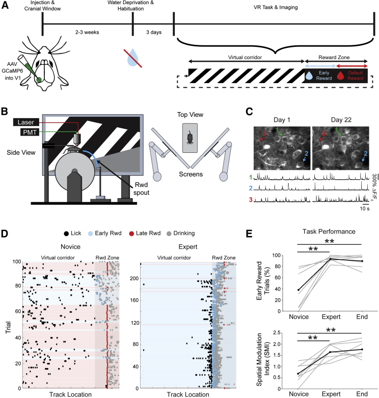
Figureu00a01
Mice Learn to Lick at a Specific Reward Location in a Visually Guided Task in a Virtual Environment (A) Experimental timeline for chronic imaging of primary visual cortex (V1). The virtual corridor ha...
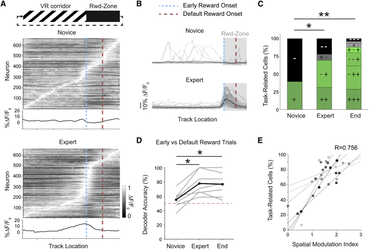
Figureu00a02
V1 Layer 2/3 Population Activity during Learning of the Visually Guided Task (A) Normalized u0394F/F 0 along the virtual corridor, plotted for all neurons on novice (top) and expert (bottom) days. Neu...
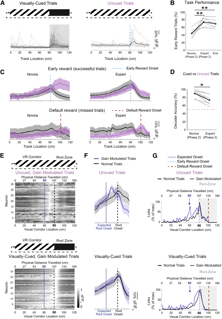
Figureu00a03
After Learning, V1 Neurons Respond to a Rewarded Spatial Location, Even in the Absence of Visual Cues (A) After mice reached expert criteria on the visually cued task (left), they were subsequently (p...
Figure images are served from the NIH/NLM PubMed Central Open Access Subset or Europe PMC; copyright remains with the publishers and authors.
💬 Discussion
0 commentsNo comments yet. Be the first to start a discussion!
Leave a Comment