Abstract
In primates, C-fibre polymodal nociceptors are broadly classified into two groups based on mechanosensitivity. Here we demonstrate that mechanically sensitive polymodal nociceptors that respond either quickly (QC) or slowly (SC) to a heat stimulus differ in responses to a mild burn, heat sensitization, conductive properties and chemosensitivity. Superficially applied capsaicin and intradermal injection of β-alanine, an MrgprD agonist, excite vigorously all QCs. Only 40% of SCs respond to β-alanine, and their response is only half that of QCs. Mechanically insensitive C-fibres (C-MIAs) are β-alanine insensitive but vigorously respond to capsaicin and histamine with distinct discharge patterns. Calcium imaging reveals that β-alanine and histamine activate distinct populations of capsaicin-responsive neurons in primate dorsal root ganglion. We suggest that histamine itch and capsaicin pain are peripherally encoded in C-MIAs, and that primate polymodal nociceptive afferents form three functionally distinct subpopulations with β-alanine responsive QC fibres likely corresponding to murine MrgprD-expressing, non-peptidergic nociceptive afferents.
🔬 Techniques
🧬 Organisms
💻 Software
✨ Fluorophores
🏭 Microscope Brands
🧪 Reagent Suppliers
💻 Software Details
💾 Data Repositories
🏛️ Research Organizations (ROR)
Affiliated research institutions:
📋 Methods
Animals
All experiments were performed in accordance with Animal Welfare Act regulations and the USPHS Policy on Humane Care and Use of Laboratory Animals and approved by the Johns Hopkins University Animal Care and Use Committee. Adult (3-10 years of age), male cynomolgus (Macaca fascicularis, n=13) and pigtail monkeys (Macaca nemestrina, n=6) were used.
Animal preparation
Animals were sedated by intramuscular ketamine (Ketaject, Phoenix, St. Joseph, MO; 12 mg/kg). A pentobarbital bolus (6 mg/kg, Nembutal, Ovation Pharmaceuticals, Inc., Deerfield, IL) was administered via an intravenous catheter to induce anesthesia. A continuous infusion of pentobarbital at 4-6 mg/kg/hr was given to maintain anesthesia. Core temperature was maintained near 38°C using feedback-controlled warm-water heating pads, and an i.v. drip of 5% dextrose was continuously given for hydration. Heart rate was monitored and depth of anesthesia was adjusted, if tachycardia occurred in response to noxious stimuli. Following intubation, paralysis was induced with pancuronium bromide (SICOR Pharmaceuticals, Irvine, CA; 0.1 mg/kg every 2 h), and animals were ventilated to maintain a pCO2 of 35-40 mmHg. A prophylactic antibacterial agent, Cefazolin (West-Ward Pharmaceutical Corp, Eatontown, NJ; 20 mg/kg/h), was administered. At the end of the experiment and on the following postoperative day, animals received buprenorphine (Buprenex Injectable, Reckitt Benckiser Pharmaceuticals Inc., Richmond, VA; 0.15 mg per injection). Recordings were made from multiple cutaneous nerves in a given animal with experiments being separated by at least 2 weeks. After all peripheral nerve recordings were completed, 3 animals were euthanized for harvesting DRG tissues for calcium imaging experiments. Using aseptic techniques, nerves were dissected under a microscope, and a standard teased-fiber technique 8 , 61 was used to record neuronal activity in single afferent fibers. Peripheral nerves innervating the hairy skin were used for recordings (saphenous, superficial peroneal, sural, superficial radial and medial antebrachial cutaneous nerves). A bundle of the dissected nerve was cut proximally and split into smaller strands on a dissecting platform which also served as a reference electrode and supported the nerve trunk. The strands were placed on the recording electrode, situated in the near vicinity of the splitting platform. About 4 cm distal to the recording electrode, a stimulating electrode was positioned on the nerve trunk to identify slowly conducting unmyelinated nerve fibers (conduction velocity 0.5 – 2m/s).
Show full methods section
Animals
All experiments were performed in accordance with Animal Welfare Act regulations and the USPHS Policy on Humane Care and Use of Laboratory Animals and approved by the Johns Hopkins University Animal Care and Use Committee. Adult (3-10 years of age), male cynomolgus (Macaca fascicularis, n=13) and pigtail monkeys (Macaca nemestrina, n=6) were used.
Animal preparation
Animals were sedated by intramuscular ketamine (Ketaject, Phoenix, St. Joseph, MO; 12 mg/kg). A pentobarbital bolus (6 mg/kg, Nembutal, Ovation Pharmaceuticals, Inc., Deerfield, IL) was administered via an intravenous catheter to induce anesthesia. A continuous infusion of pentobarbital at 4-6 mg/kg/hr was given to maintain anesthesia. Core temperature was maintained near 38°C using feedback-controlled warm-water heating pads, and an i.v. drip of 5% dextrose was continuously given for hydration. Heart rate was monitored and depth of anesthesia was adjusted, if tachycardia occurred in response to noxious stimuli. Following intubation, paralysis was induced with pancuronium bromide (SICOR Pharmaceuticals, Irvine, CA; 0.1 mg/kg every 2 h), and animals were ventilated to maintain a pCO2 of 35-40 mmHg. A prophylactic antibacterial agent, Cefazolin (West-Ward Pharmaceutical Corp, Eatontown, NJ; 20 mg/kg/h), was administered. At the end of the experiment and on the following postoperative day, animals received buprenorphine (Buprenex Injectable, Reckitt Benckiser Pharmaceuticals Inc., Richmond, VA; 0.15 mg per injection). Recordings were made from multiple cutaneous nerves in a given animal with experiments being separated by at least 2 weeks. After all peripheral nerve recordings were completed, 3 animals were euthanized for harvesting DRG tissues for calcium imaging experiments. Using aseptic techniques, nerves were dissected under a microscope, and a standard teased-fiber technique 8 , 61 was used to record neuronal activity in single afferent fibers. Peripheral nerves innervating the hairy skin were used for recordings (saphenous, superficial peroneal, sural, superficial radial and medial antebrachial cutaneous nerves). A bundle of the dissected nerve was cut proximally and split into smaller strands on a dissecting platform which also served as a reference electrode and supported the nerve trunk. The strands were placed on the recording electrode, situated in the near vicinity of the splitting platform. About 4 cm distal to the recording electrode, a stimulating electrode was positioned on the nerve trunk to identify slowly conducting unmyelinated nerve fibers (conduction velocity 0.5 – 2m/s).
Data recording Action potentials
(APs) were filtered, amplified, and digitized (Digital Acquisition Processor board, Microstar Laboratories Inc., Bellevue, WA) for storage on a personal computer. The interface was operated using DAPSYS software (v.7; Brian Turnquist, see www.dapsys.net ) for Windows. The software allowed for action potential discrimination, action potential timing, timing of events that occurred during the recordings (e.g. start baseline, stop baseline, stimulus applications, injections), and control of the laser stimulus parameters. Initial characterization of afferent nerve fibers and QC/SC classification Gentle squeeze stimuli were applied to the skin to locate receptive fields (RF) of unmyelinated afferents. The mechanosensitive RF was then mapped with an 8.9 bar (15 g) von Frey hair. Mechanosensitive spots within this RF were located with a suprathreshold von Frey hair. The spot most sensitive to a slightly suprathreshold von Frey hair was used to determine the mechanical threshold of the unit under study. After a 2 min rest, a series of von Frey hairs of increasing intensity were applied to this spot and the smallest von Frey hair that activated the unit in 2 out of 4 applications (>3 s between stimuli) was regarded as threshold. An epoxy coated brass rod and a refrigerated brass rod were applied to the RF to determine the sensitivity of the afferent to blunt pressure and cold. To determine the conduction velocity of the unit from the skin, transcutaneous electrical stimuli were applied through ball electrodes placed on the skin a few millimeters proximal to the mechanosensitive RF. Repetitive electrical stimuli (0.25 Hz) of increasing current (0.1-1 ms duration) were applied, and the smallest current intensity that could activate the afferent was regarded as electrical threshold. When current was increased beyond this threshold, the conduction latency decreased in discrete steps indicating the activation of faster conducting and deeper terminal branches in the skin 59 , 62 , 63 . The latency measured at the highest stimulation intensity (100 mA) was regarded as the shortest conduction latency and likely represents activation of the afferent fiber at the parent axon. Current intensity was then set to an intensity at which the afferent could be recorded with a stable conduction latency. Collision between electrical stimuli applied at the skin and at the nerve trunk electrode was then performed to determine the conduction latency of the afferent fiber from the nerve trunk. In some of the experiments, we then performed repetitive electrical stimulation to study the conductive properties (see methods on Conduction Properties for details below) of the unit under study. In a subset of afferents we then tested the responsiveness to suprathreshold mechanical stimuli (see methods on Suprathreshold mechanical stimulation for details below) prior to testing sensitivity to heat. The responsiveness to noxious heat was tested by applying heat stimuli using a contact-free, temperature-controlled CO 2 laser stimulator system 64 . Ten minutes after application of the test stimulus (49°C, 3 s) to classify fibers as QC or SC, a staircase heat stimulus (baseline (36°C or 38°C) for 15 s followed by temperature steps (1°C, 1s) from 37 or 39°C-49°C) was applied to determine heat threshold (lowest temperature at which the first action potential could be recorded) and to evaluate the development of the heat response with increasing temperature. The baseline temperature of 36°C was chosen for the staircase stimulus if the fibers had responded to the 38°C baseline temperature of the 49°C, 3 s stimulus. The sum of action potentials to the staircase was calculated as the total response. In experiments investigating the effects of a mild burn injury, however, the baseline of the staircase heat stimulus was set at 36°C and temperature steps ranged from 37°C-49°C (see methods on Heat Sensitization for more details below). Ten minutes following this initial battery of tests to characterize the afferent fiber, the specific protocols described below were executed in different subsets of fibers. For the classification of an afferent as QC/SC, the time of peak instantaneous discharge in response to a 49°C, 3 s heat stimulus was used. Peak instantaneous discharge and time of peak instantaneous discharge (relative to stimulus onset) were determined after 3 point median smoothing was applied to the raw instantaneous discharge frequencies of the action potentials (except the 1 st and last action potential) recorded during this heat stimulus. This procedure lessened the influence of a single action potential on the classification, thereby minimizing the potential for misclassification. Median Smoothing was performed in Microsoft Excel (2003) after raw data had been exported from DAPSYS.
Suprathreshold mechanical stimulation
Responses to suprathreshold mechanical stimulation were tested prior to applying heat stimuli in a subset of afferents. Fibers were tested, at the most sensitive spot in the RF, with three von Frey hairs exerting different mechanical pressure (4.1, 6 and 8.9 bars). With the aid of a microscope each von Frey hair was applied at that spot for 3 s and tested 3 times at an interstimulus interval of 20 s. After a 2 min rest period, the next higher von Frey hair was applied employing an identical protocol. Stimuli were applied with all audio equipment switched off so that the experimenter applying the von Frey hair was unaware of the induced responses. For analysis, the median response of the 3 applications of a given von Frey hair was used.
Cold stimulation
Responsiveness to cold stimuli was tested by applying a chilled (4°C) brass probe (19 mm diameter) to the RF for 20s. As a control, an identical brass probe of room temperature with a plastic-coated end was applied to the RF. An afferent fiber was classified as ‘cold’ responsive if the response to cold probe was at least 10 APs greater than the response to the control probe.
Punctate Chemical Stimulation
To test for responsiveness to punctate chemical stimulation, heat-inactivated cowhage spicules were coated with histamine or capsaicin 24 , 41 , 44 . Briefly, inactive spicules were soaked in histamine solution (10 mg/ml prepared in distilled water) or capsaicin solution (200 mg/ml prepared in 80% ethanol) for 12 hours, dried, and then loaded onto the tips of cotton swab applicators that had been slightly coated with a layer of compound Q (Apiezon, Manchester, UK). To establish a ‘control’ response in a given afferent, we first tested the response to inactive cowhage spicules (single trial), followed by 3 trials each for histamine- and capsaicin-coated spicules. The trial with the maximum response was used for subsequent analysis. A single trial for inactive spicules was regarded as sufficient, as these produced only a weak activation in unmyelinated afferents in a previous study 8 . Following a 1 minute period in which baseline activity was recorded, spicules were applied, during a 15 s period, to the receptive field by pressing the applicators against the skin resulting in an average of 16 spicules (range: 3-35) being inserted into the epidermis. Neuronal activity was then recorded for a minimum of 5 minutes up to a total of 20 minutes. Recordings were stopped after 5 min if no neuronal activity was recorded or at any time point thereafter when no neuronal activity was recorded for 3 consecutive minutes. After the end of a trial, spicules were picked from the skin with forceps, and the next trial was initiated after a 2 min rest period. To compare the responses induced by different chemicals, spontaneous activity encountered during the baseline was prorated and subtracted from the response observed in 5 minutes. From this response, the vehicle response was subtracted to calculate the net response. Afferents were considered ‘responders’, if this net response was 1) at least 10 APs, and 2) greater than the response to the control (uncoated, inactive spicules or vehicle injection). Heat Sensitization After a rest period (10 min) to allow recovery from fatigue induced by the initial heat stimulus (49°C for 3 s) used to classify an afferent as QC/SC, heat responsiveness was assessed by 3 consecutive heat staircase stimuli (36°C baseline for 15s followed by 37 to 49°C in 1°C, 1 s steps, 10 min interstimulus interval) applied to the RF. Ten minutes after the third staircase stimulus, a mild burn injury (48°C for 120s) was produced at the RF. In human, such injury leads to marked heat hyperalgesia without obvious skin injury (i.e. blister) 23 . To assess sensitization-induced changes in heat responses, a second block of 3 heat staircases was applied starting 10 minutes after the burn injury. For data analysis, the number of action potentials recorded at a given temperature was averaged for the 3 staircases prior to and post burn injury. Heat threshold was defined as the lowest temperature at which an action potential was recorded without correcting for conduction latency. Conduction Properties We used a stimulation paradigm (2 Hz, 3 min) that has previously been shown to differentiate functionally distinct classes of primary afferent nerve fibers in human, rat and pig 37 , 39 , 65 . After a 2 min rest period, electrical stimuli were applied transcutaneously through ball electrodes positioned slightly proximal to the mechanosensitive RF. Current was initially set to an intensity at which a stable latency could be recorded for the afferent fiber when stimulated at a frequency of 0.25 Hz. For some afferent fibers, stimulus intensity had to be increased during to the 2Hz stimulation, as the afferent became unresponsive to the originally chosen current intensity. For analysis , we determined for each action potential initiated during repetitive stimulation (2 Hz, 3 min), the conduction latency relative to the time of the initiating stimulus. We also determined the amount of total latency increase between the last and the first action potential initiated by 2 Hz stimulation. Conduction velocities and changes thereof were calculated by dividing the distance between stimulating and recording electrodes by the measured latencies. Intradermal injection studies Histamine dihydrochloride and β-alanine were purchased from Sigma-Aldrich (St Louis, MO). Solutions were prepared in extracellular fluid (ECF) according to a previous study 41 . Afferents studied with this protocol were first injected with vehicle (ECF, 10 μl) followed by an injection of β- alanine (100 mM; 90 μg/10 μl) and histamine (10 μg/10 μl). For about half the fibers tested, histamine was injected prior to β- alanine. All injections were performed at the same site within the mechanosensitive RF, i.e. the most sensitive spot to mechanical stimulation. After a baseline of 1 min to record spontaneous activity, histamine or β- alanine was injected and neuronal activity was recorded for a minimum of 5 minutes. Recordings were continued for a maximum of 20 minutes but could be aborted if no activity had been recorded for 3 consecutive minutes. Data were analyzed and fibers classified as ‘responders’ as described under Punctate Chemical Stimulation above.
Calcium imaging studies
DRG from Macaca fascicularis were collected in cold DH10 medium (90% DMEM/F-12, 10% FBS, 100 U/ml penicillin, and 100 μg/ml Streptomycin, Gibco) and treated with enzyme solution containing collagenase (1.65mg/mL, Worthington CLS I) and dispase (3.55mg/mL, GIBCO 17105-041) at 37°C for 1 hour. After trituration and centrifugation, cells were resuspended in DH10, plated on glass coverslips coated with poly-D-lysine (0.5 mg/ml, Stoughton, MA) and laminin(10 μg/ml, Invitrogen), cultured in an incubator at 37°C overnight. Those neurons were tested within 48 hours. DRG neurons were loaded with 2 μM fura 2-acetomethoxy ester (Molecular Probes) for 30 min in the dark at room temperature. After washing, cells were imaged at 340 and 380 nm excitation to detect intracellular free calcium under fluorescent microscope (Nikon Eclipse TE2000-S) and Lambda 10B shutter (Sutter Instrument). The cells were bathed in the calcium imaging buffer (pH 7.45, 130 mM NaCl, 3m M KCl, 2.5 mM CaCl 2 , 10 mM HEPES, 10 mM glucose, 1.2 mM NaHCO 3 , with sucrose adding up the osmolarity to 290 mOsm). The reagents and buffers were applied through gravity perfusion system at the rate of 2 ml/s. Images were processed and analyzed with the software NIS-Elements BR 2.30 (Nikon.) A responsive cell is defined as one that has more than 20% increment in 340/380 ratio over the baseline.
Statistical analysis
Data were analyzed using Statistica 6.1 (StatSoft Inc, Tulsa, OK). Normally distributed data were analyzed with parametric tests, otherwise non-parametric tests were used (see text for details). A p< 0.05 was regarded as significant. Data are presented as mean ± SEM or median, 10 th , 25 th , 75 th and 90 th percentile where appropriate.
Supplementary Material 1
📊 Figures

Figure 1
The response to a 49u00b0C, 3 s stepped heat stimulus separates two classes of mechano-heat sensitive C fibers
A: Temperature waveforms of the heat stimulus. The skin was first heated to a 38u00b0C, 3 s baseline temperature and then stepped (rise time < 500 ms) to a 49u00b0C, 3 s. stimulus. The starting tim...

Figure 2
SC afferents show greater sensitization to heat after a mild burn injury than QC afferents
A, B: Averaged stimulus-response functions to the heat staircase before and after the mild burn injury. The increase in response after the burn for the QC afferents ( A ) reached a plateau at the high...
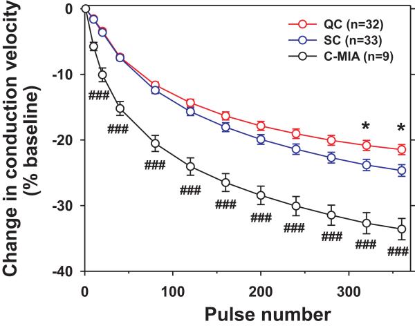
Figure 3
SC afferents exhibit greater activity-dependent slowing of conduction than QC afferents
The overall decrease in conduction velocity induced by suprathreshold electrical stimulation (2Hz for 3 min) at the receptive field differed significantly between fiber classes (F (2,73) = 29.7, p<...

Figure 4
QC- and SC fibers differ in their responses to chemical stimuli
A: QC afferents responded more vigorously than SC afferents to heat-inactivated cowhage spicules coated either with histamine (10 mg/ml) or capsaicin (200 mg/ml) that were inserted into the superficia...

Figure 5
C-MIAs are vigorously activated by histamine and capsaicin
A: Intradermal injection of histamine produced a significantly greater net response in C-MIAs than in CMHs (* p<0.05, Mann-Whitney U test). Dashed line indicates net response of 10 action potential...

Figure 6
Histamine and u03b2-alanine activate different populations of DRG neurons
A: Specimen fluorescent picture of cultured DRG neurons investigated in calcium imaging studies. The intracellular calcium levels of the neurons numbered are shown in the traces in B. (Scale bar: 100 ...
Figure images are served from the NIH/NLM PubMed Central Open Access Subset or Europe PMC; copyright remains with the publishers and authors.
💬 Discussion
0 commentsNo comments yet. Be the first to start a discussion!
Leave a Comment