Abstract
BACKGROUND: Cough is the most frequent reason for consultation with a family doctor, or with a general or respiratory physician. Treatment options are limited and a recent meta-analysis concluded that over-the-counter remedies are ineffective and there is increasing concern about their use in children. Endogenous inflammatory mediators such as prostaglandin E2 (PGE2) and bradykinin (BK), which are often elevated in respiratory disease states, are also known to cause cough by stimulating airway sensory nerves. However, how this occurs is not understood. METHODS: We hypothesised that the transient receptor potential (TRP) channels, TRPA1 and TRPV1, may have a role as 'common effectors' of tussive responses to these agents. We have employed a range of in vitro imaging and isolated tissue assays in human, murine and guinea pig tissue and an in vivo cough model to support this hypothesis. RESULTS: Using calcium imaging we demonstrated that PGE2 and BK activated isolated guinea pig sensory ganglia and evoked depolarisation (activation) of vagal sensory nerves, which was inhibited by TRPA1 and TRPV1 blockers (JNJ17203212 and HC-030031). These data were confirmed in vagal sensory nerves from TRPA1 and TRPV1 gene deleted mice. TRPV1 and TRPA1 blockers partially inhibited the tussive response to PGE2 and BK with a complete inhibition obtained in the presence of both antagonists together in a guinea pig conscious cough model. CONCLUSION: This study identifies TRPA1 and TRPV1 channels as key regulators of tussive responses elicited by endogenous and exogenous agents, making them the most promising targets currently identified in the development of anti-tussive drugs.
🔬 Techniques
🧪 Sample Preparation
🔬 Cell Lines
💾 Data Repositories
🏛️ Research Organizations (ROR)
Affiliated research institutions:
📋 Methods
We hypothesised that the transient receptor potential (TRP) channels, TRPA1 and TRPV1, may have a role as ‘common effectors’ of tussive responses to these agents. We have employed a range of in vitro imaging and isolated tissue assays in human, murine and guinea pig tissue and an in vivo cough model to support this hypothesis.
Methods
Isolated vagal ganglia Intracellular free calcium ([Ca 2+ ] i ) measurements were performed in dissociated jugular and nodose neurons. These studies were performed on all isolated vagal neurons (not airway specific), with the concentration–response data representing an overview of responding and non-responding cells. For subsequent antagonist studies only responding cells were analysed, with the criteria for a ‘responsive cell’ judged as an increase in [Ca 2+ ] i of ≥10% of the K 50 response. In each case, N = number of animals and n = number of cells tested. Comprehensive methods are detailed in online supplementary text.
Isolated vagus nerve preparation
Guinea pigs or mice (C57BL/6, Trpa1 −/− and Trpv1 −/− ) were sacrificed by injection of sodium pentobarbitone (200 mg/kg intraperitoneal injection). The vagus nerves were removed and experiments conducted in our fully characterised isolated vagus preparation, as described in previous publications. 10 26 Human vagal tissue (n=6, two men, 27–72-year-old donors with no respiratory disease) was obtained from two sources—transplant tissue surplus to requirements (Harefield Hospital, UK); and purchased from the International Institute for the Advancement of Medicine (Edison, New Jersey, USA). In all cases, the tissue was consented for use in scientific research and ethics approval obtained from the Royal Brompton & Harfield Trust. See online supplementary text for full methods.
Show full methods section
We hypothesised that the transient receptor potential (TRP) channels, TRPA1 and TRPV1, may have a role as ‘common effectors’ of tussive responses to these agents. We have employed a range of in vitro imaging and isolated tissue assays in human, murine and guinea pig tissue and an in vivo cough model to support this hypothesis.
Methods
Isolated vagal ganglia Intracellular free calcium ([Ca 2+ ] i ) measurements were performed in dissociated jugular and nodose neurons. These studies were performed on all isolated vagal neurons (not airway specific), with the concentration–response data representing an overview of responding and non-responding cells. For subsequent antagonist studies only responding cells were analysed, with the criteria for a ‘responsive cell’ judged as an increase in [Ca 2+ ] i of ≥10% of the K 50 response. In each case, N = number of animals and n = number of cells tested. Comprehensive methods are detailed in online supplementary text.
Isolated vagus nerve preparation
Guinea pigs or mice (C57BL/6, Trpa1 −/− and Trpv1 −/− ) were sacrificed by injection of sodium pentobarbitone (200 mg/kg intraperitoneal injection). The vagus nerves were removed and experiments conducted in our fully characterised isolated vagus preparation, as described in previous publications. 10 26 Human vagal tissue (n=6, two men, 27–72-year-old donors with no respiratory disease) was obtained from two sources—transplant tissue surplus to requirements (Harefield Hospital, UK); and purchased from the International Institute for the Advancement of Medicine (Edison, New Jersey, USA). In all cases, the tissue was consented for use in scientific research and ethics approval obtained from the Royal Brompton & Harfield Trust. See online supplementary text for full methods.
Conscious guinea pig cough model
Conscious unrestrained guinea pigs were placed in individual plastic transparent whole-body plethysmograph chambers (Buxco, Wilmington, North Carolina, USA) and cough detected as previously described. 10 26 Data analysis and statistics For imaging, RM is the maximum response observed expressed as a percentage of the K 50 response. EC 50 values quoted in the imaging studies are the concentrations of drug that produced 50% of the maximum response obtained. Inhibition of agonist responses in the isolated vagus nerve preparation was analysed by two-tailed paired t test, comparing responses to the agonist in the absence and presence of an antagonist in the same piece of nerve. Inhibition of cough by TRPA1 and TRPV1 antagonists in vivo was analysed by Kruskal–Wallis test for multiple comparisons with Dunn's post hoc test, comparing responses from each group of antagonist/vehicle combination to the vehicle-only control. Data are presented as median ± IQR, with statistical significance set at p
📊 Figures
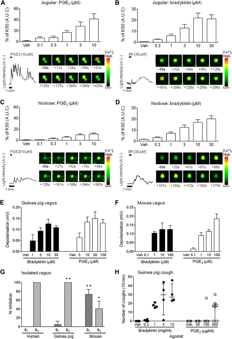
Figure 1
Establishing concentration responses for prostaglandin (PGE 2 ) and bradykinin (BK) in the in vitro preparations and in vivo cough model. (Au2013D) Concentration responses showing increases in intrace...
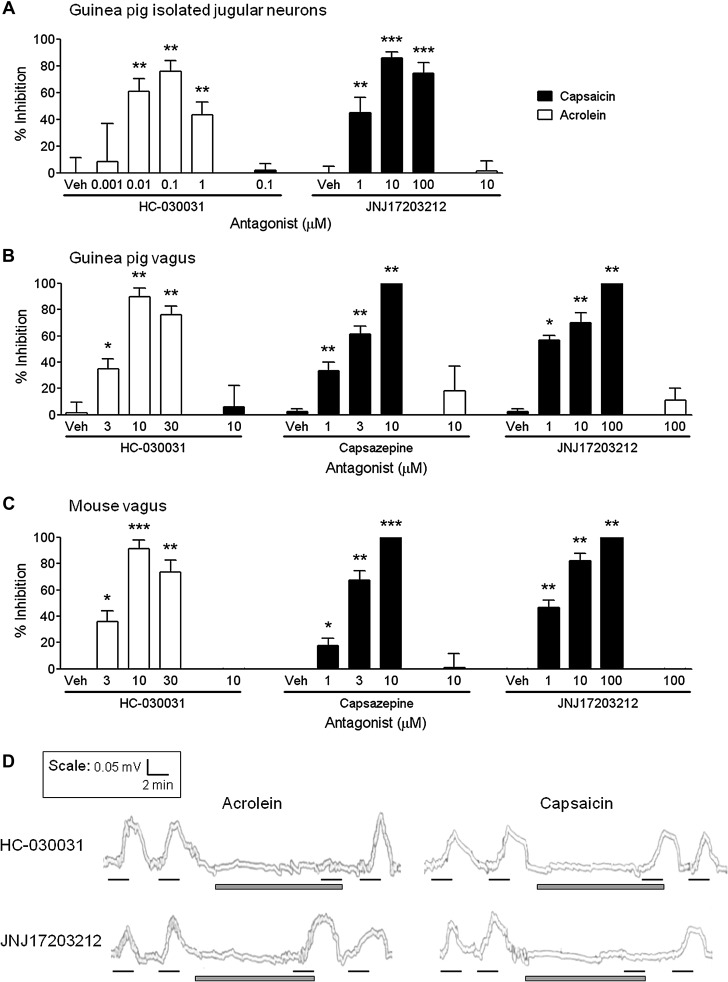
Figure 2
Characterisation of transient receptor potential channel A1 (TRPA1)-selective and TRPV1-selective antagonists in the in vitro primary ganglia and isolated vagus nerve preparations. The TRPA1 antagonis...
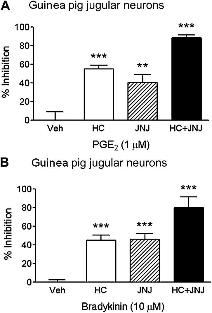
Figure 3
Determining the role of transient receptor potential channel A1 (TRPA1) and TRPV1 in prostaglandin E 2 (PGE 2 ) and bradykinin (BK) induced isolated primary jugular neurons. The TRPA1 antagonist HC-03...
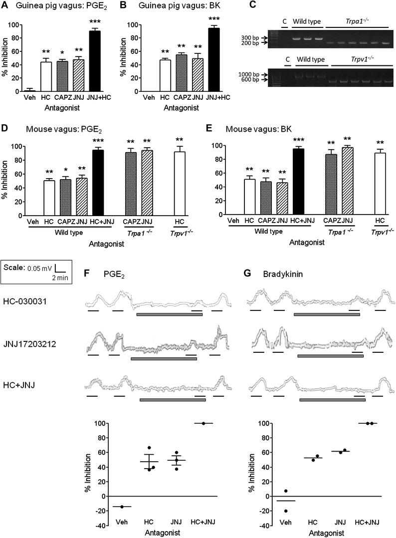
Figure 4
Determining the role of transient receptor potential channel A1 (TRPA1) and TRPV1 in prostaglandin E 2 (PGE 2 ) and bradykinin (BK) induced sensory nerve activation. The TRPA1 antagonist HC-030031 (HC...
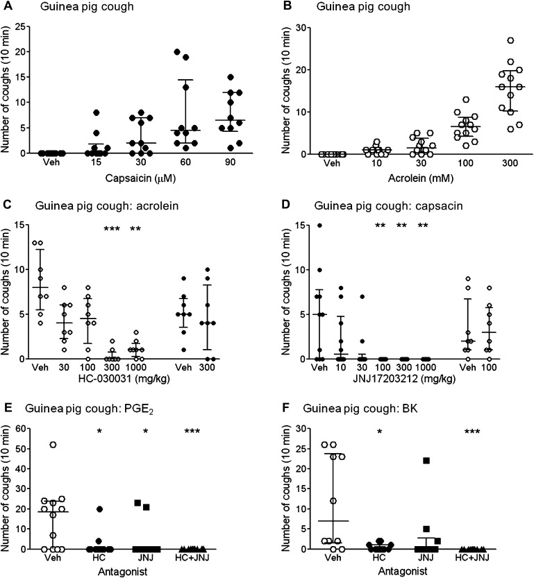
Figure 5
Determining the role of transient receptor potential channel A1 (TRPA1) and TRPV1 in prostaglandin E 2 (PGE 2 ) and bradykinin (BK)-induced cough in conscious guinea pigs. (A) Capsaicin and (B) acrole...
Figure images are served from the NIH/NLM PubMed Central Open Access Subset or Europe PMC; copyright remains with the publishers and authors.
💬 Discussion
0 commentsNo comments yet. Be the first to start a discussion!
Leave a Comment