Abstract
Intense exercise is a physiological stress capable of inducing the interaction of neutrophils with muscle endothelial cells and their transmigration into tissue. Mechanisms driving this physiological inflammatory response are not known. Here, we investigate whether production of reactive oxygen species is relevant for neutrophil interaction with endothelial cells and recruitment into the quadriceps muscle in mice subjected to the treadmill fatiguing exercise protocol. Mice exercised until fatigue by running for 56.3±6.8 min on an electric treadmill. Skeletal muscle was evaluated by intravital microscopy at different time points after exercise, and then removed to assess local oxidative stress and histopathological analysis. We observed an increase in plasma lactate and creatine kinase (CK) concentrations after exercise. The numbers of monocytes, neutrophils, and lymphocytes in blood increased 12 and 24 hours after the exercise. Numbers of rolling and adherent leukocytes increased 3, 6, 12, and 24 hours post-exercise, as assessed by intravital microscopy. Using LysM-eGFP mice and confocal intravital microscopy technology, we show that the number of transmigrating neutrophils increased 12 hours post-exercise. Mutant gp91phox-/- (non-functional NADPH oxidase) mice and mice treated with apocynin showed diminished neutrophil recruitment. SOD treatment promoted further adhesion and transmigration of leukocytes 12 hours after the exercise. These findings confirm our hypothesis that treadmill exercise increases the recruitment of leukocytes to the postcapillary venules, and NADPH oxidase-induced ROS plays an important role in this process.
🔬 Techniques
✨ Fluorophores
🧪 Sample Preparation
🔬 Cell Lines
🏭 Microscope Brands
🧪 Reagent Suppliers
🏛️ Research Organizations (ROR)
Affiliated research institutions:
📋 Methods
Experimental protocol Figure 1A provides a detailed overview of the experimental protocol used in our study. First, animals were subjected to the exercise protocol and oxygen consumption (VO 2 ) was measured during time of running. Drug treatment was performed 30 min before the fatiguing exercise protocol. Lactate and glucose were assessed in blood collected immediately before and after the exercise protocol (without anesthesia). At different time-points after exercise, animals were anesthetized and prepared for intravital microscopy, when quadriceps muscle was collected for GSH assay. At 6, 12 and 24h after exercise, blood was collected from the brachial plexus for leukocyte counts and analyses of CK activity. At 12 hour (at the peak of leukocyte accumulation) muscle was collected for histopathological analyses and expression of adhesion molecules by real-time PCR. Finally, we also conducted experiments in mice exercised to 60% of duration or 60% of the intensity to generate a different exercise overload. After intravital microscopy all mice were euthanized by overdose of anesthesia. 10.1371/journal.pone.0096464.g001 Figure 1 Oxygen consumption, lactate and creatine kinase (CK) levels in the blood. Overall experimental procedures (A). Fatigue was induced by spontaneous running on an electric treadmill that elevated the oxygen consumption (B). At different time-points after the fatiguing exercise blood samples were collected for measurement of lactate (C), creatine kinase (D) and glucose level (E). In this study WT C57Bl/6 mice were used. These results C, D and E are presented as the mean ± SEM (n = 5). *P
📊 Figures
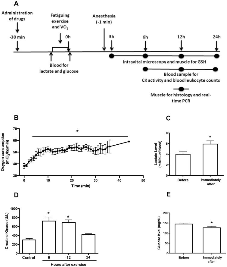
Figure 1
Oxygen consumption, lactate and creatine kinase (CK) levels in the blood.
Overall experimental procedures (A). Fatigue was induced by spontaneous running on an electric treadmill that elevated the oxygen consumption (B). At different time-points after the fatiguing exercise...
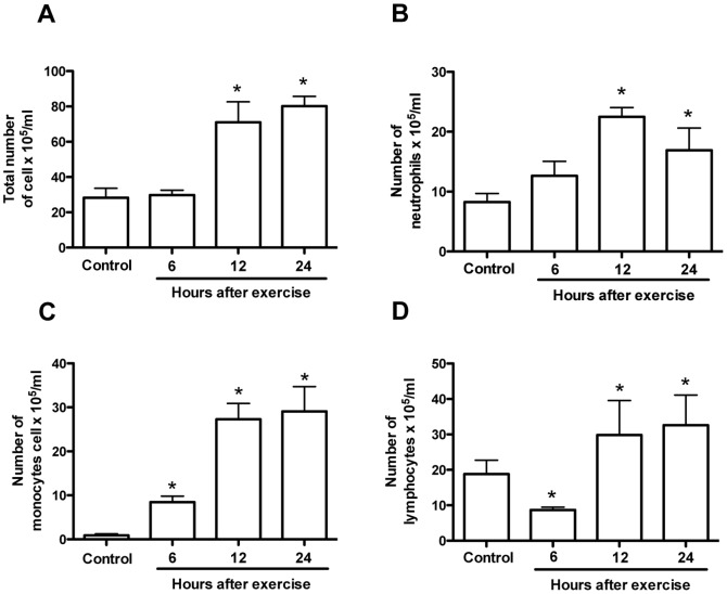
Figure 2
Number of leukocytes in the peripheral blood.
Fatigue was induced by spontaneous running on an electric treadmill. At different time-points after the exercise protocol blood samples were collected and numbers of total cell (A) neutrophils (B), mo...
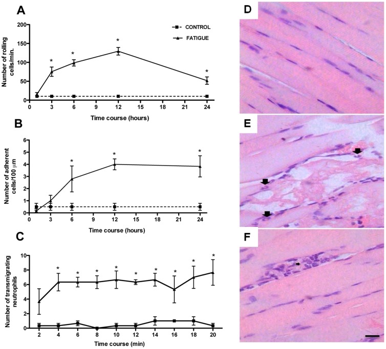
Figure 3
Recruitment of neutrophil into quadriceps muscle.
Fatigue was induced by spontaneous running on an electric treadmill. At different time-points after the exercise protocol, mice were anesthetized and muscle postcapillary venules selected for counting...
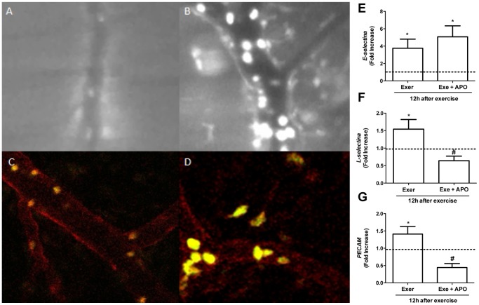
Figure 4
Leukocytes-endothelium interaction in quadriceps muscle after exercise and adhesion molecules expression.
Fatigue was induced by spontaneous running on an electric treadmill. Quadriceps muscle was exposed to visualize the vasculature 12(A) and exercised wild-type mice (B). Confocal intravital microscopy i...
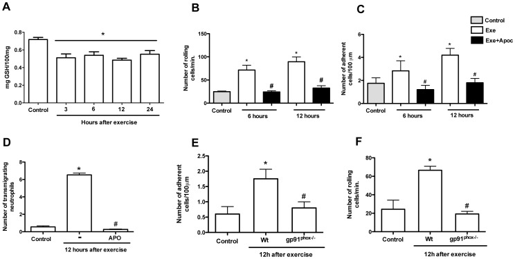
Figure 5
ROS production from NADPH oxidase on leukocyte recruitment.
Fatigue was induced by spontaneous running on an electric treadmill. GSH levels were measured at different time-points after exercise protocol (A). Mice received an injection of apocynin (10 mg/kg, i....
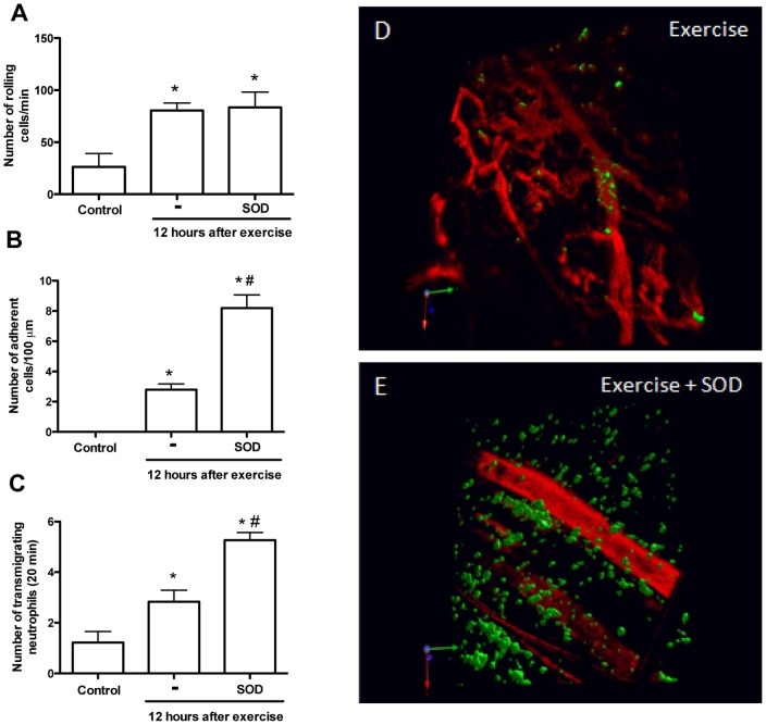
Figure 6
Superoxide dismutase (SOD) exacerbated adhesion and transmigration of neutrophil.
Fatigue was induced by spontaneous running on an electric treadmill. Superoxide dismutase (3 mg/kg, i.p.) was injected 30 minutes before exercise. Using LysM-eGFP mice, numbers of rolling (A), adheren...
Figure images are served from the NIH/NLM PubMed Central Open Access Subset or Europe PMC; copyright remains with the publishers and authors.
💬 Discussion
0 commentsNo comments yet. Be the first to start a discussion!
Leave a Comment