Abstract
Wide-field temporal focusing is a novel technique that provides optical sectioning for imaging without the need for beam scanning. However, illuminating over large areas greatly reduces the photon density which limits the technique applicability to small regions, precluding functional imaging of cellular networks. Here we present a strategy that combines beam shaping and temporal focusing of amplified pulses (>1 µJ/pulse) for fast imaging of cells from the central nervous system in acute slices. Multiphoton video-rate imaging over total areas as wide as 4800 µm(2) with an optical sectioning under 10 µm at 800 nm is achieved with our setup, leading to imaging of calcium dynamics of multiple cells simultaneously in thick tissue.
🔬 Techniques
✨ Fluorophores
🧪 Sample Preparation
🔬 Cell Lines
🏭 Microscope Brands
🧪 Reagent Suppliers
📷 Detectors
💻 Software Details
🏛️ Research Organizations (ROR)
Affiliated research institutions:
📋 Methods
2.1.
Microscopy setup for fluorescence imaging
The microscopy setup is shown in Fig. 1(a) . The laser source is a regenerative laser amplifier (Coherent, Inc. RegA 9000, California) pumped by a Titanium:sapphire laser (Coherent, Inc. Mira 900-F, California). The RegA (800 nm, >1 µ J,
📊 Figures
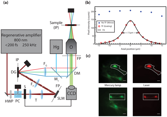
Fig. 1.
(a) Microscopy Setup. HWP: half-wave plate, PC: polarizer cube, S: camera-controlled shutter, T: telescope comprised of two achromatic lenses (f = u221275 mm and +125 mm), SLM: spatial light modulator...
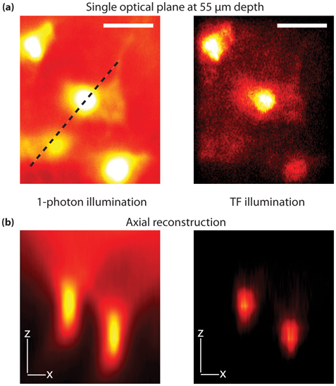
Fig. 2.
Comparison between one-photon and TF imaging of astrocytes in 80 u00b5 m spinal cord slice immunolabeled for glial fibrillary acidic protein (GFAP), revealed with AlexaFluo 594 secondary antibody. (a)...
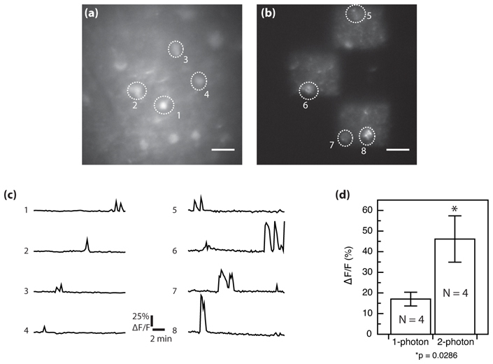
Fig. 3.
Comparison between one-photon and TF functional imaging in brain slice from two independent spontaneous activity measurements. Fluorescence from Fluo-4 AM calcium indicator loaded in a P8 300 u00b5 m ...
Figure images are served from the NIH/NLM PubMed Central Open Access Subset or Europe PMC; copyright remains with the publishers and authors.
💬 Discussion
0 commentsNo comments yet. Be the first to start a discussion!
Leave a Comment