Abstract
Abstract The first transition‐metal complex‐based two‐photon absorbing luminescence lifetime probes for cellular DNA are presented. This allows cell imaging of DNA free from endogenous fluorophores and potentially facilitates deep tissue imaging. In this initial study, ruthenium(II) luminophores are used as phosphorescent lifetime imaging microscopy (PLIM) probes for nuclear DNA in both live and fixed cells. The DNA‐bound probes display characteristic emission lifetimes of more than 160 ns, while shorter‐lived cytoplasmic emission is also observed. These timescales are orders of magnitude longer than conventional FLIM, leading to previously unattainable levels of sensitivity, and autofluorescence‐free imaging.
🔬 Techniques
💻 Software
🧪 Sample Preparation
🔬 Cell Lines
🏭 Microscope Brands
🧪 Reagent Suppliers
💻 Software Details
💾 Data Repositories
🏛️ Research Organizations (ROR)
Affiliated research institutions:
📋 Methods
1 and 2 were synthesized and characterized as described previously[ 29a ] and used as their chloride salts. Each compound was used as a mixture of enantiomers. 2P-PLIM imaging was performed using Becker & Hickl GmbH combined FLIM/PLIM apparatus connected to a Zeiss-510 Meta Microscope. PLIM data was processed using SPCImage software. See the Supporting Information for full details of experimental apparatus, conditions, and procedures.
Supporting Information As a service to our authors and readers, this journal provides supporting information supplied by the authors. Such materials are peer reviewed and may be re-organized for online delivery, but are not copy-edited or typeset. Technical support issues arising from supporting information (other than missing files) should be addressed to the authors. miscellaneous_information
📊 Figures
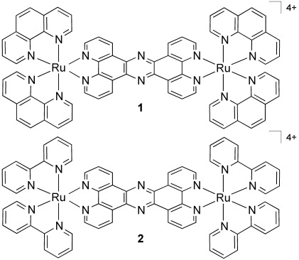
Scheme 1
Structures of the complexes used in this study.
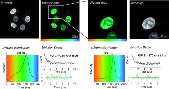
Figure 1
PLIM imaging of live MCF-7 cells pretreated with complex 1 (500 u03bc m , 1 h, serum-free media).
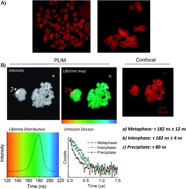
Figure 2
A) Metaphase spreads of HeLa chromosomes stained with 2 (left) or 1 (right) (100 u03bc m , 30 min) and imaged by confocal microscopy; B) 2P-PLIM (left) and confocal (right) imaging of HeLa metaphase s...
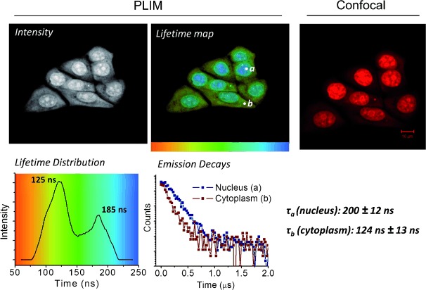
Figure 3
PLIM (left) and confocal (right) comparison of fixed, permeabilized MCF7 cells treated with complex 1 (100 u03bc m , 45 min, PBS buffer).
Figure images are served from the NIH/NLM PubMed Central Open Access Subset or Europe PMC; copyright remains with the publishers and authors.
💬 Discussion
0 commentsNo comments yet. Be the first to start a discussion!
Leave a Comment