Abstract
Anti-angiogenetic cancer therapy is a potential new form for treatment of solid tumours. The alpha(v)-integrins (alpha(v)beta3, alpha(v)beta5) mediate the contact of activated endothelial cells to proteins of the extracellular matrix during tumour angiogenesis as a prerequisite for survival of endothelial cells. The aim of this study was to investigate the effects of application of a methylated cyclic RGD-peptide as an alpha(v)-integrin antagonist on angiogenesis, microcirculation, growth and metastasis formation of a solid tumour in vivo. Experiments were performed in the dorsal skinfold preparation of Syrian Golden hamsters bearing the amelanotic hamster melanoma A-Mel-3. Animals were injected intraperitoneally with a methylated cyclic RGD-peptide every 12 h, the control group received an inactive peptide. Microcirculatory parameters of tumour angiogenesis including functional vessel density, red blood cell velocity, vessel diameter and leucocyte-endothelium interaction were analysed using intravital microscopy. In an additional study the effects on growth and metastasis of subcutaneous A-Mel-3 were quantified. Functional vessel density was markedly reduced on day 3 in treated animals compared to controls (37.2 +/- 12.1 vs 105.2 +/- 11.2 cm(-1); mean +/- s.e.m.; P<0.05) and increased subsequently in both groups. Red blood cell velocity at day 3 was below values of controls (0.026 +/- 0.01 vs 0.12 +/- 0.03 mm x s(-1); P<0.05). No differences were observed in vessel diameters and leucocyte-endothelium interaction was almost absent in both groups. Furthermore, growth and metastasis of subcutaneous tumours after administration of the cyclic RGD-peptide was significantly delayed in comparison to controls (P<0.05). Inhibition of alpha(v)-integrins by a cyclic RGD-peptide resulted in significant reduction of functional vessel density, retardation of tumour growth and metastasis in vivo. Taken together, these results implicate RGD-peptides as agents which have anti-tumour and anti-metastatic activity in vivo.
🔬 Techniques
✨ Fluorophores
🔬 Cell Lines
🏭 Microscope Brands
🧪 Reagent Suppliers
📷 Detectors
💾 Data Repositories
🏛️ Research Organizations (ROR)
Affiliated research institutions:
📋 Methods
Experiments were performed with male Syrian Golden Hamsters (6–8 weeks old, 50–60 g body weight (bw)) in accordance with institutional guidelines after approval of the animal committee of the Bavarian state. The animals were housed one per cage and had free access to tap water and standard laboratory food throughout the experiments. Animals were inspected at least two times a day by specialised animal colony staff to assure a normal clinical condition (including appearance, posture, behaviour and physiological responses). Particular attention was given to body weight and any signs of discomfort, ulceration or inflammation. All surgical procedures were performed under pentobarbital anaesthesia (50 mg kg −1 b.w., i.p.; Nembutal, Sanofia-Leva, Hannover, Germany). The experiments met all the standards required by the UKCCCR guidelines for the welfare of animals in experimental neoplasia (United Kingdom Co-ordinating Committee on Cancer Research ( UKCCCR), 1998 ).
Dorsal skinfold chamber preparation
For quantification of tumour angiogenesis, a dorsal skinfold chamber preparation consisting of two symmetrical titanium frames was surgically implanted into the dorsal skin as described earlier in detail ( Asaishi et al , 1981 ; Endrich et al , 1982 ). This is considered to cause the least distress upon the animal because the dorsal skin is extremely expansible. Following implantation of the transparent chamber and a recovery period of 48 h from anaesthesia and microsurgery, preparations fulfilling the criteria of an intact microcirculation were implanted with 2×10 5 cells of the amelanotic melanoma of the hamster A-Mel-3 ( Fortner et al , 1961 ). Fine polyethylene catheters (PE10, inner diameter 0.28 mm) were permanently inserted into the right jugular vein 48 h before first measurement to allow injection of fluorescent agents for intravital fluorescence microscopy. The animals tolerated the dorsal skinfold chamber well and showed no signs of discomfort.
Show full methods section
Experiments were performed with male Syrian Golden Hamsters (6–8 weeks old, 50–60 g body weight (bw)) in accordance with institutional guidelines after approval of the animal committee of the Bavarian state. The animals were housed one per cage and had free access to tap water and standard laboratory food throughout the experiments. Animals were inspected at least two times a day by specialised animal colony staff to assure a normal clinical condition (including appearance, posture, behaviour and physiological responses). Particular attention was given to body weight and any signs of discomfort, ulceration or inflammation. All surgical procedures were performed under pentobarbital anaesthesia (50 mg kg −1 b.w., i.p.; Nembutal, Sanofia-Leva, Hannover, Germany). The experiments met all the standards required by the UKCCCR guidelines for the welfare of animals in experimental neoplasia (United Kingdom Co-ordinating Committee on Cancer Research ( UKCCCR), 1998 ).
Dorsal skinfold chamber preparation
For quantification of tumour angiogenesis, a dorsal skinfold chamber preparation consisting of two symmetrical titanium frames was surgically implanted into the dorsal skin as described earlier in detail ( Asaishi et al , 1981 ; Endrich et al , 1982 ). This is considered to cause the least distress upon the animal because the dorsal skin is extremely expansible. Following implantation of the transparent chamber and a recovery period of 48 h from anaesthesia and microsurgery, preparations fulfilling the criteria of an intact microcirculation were implanted with 2×10 5 cells of the amelanotic melanoma of the hamster A-Mel-3 ( Fortner et al , 1961 ). Fine polyethylene catheters (PE10, inner diameter 0.28 mm) were permanently inserted into the right jugular vein 48 h before first measurement to allow injection of fluorescent agents for intravital fluorescence microscopy. The animals tolerated the dorsal skinfold chamber well and showed no signs of discomfort.
Assessment of tumour angiogenesis and growth by intravital microscopy
Quantification of tumour angiogenesis using intravital fluorescence microscopy have been described in detail elsewhere ( Asaishi et al , 1981 ; Dellian et al , 1995 , 1996 ). In brief, the awake animal was trained to crawl into a transparent plastic tube and was placed on a custom-made stage under a modified Leitz microscope (Orthoplan; Leitz, Munich, Germany). FITC-labelled dextran (Sigma, Deisenhofen, Germany; MW 500000; 0.05–0.1 ml of a 5% solution in 0.9% NaCl) as a plasma marker and rhodamine 6G (Molecular Probes, Eugene, OR, USA; 0.04 ml of a 0.05% solution in 0.9% NaCl) to label white blood cells in vivo , were injected i.v. to visualise microcirculation and leucocyte–endothelium interaction, respectively. The used dosage of the applied fluorescence dye does not have any toxic effect upon the animal. Selective observation of FITC-labelled plasma was possible using epi-illumination with a 100 W mercury lamp attached to a Ploemopack illuminator with a Leitz I2/3 filter block (excitation 450–490 nm, emission ⩽515 nm) and rhodamine 6G-stained leucocytes were visualised using a Leitz N2 filter block (excitation 530–560 nm, emission ⩽580 nm). Intravital microscopy was performed 3, 5, 9 and 13 days after implantation of the tumour cells. At least 3–5 sites of interest per animal were randomly selected in centre and periphery of the tumour. Images were acquired by a SIT video camera (C2400-08; Hamamatsu Herrsching, Germany) and recorded on S-VHS video tape (Sony) for subsequent analysis. Parallel to microvascular observations the area covered by tumour in the chamber preparation was registered on videotape using a Leitz macroscope with video camera (XC-77; Sony) to evaluate tumour growth. The tumour area (mm 2 ) was quantified from videotape by digital image analysis. Analysis of microcirculatory parameters was performed off-line from video tape by an image analysis system (Cap Image; Zeintl, Heidelberg, Germany). This system described in detail by Zeintl et al (1989) and Klyscz et al (1997) allows measurement of functional vessel density (FVD) as a parameter of angiogenic activity ( Dellian et al , 1996 ). FVD is defined as the total length of perfused microvessels per unit area of observation and is given in cm −1 . Red blood cell velocity (vRBC) was quantified with the line shift diagram method according to Klyscz et al (1997) in mm s −1 . Rolling leucocytes were defined as population of cells temporarily interacting with the vessel wall and thus having a velocity at least 50% below vRBC in the same vessel. Adherent leucocytes were given as the number of leucocytes remaining stationary for at least 30 s per square millimetre of vessel wall surface ( Atherton and Born, 1972 ; Dellian et al , 1996 ).
Evaluation of tumour growth and metastasis Male Syrian Golden
Hamsters (weight, age and housing as described above) were anaesthetised with pentobarbital and the dorsal skin was shaven and chemically depilated (Pilcamed, Schwarzkopf, Germany). Cells (4 to 6×10 6 ) of the A-Mel-3 were suspended in a 10 μl volume and injected s.c. over the lumbosacral region of the dorsal skin. This is considered to cause the least distress upon the animal because the dorsal skin is extremely expansible. Starting on day 5 after tumour cell implantation, the longer (l) and shorter (w) perpendicular axes and the height (h) of each tumour nodule were measured with callipers every other day. Metastases of the animals were determined by palpation of axillar and inguinal lymph nodes. The day when metastases were first palpable the animal was defined as metastasised. To minimise the pain and distress of the animals no biopsies were taken from the lymph nodes during the observation time. This was due to the known fact that the axillar or inguinal lymph nodes were the most frequent sites of metastasis formation ( Fortner et al , 1961 ; Weiss et al , 1990 ). After the end of the experiments when animals were euthanised, biopsies were taken to confirm histologically the invasion of the lymph nodes. Tumour volume was calculated according to the formula V t =0.837×l×w×h ( Weiss et al , 1990 ). Animals were observed for 15 to 17 days until tumours reached a maximum volume of 7 cm. The A-Mel-3 tumour does not have severe side effects during this early period of growth. Treatment and experimental groups The cyclic integrin α v antagonist EMD121974 (cyclic Arg-Gly-Asp-D-Phe(N-methyl)Val) ( Dechantsreiter et al , 1999 ) and the control peptide EMD135981 (cyclic Arg-β-Ala-Asp-D-Phe(N-methyl)Val) were synthesised and characterised at Merck KgAa (Darmstadt, Germany). In pilot studies pharmacokinetics of the peptides were compared after intravenous and intraperitoneal injection in hamsters. Plasma half-life time of the cyclic RGD-peptide after intraperitoneal injection was 47 min, and the area under the curve was similar after intraperitoneal and intravenous administration. A subsequent pilot study demonstrated a slightly higher effect on tumour growth after administration of 30 mg kg −1 every 12 h compared to 30 mg every 2 days. Therefore, we chose the following set-up for administration: Peptides were dissolved in PBS (10 mg ml −1 ). One day after implantation of tumour cells, animals were randomly assigned to two groups ( n =6) and RGD-peptide EMD121974 or control-peptide EMD135981 were given i.p. every 12 h (30 mg kg −1 b.w.) until the end of the experiments.
Statistical analysis
Results are presented as mean±s.e.m. Data were evaluated using Wilcoxon–Mann–Whitney U and Kruskal–Wallis test, respectively (SigmaStat; Jandel Scientific, San Rafael, CA, USA). Metastasis analysis was performed according to the Kaplan–Meier method ( Kaplan and Meier, 1958 ) and the differences were compared for statistical significance using the Cox- F test ( Lee, 1975 , 1980 ) with the statistical software program Statistica (StatSoft, Inc, 1997, Tulsa, OK, USA). P values smaller than 5% were considered to be significant.
Treatment and experimental groups The cyclic integrin α v antagonist EMD121974 (cyclic Arg-Gly-Asp-D-Phe(N-methyl)Val) ( Dechantsreiter et al , 1999 ) and the control peptide EMD135981 (cyclic Arg-β-Ala-Asp-D-Phe(N-methyl)Val) were synthesised and characterised at Merck KgAa (Darmstadt, Germany). In pilot studies pharmacokinetics of the peptides were compared after intravenous and intraperitoneal injection in hamsters. Plasma half-life time of the cyclic RGD-peptide after intraperitoneal injection was 47 min, and the area under the curve was similar after intraperitoneal and intravenous administration. A subsequent pilot study demonstrated a slightly higher effect on tumour growth after administration of 30 mg kg −1 every 12 h compared to 30 mg every 2 days. Therefore, we chose the following set-up for administration: Peptides were dissolved in PBS (10 mg ml −1 ). One day after implantation of tumour cells, animals were randomly assigned to two groups ( n =6) and RGD-peptide EMD121974 or control-peptide EMD135981 were given i.p. every 12 h (30 mg kg −1 b.w.) until the end of the experiments.
📊 Figures
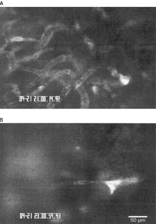
Figure 1
Images of tumour microcirculation 3 days after tumour cell implantation acquired by intravital microscopy after injection of the plasma marker FITC-Dextran. ( A ) Tumour vasculature of animals treated...
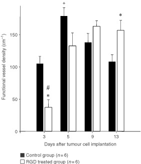
Figure 2
Functional vessel density (meanu00b1s.e.m.) of control group and RGD treated group. Measurements were performed 3, 5, 9 and 13 days after tumour cell implantation by intravital microscopy. Animals wer...
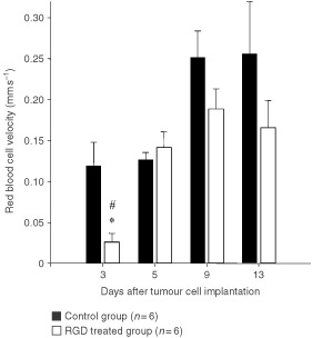
Figure 3
Red blood cell velocity (meanu00b1s.e.m.) of control group and RGD treated group. * P <0.05 vs corresponding controls; # P <0.05 vs treatment day 9 and day 13.
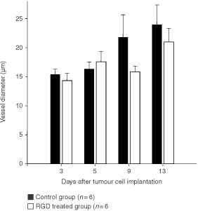
Figure 4
Vessel diameters (meanu00b1s.e.m.) as measured by intravital microscopy of control group and RGD treated group.
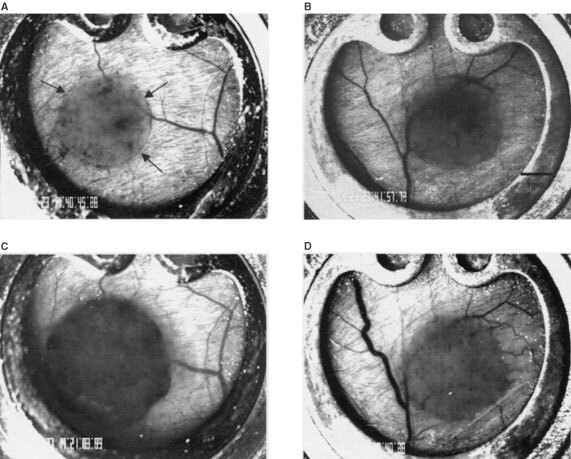
Figure 5
Images of the A-Mel-3 tumour in the dorsal skinfold chamber acquired by video macroscopy. ( A ) Tumour of control group 5 days after tumour cell implantation. Arrows define the tumour margin. ( C ) Th...
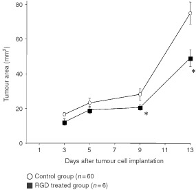
Figure 6
Area of the A-Mel-3 tumour of the control group and the RGD treated group in the dorsal skinfold chamber measured on day 3, 5, 9 and 13 after tumour cell implantation (meanu00b1s.e.m.). * P <0.05 v...
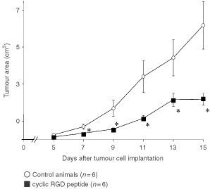
Figure 7
Tumour growth curves of animals with subcutaneously implanted solid A-Mel-3 tumours. Changes in tumour volume are presented for control animals and following application of cyclic RGD peptide. Values ...
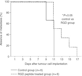
Figure 8
Metastasis formation presented as a Kaplanu2013Meier curve. Animals of the control group and animals of the RGD peptide treated group were examined for axillar and inguinal lymph node metastases durin...
Figure images are served from the NIH/NLM PubMed Central Open Access Subset or Europe PMC; copyright remains with the publishers and authors.
💬 Discussion
0 commentsNo comments yet. Be the first to start a discussion!
Leave a Comment