Abstract
This review analyses the literature concerning non-fluorescent and fluorescent probes for nucleic acid imaging in fixed and living cells from the point of view of their suitability for imaging intracellular native RNA and DNA. Attention is mainly paid to fluorescent probes for fluorescence microscopy imaging. Requirements for the target-binding part and the fluorophore making up the probe are formulated. In the case of native double-stranded DNA, structure-specific and sequence-specific probes are discussed. Among the latest, three classes of dsDNA-targeting molecules are described: (i) sequence-specific peptides and proteins; (ii) triplex-forming oligonucleotides and (iii) polyamide oligo(N-methylpyrrole/N-methylimidazole) minor groove binders. Polyamides seem to be the most promising targeting agents for fluorescent probe design, however, some technical problems remain to be solved, such as the relatively low sequence specificity and the high background fluorescence inside the cells. Several examples of fluorescent probe applications for DNA imaging in fixed and living cells are cited. In the case of intracellular RNA, only modified oligonucleotides can provide such sequence-specific imaging. Several approaches for designing fluorescent probes are considered: linear fluorescent probes based on modified oligonucleotide analogs, molecular beacons, binary fluorescent probes and template-directed reactions with fluorescence probe formation, FRET donor-acceptor pairs, pyrene excimers, aptamers and others. The suitability of all these methods for living cell applications is discussed.
🔬 Techniques
🧬 Organisms
✨ Fluorophores
🧪 Sample Preparation
🔬 Cell Lines
🏛️ Research Organizations (ROR)
Affiliated research institutions:
📊 Figures
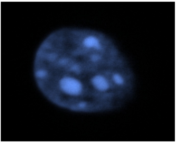
Figure 1
DNA visualization in fixed murine 3T3 cell nucleus using minor groove binder 4',6-diamino-2-phenylindole (DAPI). The image was kindly provided by Dr. C. Escudu00e9 (CNRS, UMR 7196, Paris, France).
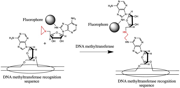
Figure 2
DNA sequence-specific labeling using DNA methyltransferase and fluorescent aziridinyl derivative of adenosine as its substrate instead of S-adenosylmethionine [ 36 , 37 , 38 ].
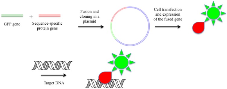
Figure 3
DNA imaging in living cells with fused proteins containing GFP and site-specific or sequence-specific proteins.
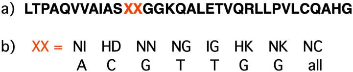
Figure 4
( a ) Primary structure of the TALE element recognizing one base pair. 12-th and 13-th amino acids (XX in red) determine the element specificity. ( b ) Recognition code of the TALE elements [ 55 ].
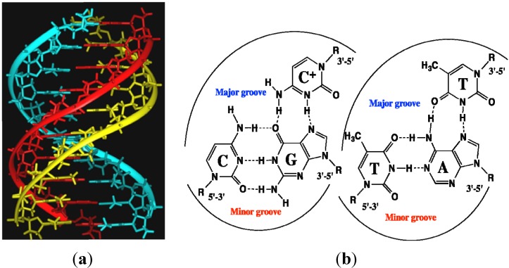
Figure 5
( a ) DNA triple helix: polypurine strand (red), polypyrimidine strand (cyan) and third polypyrimidine strand (yellow) in a major groove of the duplex. ( b ) Examples of Hoogsteen triplets C:G-C and T...
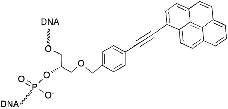
Figure 6
TINA (Twisted Intercalating Nucleic Acid) monomer.
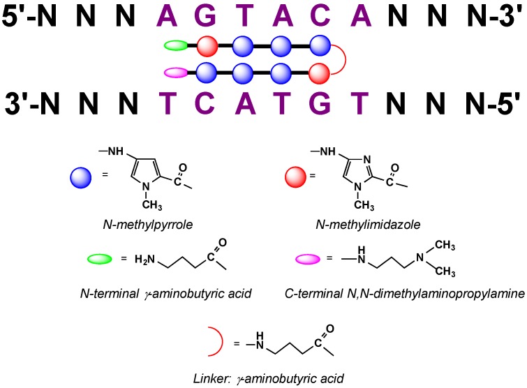
Figure 7
Structure of hairpin N-methylpyrrole/N-methylimidazole polyamides and code of DNA recognition upon their binding to DNA minor groove [ 79 ].
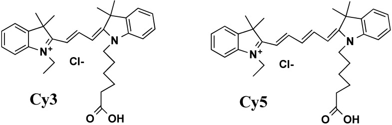
Figure 8
Cyanine fluorophores Cy3 and Cy5.

Figure 9
Fluorescence Resonance Energy Transfer (FRET).
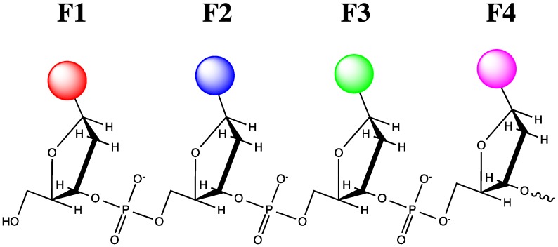
Figure 10
u201cOligodeoxyfluorosidesu201d (F1, F2, F3, F4, u2026 u2014different fluorophore residues).
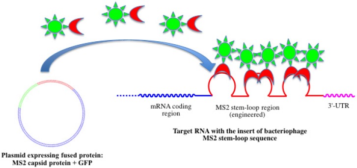
Figure 11
Bacteriophage MS2 genetic encoding system for RNA detection using genetically modified RNA and GFP fused with MS2 capsid protein.

Figure 12
Linear complementary fluorescent probe with fluorophore and quencher at the 5'-terminus. When the quencher intercalates into duplex formed upon hybridization, the fluorescence appears [ 165 ].
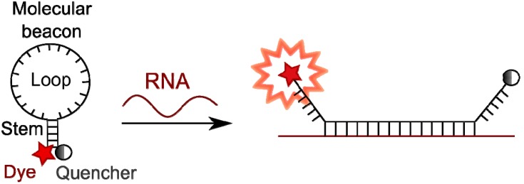
Figure 13
Mode of functioning of molecular beacons.
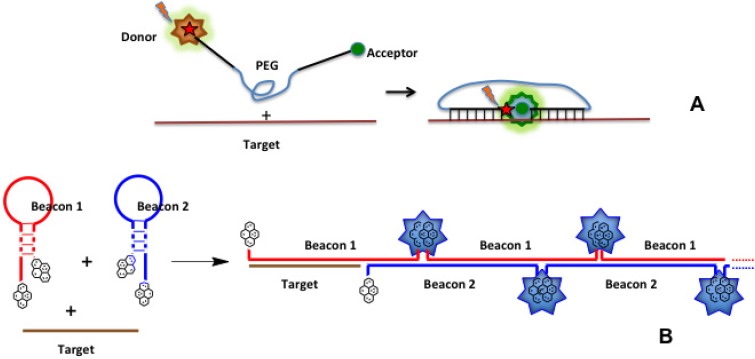
Figure 14
New types of molecular beacons. ( A ) hybrid molecular probe with two antiparallel oligonucleotide strands linked by a long poly(ethyleneglycol) chain [ 204 ]. Oligonucleotides complementary to adjace...
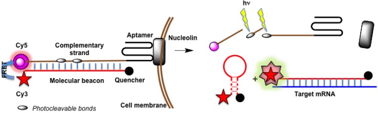
Figure 15
A targeted, self-delivered and photocontrolled aptamer-based molecular beacon [ 216 ].

Figure 16
Quenched autoligation probes [ 173 ].
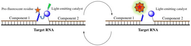
Figure 17
Template-directed light-induced catalyzed cleavage of a linker in pro-fluorophore-conjugated probe with the release of fluorescent molecule.
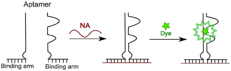
Figure 18
Fluorophore-binding aptamers as binary probes.
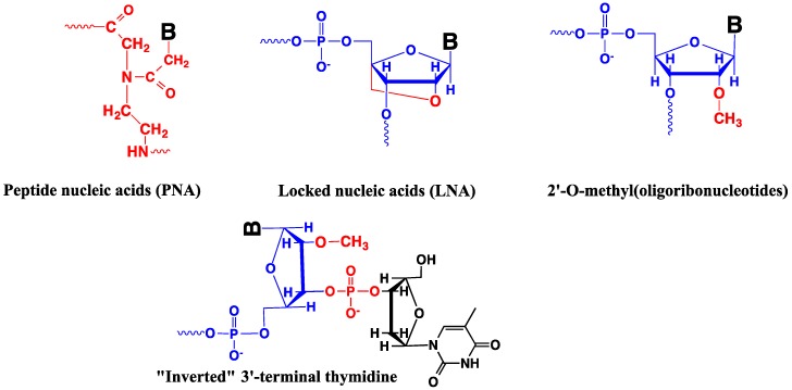
Figure 19
Modified oligonucleotides used for probe design. Bu2014nucleobase. Modifications are indicated in red.
Figure images are served from the NIH/NLM PubMed Central Open Access Subset or Europe PMC; copyright remains with the publishers and authors.
💬 Discussion
0 commentsNo comments yet. Be the first to start a discussion!
Leave a Comment