Abstract
Direct targeting to the kidneys is a promising strategy to improve drug therapeutic index for the treatment of kidney diseases. We sought to investigate the renal selectivity and safety of kidney-targeted mesoscale nanoparticle technology. We found that direct intravenous administration of these particles resulted in 26-fold renal selectivity and localized negligibly in the liver or other organs. The nanoparticles targeted the renal proximal tubular epithelial cells, as evidenced by intravital microscopy and ex vivo imaging. Mice treated with the nanoparticles exhibited no negative systemic consequences, immune reaction, liver impairment, or renal impairment. The localization of material selectively to the renal tubules is uncommon, and this work portends the development of renal-targeted drugs for the treatment of kidney diseases.
🔬 Techniques
✨ Fluorophores
🧪 Sample Preparation
🔬 Cell Lines
🏭 Microscope Brands
🧪 Reagent Suppliers
🎨 Filters
💻 Software Details
🏛️ Research Organizations (ROR)
Affiliated research institutions:
📋 Methods
Particle Formulation and Characterization
Mesoscale nanoparticles (MNPs) were formed from poly(lactic- co -glycolic acid) conjugated to polyethylene glycol (PLGA-PEG). The block copolymer was conjugated as we previously described 17 . Briefly, 5 g (90–130 μmol) carboxylic acid-terminated PLGA (50:50; MW 28–54 kDa) (Aldrich; St. Louis, MO) was dissolved in methylene chloride with 1.2 mmol N -hydroxysuccinimide (NHS) and 1.2 mmol 1-ethyl-3-(3-(dimethylamino)propyl)-carbodiimide (EDC) and stirred for approximately 30 minutes. PLGA-NHS was then precipitated with ethyl ether and washed with 50:50 ethyl ether:methanol and dried under vacuum. PLGA-NHS (1 g, 18–26 μmol) was mixed with 50 μmol amine-PEG-carboxylic acid (MW 5 kDa) (Nanocs; New York, NY) in chloroform and 220 μmol N,N -diisopropylethylamine overnight. Conjugated PLGA-PEG was precipitated and washed with cold methanol then dried under vacuum. 1 H NMR was used to confirm conjugation as previously described 19 . Fluorescent MNPs were formed from PLGA-PEG and 3,3′-diethylthiadicarbocyanine iodide (DEDC) (Acros Organics; Geel, Belgium) as we previously described via nanoprecipitation 17 . 100 mg PLGA-PEG was dissolved with 10 mg DEDC in 2 mL acetonitrile and added dropwise to 4 mL water with 100 μL 10% Pluronic F-68 (Gibco; Grand Island, NY). Alternatively, we formulated MNPs encapsulating a random double-stranded DNA duplex 5′-AGTCGTCAGTACGATGCAGAC/3Cy5/3′ with a molecular weight of 13,523.1 g/mol. After stirring for 2 hours, particles were centrifuged at 7356 RCF for 15 minutes and washed before lyophilization in a 2% sucrose solution. Freeze-dried particles were analyzed for size by dynamic light scattering (DLS) in phosphate-buffered saline and ζ-potential in water by electrophoretic light scattering (ELS) (Malvern; Worcestershire, United Kingdom). Total DEDC encapsulation was measured by UV-Vis absorbance (Jasco; Easton, MD).
Show full methods section
Particle Formulation and Characterization
Mesoscale nanoparticles (MNPs) were formed from poly(lactic- co -glycolic acid) conjugated to polyethylene glycol (PLGA-PEG). The block copolymer was conjugated as we previously described 17 . Briefly, 5 g (90–130 μmol) carboxylic acid-terminated PLGA (50:50; MW 28–54 kDa) (Aldrich; St. Louis, MO) was dissolved in methylene chloride with 1.2 mmol N -hydroxysuccinimide (NHS) and 1.2 mmol 1-ethyl-3-(3-(dimethylamino)propyl)-carbodiimide (EDC) and stirred for approximately 30 minutes. PLGA-NHS was then precipitated with ethyl ether and washed with 50:50 ethyl ether:methanol and dried under vacuum. PLGA-NHS (1 g, 18–26 μmol) was mixed with 50 μmol amine-PEG-carboxylic acid (MW 5 kDa) (Nanocs; New York, NY) in chloroform and 220 μmol N,N -diisopropylethylamine overnight. Conjugated PLGA-PEG was precipitated and washed with cold methanol then dried under vacuum. 1 H NMR was used to confirm conjugation as previously described 19 . Fluorescent MNPs were formed from PLGA-PEG and 3,3′-diethylthiadicarbocyanine iodide (DEDC) (Acros Organics; Geel, Belgium) as we previously described via nanoprecipitation 17 . 100 mg PLGA-PEG was dissolved with 10 mg DEDC in 2 mL acetonitrile and added dropwise to 4 mL water with 100 μL 10% Pluronic F-68 (Gibco; Grand Island, NY). Alternatively, we formulated MNPs encapsulating a random double-stranded DNA duplex 5′-AGTCGTCAGTACGATGCAGAC/3Cy5/3′ with a molecular weight of 13,523.1 g/mol. After stirring for 2 hours, particles were centrifuged at 7356 RCF for 15 minutes and washed before lyophilization in a 2% sucrose solution. Freeze-dried particles were analyzed for size by dynamic light scattering (DLS) in phosphate-buffered saline and ζ-potential in water by electrophoretic light scattering (ELS) (Malvern; Worcestershire, United Kingdom). Total DEDC encapsulation was measured by UV-Vis absorbance (Jasco; Easton, MD).
Serum Stability Assay
Nanoparticle stability was measured in complete mouse serum obtained from healthy C57BL/6 mice (Charles River; Troy, NY). 1 mg/mL MNPs were suspended in 500 μL serum and incubated at room temperature. At 2, 4, 6, 24, 48, and 72 hours, 100 μL of the sample was removed for DLS measurement and subsequently replaced into the sample. 10 mg/mL MNPs were suspended in 100 μL serum and incubated at room temperature. At 2, 4, 6, 24, 48, and 72 hours, the sample was centrifuged at 4286 RCF for 15 minutes, the supernatant was removed, and the pellet was resuspended in serum and incubated at room temperature. Absorbance measurements of the dye in the supernatant and pellet were obtained using a Tecan Infinite M1000Pro (Mannedorf, Switzerland) in a 96-well plate at 650 nm. Dye release (%) was calculated as the amount of dye in each sample divided by total dye combined from supernatants at each time point and 72 hour pellet measurements. Administration Route Investigation All animal experiments were approved by and carried out in accordance with IACUC guidelines at Memorial Sloan Kettering Cancer Center and in accordance with the NIH Guide for the Care and Use of Laboratory Animals. Healthy female 4–8 week hairless mice with intact immune systems (Crl:SKH1- Hr Hr ) were used (Charles River. Mice were fed irradiated 5V75 alfalfa-free diet (LabDiet; St. Louis, MO) to reduce fluorescent imaging background. Groups of 3 mice each were dosed with 25 mg/kg MNPs via the following administration routes: oral gavage (per os, PO), intravenous via the retroorbital vein (RO), intravenous via the tail vein (IV), subcutaneous flank (SQ), and intraperitoneal (IP). Live mice were imaged five at a time (one per group) to determine whole-animal biodistribution at the following post-injection time-points: 30 minutes, 4 hours, 24 hours, 48 hours, and 72 hours. Imaging was performed using an IVIS Spectrum Preclinical In Vivo Imaging System (Perkin Elmer; Waltham, MA) using 640/680 nm excitation/emission filters. Following the 72 hour imaging time-point, mice were euthanized and the following organs were harvested and fluorescently imaged: heart, lungs, liver, spleen, and kidneys. Organs and regions of interest (ROI) in live mice were selected using Living Image Software v4.3 (Perkin Elmer) to quantify average fluorescence efficiency per square centimeter in each ROI. Organ fluorescence measurements were calculated as the fluorescence efficiency normalized by the fluorescence efficiency of PBS-injected control organs. Mean and standard deviation of organ fluorescence were calculated for each group of 3 mice. Dose Investigation Healthy female 4–8 hairless mice with intact immune systems were used (Crl:SKH1- Hr Hr ). Three mice were injected IV with 25 mg/kg DEDC MNPs and one mouse was injected with phosphate buffered saline (PBS) as a vehicle control. Separately, three mice were injected IV with 5 mg/kg DEDC MNPs and one mouse was injected with PBS as a control. Imaging was performed as described above at 30 minutes, 24 hours, 48 hours, and 72 hours post injection. Mice were euthanized after 72 hours with organs extracted, imaged, and analyzed as above. Background-subtracted average fluorescence efficiency was reported. Where denoted, normalized total fluorescence efficiency was reported to account for the full volume of each organ. Normalized total fluorescence efficiency was obtained by dividing each organ’s average fluorescence by the total weight of that organ and subtracting that of a control animal. Results were compared with the results from a 50 mg/kg IV administration of anionic MNPs as previously described 17 . For investigations with Cy5-dsDNA MNPs, three mice were injected with nanoparticles at 50 mg/kg IV with a single PBS-injected control mouse. The mice were euthanized 24 hours after injection; organs were extracted, imaged, and analyzed as above. In Vivo Imaging Healthy Cx3cr gfp/+ C57BL/6 mice, with green fluorescent protein (GFP)-expressing renal macrophages were used 20 , 21 . An intravital confocal microscopy imaging setup was used to perform superficial renal cortex imaging 21 . Mice were IV injected via the tail vein with either 25 mg/kg DEDC MNPs or a matched dose of free DEDC. Mice were imaged at 72 hours post-injection. Perfused Animal Ex Vivo imaging Healthy 4–8 week hairless mice with intact immune systems were used. One mouse was injected with 25 mg/kg MNPs and one with a matched dose of free DEDC. At 72 hours post-injection, cardiac perfusions were performed with 4% paraformaldehyde (PFA). Organs were removed and fixed in 4% PFA, dehydrated, and embedded in paraffin. 5 μm sections were placed onto glass slides, de-paraffinized and prepared for immunofluorescence imaging. Slides were stained with 4′,6-diamidino-2-phylindole (DAPI) to stain nuclei and then stained with either an anti-CD31 antibody for endothelial cells (Dianova; Hamburg, Germany) or anti-E-cadherin to stain epithelial cells (BD Bioscience; San Jose, CA). Slides were imaged with an Olympus IX51 inverted light microscope (Olympus; Center Valley, PA) and an Olympus XM10 monochrome camera after excitation with a X-Cite 120Q lamp (Lumen Dynamics; Ontario, Canada). Appropriate filter cubes for DAPI, AlexaFluor488, and Cy5 were used with consistent exposure times for each channel and analyzed in ImageJ (NIH; Bethesda, MD) with consistent brightness values. Long-term Biodistribution and Safety Healthy 4–8 week female BALB/c mice (BALB/cAnNCrl) were used (Charles River). Twelve mice were IV injected with 25 mg/kg DEDC MNPs and separated into groups of three. One group was euthanized at each of the following time-points: 1 day, 3 days, 7 days, and 28 days post-injection. Separately, three mice were injected with a PBS vehicle control and sacrificed on day 1. Prior to sacrifice, blood was collected retroorbitally and urine was collected following excretion (urine collection failed at day 28). Organs were collected, imaged, and analyzed as described above. A complete renal panel was performed on serum samples, including: blood urea nitrogen (BUN), creatinine, total protein, albumin, globulin, phosphorous, calcium, total CO 2 , sodium, potassium, chloride, and anion gap. Urine chemistry was performed to obtain BUN, creatinine, and micro total protein. Whole blood was used to perform a complete blood count for each mouse, including: white blood cells, neutrophils, lymphocytes, monocytes, eosinophils, red blood cells (RBCs), nucleated RBCs, hemoglobin, hematocrit, mean corpuscular volume, mean corpuscular hemoglobin, mean corpuscular hemoglobin concentration, red blood cell distribution width, reticulocytes, and platelets. Histological analyses were performed on tissues of 4–8 week SKH1 mice injected with 50 mg/kg DEDC MNPs and sacrificed at three and seven days post-injection. As a control, mice were injected with an equal amount of PBS and sacrificed at seven days post-injection. Kidneys, lungs, spleen, heart, and liver were obtained and fixed in 4% paraformaldehyde overnight. Fixed organs were dehydrated and paraffin-embedded before 5 μm sections were placed on glass slides. Paraffin was removed and slides were stained with haematoxylin and eosin (H&E) for basic histology. Immunohistochemistry was performed using a Discovery XT processor (Ventana Medical Systems; Oro Valley, AZ) by first blocking for 30 minutes with 10% normal rabbit serum in PBS + 2% BSA. A 2 μg/mL aliquot of an anti-F4/80 antibody (Abcam; Cambridge, UK; Cat # ab6640) was applied to sections and incubated for 3 hours, followed by a 60 minute incubation with biotinylated rabbit anti-rat IgG (Vector Labs; Burlingame, CA; Cat BA-400) at 1:200 dilution. The assay was performed with a DAB detection kit (Ventana Medical Systems) according to manufacturer instructions. Slides were counterstained with hematoxylin (Ventana Medical Systems). Slides were imaged with an Olympus IX51 inverted light microscope (Olympus; Center Valley, PA) outfitted with an Olympus DP73 digital color camera.
Supplementary Material Online Supplement
Summary We found that renal-selective (26-fold more than other organs) mesoscale nanoparticles target the renal tubules, degrade over the course of a month, and are safe both renally and systemically.
📊 Figures
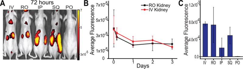
Figure 1
Investigation of route of nanoparticle administration. A) In vivo near-infrared fluorescence images of nanoparticle-encapsulated dye in mice 72 hours following administration via different administrat...
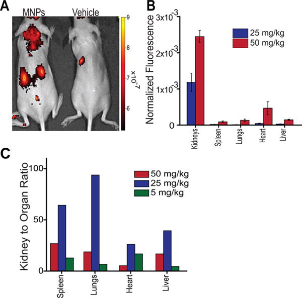
Figure 2
Dependence of nanoparticle dose on renal targeting efficiency. A) Dorsal in vivo fluorescence images of mice injected IV with 25 mg/kg nanoparticles or PBS vehicle control. B) Average fluorescence eff...
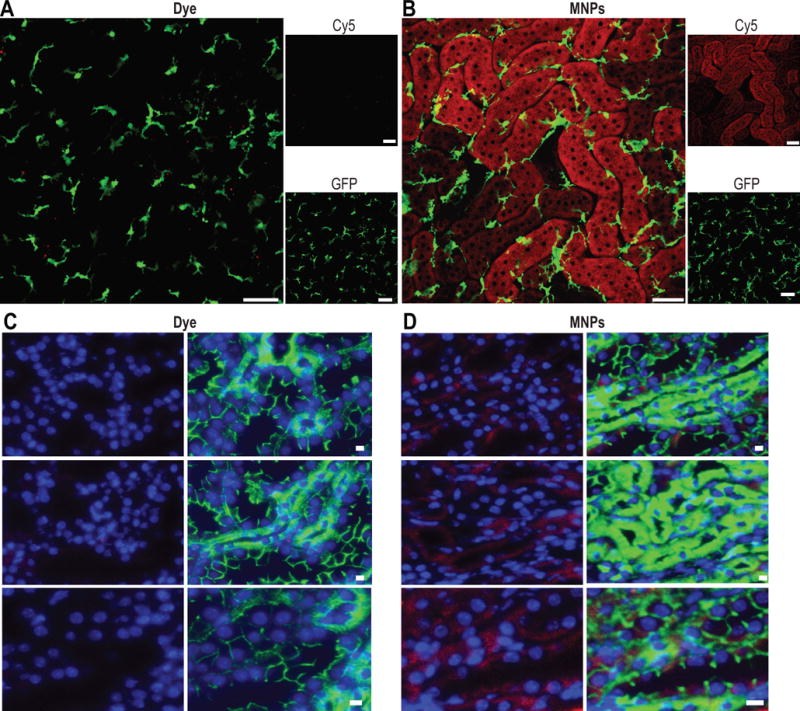
Figure 3
Renal tissue imaging. Intravital microscopy of mice injected IV with A) fluorescent dye (equal to amount found in particles) or B) 25 mg/kg MNPs. Animals were imaged at 72 hours following injection. R...
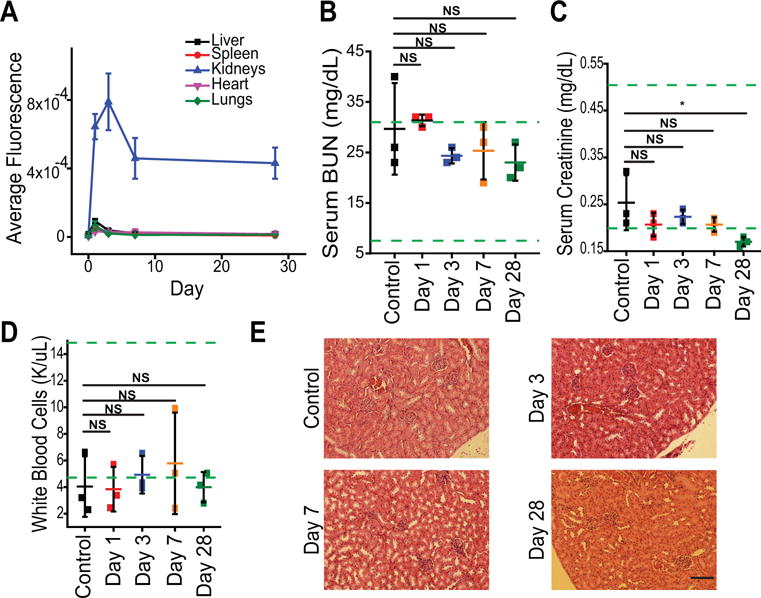
Figure 4
Nanoparticle safety studies. A) Average organ fluorescence measured following sacrifice of mice at each time point after IV tail vein injection of 25 mg/kg MNPs. Data represent mean u00b1 standard dev...
Figure images are served from the NIH/NLM PubMed Central Open Access Subset or Europe PMC; copyright remains with the publishers and authors.
💬 Discussion
0 commentsNo comments yet. Be the first to start a discussion!
Leave a Comment