Abstract
Activity-dependent refinement of neural circuits is a fundamental principle of neural development. This process has been well studied at retinogeniculate synapses-synapses that form between retinal ganglion cells (RGCs) and relay cells within the dorsal lateral geniculate nucleus. Physiological studies suggest that shortly after birth, inputs from ∼20 RGCs converge onto relay cells. Subsequently, all but just one to two of these inputs are eliminated. Despite widespread acceptance, this notion is at odds with ultrastructural studies showing numerous retinal terminals clustering onto relay cell dendrites in the adult. Here, we explored this discrepancy using brainbow AAVs and serial block face scanning electron microscopy (SBFSEM). Results with both approaches demonstrate that terminals from numerous RGCs cluster onto relay cell dendrites, challenging the notion that only one to two RGCs innervate each relay cell. These findings force us to re-evaluate our understanding of subcortical visual circuitry.
🔬 Techniques
🔭 Microscopes
✨ Fluorophores
🧪 Sample Preparation
🏭 Microscope Brands
🧪 Reagent Suppliers
🏛️ Research Organizations (ROR)
Affiliated research institutions:
📋 Methods
Mice Wild-type C57 mice were obtained from Charles River. Calb2-cre mice were obtained from Jackson Laboratory (stock #010774). All analyses conformed to NIH guidelines and protocols approved by the Virginia Polytechnic Institute and State University Institutional Animal Care and Use Committees. Intraocular Injections of Brainbow AAVs The following brainbow AAVs were obtained from the University of Pennsylvania Vector Core ( http://www.med.upenn.edu/gtp/vectorcore/ ): AAV9.hEF1a. lox.TagBFP.lox.eYFP.lox.WPRE.hGH-InvBYF (lot #V3809TI-R) and AAV9. hEF1a.lox.mCherry.lox.mTFP1.lox.WPRE.hGH-InvCheTF (lot #V3530TI-R). Each brainbow AAV is capable of driving the expression of two different fluorescent proteins (see Figure 1A ). Intraocular injection of brainbow AAVs was performed as described previously for the intraocular delivery of cholera toxin subunit B ( Jaubert-Miazza et al., 2005 ; Su et al., 2011 ). Briefly, mice were anesthetized with isoflurane vapors at P12-14. The sclera was pierced with a sharp-tipped glass pipette, and excess vitreous was drained. Another pipette, filled with a 1:1 mixture of both brainbow AAVs, was inserted into the hole made by the first pipette. The pipette containing the AAVs was attached to a Picospritzer and a prescribed volume (3–5 μl) of solution was injected into the eye. After 21 days, mice were euthanized and transcardially perfused with PBS and 4% paraformaldehyde, and retinas and brains were post-fixed in 4% paraformaldehyde for 12 hr. Fixed brains were coronally sectioned (80–100 μm) on a vibratome (Microm HM 650V, Thermo Scientific) and mounted in VectaShield (Vector Laboratories). Fixed retinas were either prepared as whole mounts or were sectioned on a Leica CM1850 cryostat (16-μm cross-sections) and in either case were mounted in VectaShield (Vector Laboratories) ( Su et al., 2011 ). RGCs and retinal projections were analyzed from six animals. Images were acquired on a Zeiss LSM 700 confocal microscope and color analysis of maximum projections images was performed in Photoshop.
Show full methods section
Mice Wild-type C57 mice were obtained from Charles River. Calb2-cre mice were obtained from Jackson Laboratory (stock #010774). All analyses conformed to NIH guidelines and protocols approved by the Virginia Polytechnic Institute and State University Institutional Animal Care and Use Committees. Intraocular Injections of Brainbow AAVs The following brainbow AAVs were obtained from the University of Pennsylvania Vector Core ( http://www.med.upenn.edu/gtp/vectorcore/ ): AAV9.hEF1a. lox.TagBFP.lox.eYFP.lox.WPRE.hGH-InvBYF (lot #V3809TI-R) and AAV9. hEF1a.lox.mCherry.lox.mTFP1.lox.WPRE.hGH-InvCheTF (lot #V3530TI-R). Each brainbow AAV is capable of driving the expression of two different fluorescent proteins (see Figure 1A ). Intraocular injection of brainbow AAVs was performed as described previously for the intraocular delivery of cholera toxin subunit B ( Jaubert-Miazza et al., 2005 ; Su et al., 2011 ). Briefly, mice were anesthetized with isoflurane vapors at P12-14. The sclera was pierced with a sharp-tipped glass pipette, and excess vitreous was drained. Another pipette, filled with a 1:1 mixture of both brainbow AAVs, was inserted into the hole made by the first pipette. The pipette containing the AAVs was attached to a Picospritzer and a prescribed volume (3–5 μl) of solution was injected into the eye. After 21 days, mice were euthanized and transcardially perfused with PBS and 4% paraformaldehyde, and retinas and brains were post-fixed in 4% paraformaldehyde for 12 hr. Fixed brains were coronally sectioned (80–100 μm) on a vibratome (Microm HM 650V, Thermo Scientific) and mounted in VectaShield (Vector Laboratories). Fixed retinas were either prepared as whole mounts or were sectioned on a Leica CM1850 cryostat (16-μm cross-sections) and in either case were mounted in VectaShield (Vector Laboratories) ( Su et al., 2011 ). RGCs and retinal projections were analyzed from six animals. Images were acquired on a Zeiss LSM 700 confocal microscope and color analysis of maximum projections images was performed in Photoshop.
Serial Block Face Scanning Electron Microscopy
Mice were transcardially perfused sequentially with PBS and 4% paraformaldehyde/2% glutaradehyde in 0.1 M cacodylate buffer. Brains were immediately removed and vibratomed (300-μm coronal sections) and dLGN were dissected. Tissues were then stained, embedded, sectioned, and imaged by Renovo Neural. Images were acquired at a resolution of 5 nm/pixel and image sets included >200 serial sections (with each section representing 75 nm in the z axis). SBFSEM data sets were 40 μm × 40 μm × 12–20 μm. Four data sets were analyzed (from a total of three P42 wild-type mice). Data sets were traced and analyzed in TrakEM2 ( Cardona et al., 2012 ). Retinal terminals were identified (and distinguished from non-retinal terminals) by the presence of synaptic vesicles and pale mitochondria as previously described ( Lund and Cunningham 1972 ; Bickford et al., 2010 ; Hammer et al., 2014 ). Synaptic sites were identified by the presence of active zones and postsynaptic densities. Analysis of data sets was performed independently by three researchers to ensure unbiased results.
Supplementary Material Supplemental material
📊 Figures
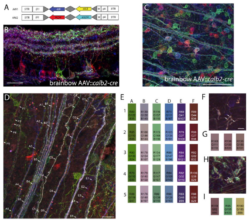
Figure 1
Labeling of RGCs and Retinal Axons with Brainbow AAVs
(A) Schematic representing the constructs of each of the two brainbow AAVs used in these studies. Following Cre recombination, these two constructs generate either farnesylated Tag-blue fluorescent pr...
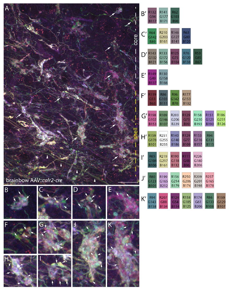
Figure 2
Clusters of Retinal Terminals in dLGN Contain Boutons from Multiple Retinal Axons
(A) Maximum projection, confocal image of retinal axons, and terminals labeled with brainbow AAVs in the u201ccoreu201d and u201cshellu201d region of dLGN of P35 calb2-cre mice. White and yellow dashe...
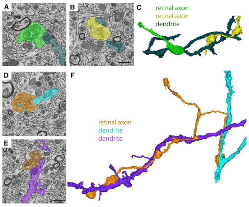
Figure 3
Ultrastructural Analysis and Reconstruction of Retinal Axons Contributing to u201cSimple Encapsulatedu201d Retinogeniculate Synapses in dLGN
(A and B) SBFSEM images of two retinal terminals synapsing onto the same relay cell dendrite in the u201cshellu201d region of dLGN. (C) 3D reconstruction of the two RGC terminal boutons from (A) and (...
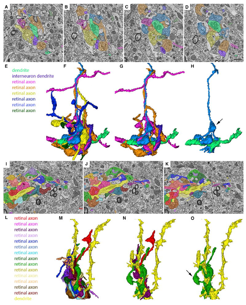
Figure 4
Ultrastructural Analysis and Reconstruction of Retinal Axons Contributing to u201cComplex Encapsulatedu201d Retinogeniculate Synapses in dLGN
(Au2013D) SBFSEM images of six retinal terminals synapsing onto the same relay cell dendrite (pseudo-colored in bright green) in the u201cshellu201d region of dLGN. (E) Key indicates the types of cell...
Figure images are served from the NIH/NLM PubMed Central Open Access Subset or Europe PMC; copyright remains with the publishers and authors.
💬 Discussion
0 commentsNo comments yet. Be the first to start a discussion!
Leave a Comment