Abstract
The RecBCD complex plays key roles in phage DNA degradation, CRISPR array acquisition (adaptation) and host DNA repair. The switch between these roles is regulated by a DNA sequence called Chi. We report cryo-EM structures of the Escherichia coli RecBCD complex bound to several different DNA forks containing a Chi sequence, including one in which Chi is recognized and others in which it is not. The Chi-recognized structure shows conformational changes in regions of the protein that contact Chi and reveals a tortuous path taken by the DNA. Sequence specificity arises from interactions with both the RecC subunit and the sequence itself. These structures provide molecular details for how Chi is recognized and insights into the changes that occur in response to Chi binding that switch RecBCD from bacteriophage destruction and CRISPR spacer acquisition to constructive host DNA repair.
🔬 Techniques
🧬 Organisms
💻 Software
✨ Fluorophores
🧪 Sample Preparation
🔬 Cell Lines
🏭 Microscope Brands
🧪 Reagent Suppliers
📷 Detectors
💻 Software Details
💾 Data Repositories
🏛️ Research Organizations (ROR)
Affiliated research institutions:
📋 Methods
Purification of RecBCD
To prevent digestion of the single-stranded tails of the DNA substrate, a nuclease-deficient E. coli RecBCD mutant (D1080A in RecB) was used in this work. The complex was produced from three plasmids, pETduet-His6-TEVsite-recBD1080A, pRSFduet-recC and pCDFduet-recD in a ΔrecBD E. coli strain as described previously ( 12 ). His-tagged RecBCD complex was expressed and purified as described previously ( 16 , 36 ) with the His-tag removed by TEV protease. The purified protein was flash-frozen with 15% glycerol and stored at -80 °C until use.
Preparation of forked DNA substrates
The forked DNA substrates used during this work were made by annealing equal molar quantities of two respective ssDNA oligonucleotides to generate forks with 25 bp duplex, 15 base single-stranded 5’-tail and a 20 base single-stranded 3’-tail. The same upper strand, responsible for generating the 5’-tail, was used in all cases and had the sequence: 5’ TTTTTTTTTTTTTTTgagcgactgcactacaacagaacca 3’ (lower case represents the bases that form the duplex region). The variable lower strand, responsible for presenting Chi (GCTGGTGG), contained the sequence: 5’ tggttctgttgtagtgcagtcgctc(Y)GCTGGTGG(Z) 3’. Y and Z represent the variable number of T bases used to alter the position of the Chi sequence whilst keeping the total number of unpaired bases to 20 (see Figure 1 ). In the Chi-minus negative control substrate, the Chi sequence was replaced by eight T bases to give a 3’-tail of twenty unpaired T bases.
Native gel mobility assays
For native gel mobility assays, all DNA substrates contained the upper strand labelled with fluorophore (Cy5) at the 5’-terminus and for the lower strand Y values of 1-10 with corresponding Z values of 11-2 were used. Typically, 500 nM RecB D1080A CD was incubated with 500 nM DNA substrate in a 5μl binding solution containing 50 mM Tris-HCl (pH 8.0), 100 mM NaCl, 0.5 mM TCEP and 5% glycerol, which was incubated on ice for 10 min. Samples were separated on 5% native polyacrylamide gels in 1 × TB buffer. Gels were scanned in fluorescence mode (Cy5) on a BIO-RAD ChemiDocTM MP imaging system.
Show full methods section
Purification of RecBCD
To prevent digestion of the single-stranded tails of the DNA substrate, a nuclease-deficient E. coli RecBCD mutant (D1080A in RecB) was used in this work. The complex was produced from three plasmids, pETduet-His6-TEVsite-recBD1080A, pRSFduet-recC and pCDFduet-recD in a ΔrecBD E. coli strain as described previously ( 12 ). His-tagged RecBCD complex was expressed and purified as described previously ( 16 , 36 ) with the His-tag removed by TEV protease. The purified protein was flash-frozen with 15% glycerol and stored at -80 °C until use.
Preparation of forked DNA substrates
The forked DNA substrates used during this work were made by annealing equal molar quantities of two respective ssDNA oligonucleotides to generate forks with 25 bp duplex, 15 base single-stranded 5’-tail and a 20 base single-stranded 3’-tail. The same upper strand, responsible for generating the 5’-tail, was used in all cases and had the sequence: 5’ TTTTTTTTTTTTTTTgagcgactgcactacaacagaacca 3’ (lower case represents the bases that form the duplex region). The variable lower strand, responsible for presenting Chi (GCTGGTGG), contained the sequence: 5’ tggttctgttgtagtgcagtcgctc(Y)GCTGGTGG(Z) 3’. Y and Z represent the variable number of T bases used to alter the position of the Chi sequence whilst keeping the total number of unpaired bases to 20 (see Figure 1 ). In the Chi-minus negative control substrate, the Chi sequence was replaced by eight T bases to give a 3’-tail of twenty unpaired T bases.
Native gel mobility assays
For native gel mobility assays, all DNA substrates contained the upper strand labelled with fluorophore (Cy5) at the 5’-terminus and for the lower strand Y values of 1-10 with corresponding Z values of 11-2 were used. Typically, 500 nM RecB D1080A CD was incubated with 500 nM DNA substrate in a 5μl binding solution containing 50 mM Tris-HCl (pH 8.0), 100 mM NaCl, 0.5 mM TCEP and 5% glycerol, which was incubated on ice for 10 min. Samples were separated on 5% native polyacrylamide gels in 1 × TB buffer. Gels were scanned in fluorescence mode (Cy5) on a BIO-RAD ChemiDocTM MP imaging system.
Cryo-electron microscopy grid preparation and data collection
Prior to preparing grids, RecBCD was thawed and desalted into 20 mM Tris-HCl (pH 8.0), 50mM NaCl, 0.5 mM TCEP using Sephadex G25 spintrap columns (GE Healthcare). The protein was mixed with a 1.5 fold excess of DNA substrate for 10 min at room temperature. Final concentrations were 1 μM RecBCD and 1.5 μM DNA substrate. A RecBCD-Chi complex was prepared using a Chi-containing DNA substrate with a Y spacing of 8 and a Z spacing of 4 (see Figure 1 ). For the Chi-minus dataset, a complex was prepared using the negative control substrate lacking Chi. For the Chi-minus 2 dataset, a complex was prepared using a Chi-containing DNA substrate with a Y spacing of 6 and a Z spacing of 4 ( Figure 1 ). For the Chi-plus 2 dataset, a complex was prepared using a Chi-containing DNA substrate with a Y spacing of 10 and a Z spacing of 4 ( Figure 1 ). Quantifoil R2/2 μm holey carbon film grids (300 mesh) were treated by plasma cleaning for 30 s before being covered with graphene oxide sheets. In order to improve grid preparation reproducibility and hydrophilicity of the graphene oxide, the following method was used: 5μl of graphene oxide solution (Aldrich 763705, 2mg/ml) were mixed with 5μl of 1.5% w/v solution of nonionic detergent n-Dodecyl-β-D-Maltoside. The mixture was diluted 100 times with water and 5ul was applied to the carbon side of the grid. A sharp edge of a piece of filter paper was applied to the centre of the opposite surface of the grid to pull the graphene oxide solution through the grid. Grids were used within 30 minutes of graphene oxide application. Sample (4 μL) was evenly applied to the graphene oxide-coated side of the grid, followed by a 5 s wait time, 1 s blot time, and freezing in liquid ethane using a Vitrobot Mark IV (FEI). The Vitrobot chamber was maintained at close to 100% humidity at 4°C. The dataset with the Chi substrate was collected using a Titan Krios microscope operated at 300 KV at eBIC, Diamond, UK. Zero loss energy images were collected automatically using EPU (FEI) on a Gatan K2-Summit detector in counting mode with a pixel size of 1.047 Å. A total of 3,721 images were collected with a nominal defocus range of −1.3 to −2.5 μm in 0.3 μm increments. Each image consisted of a movie stack of 40 frames with a total dose of 45 e-/Å 2 over 10 s corresponding to a dose rate of 5 e-/pixel/s. The dataset with the Chi-minus control substrate was collected using a similar collection strategy again with a Gatan K2-Summit detector and Titan Krios microscope at eBIC, Diamond, UK. The pixel size was 1.048 Å and a total of 788 images were collected with a similar defocus range to the above. A total dose of 45 e-/Å 2 was split into 40 frames over 7 s, corresponding to a dose rate of 7 e-/pixel/s. The datasets with the Chi-minus 2 and Chi-plus 2 were collected using a Titan Krios microscope operated at 300 KV at eBIC, Diamond, UK. Zero loss energy images were collected automatically using EPU (FEI) on a Falcon3 detector in integrating mode with a pixel size of 1.085 Å. A total of 4,013 images (Chi-minus 2 substrate) and 3,613 images (Chi-plus 2 substrate) were collected with a nominal defocus range of −1.2 to −2.7 μm in 0.3 μm increments. Each image consisted of a movie stack of 39 frames with a total dose of 76 e - /Å 2 over 1 s corresponding to a dose rate of 89 e - /pixel/s.
Data processing – The Chi-containing dataset
Movie stacks were aligned and summed using Motioncor2 ( 37 ). Template-free particle picking was done in Gautomatch, using a circular diameter of 150 Å and CTF parameters were estimated for each micrograph using Gctf ( 38 ). A total of 1,053,451 picked particles were extracted 2x binned from 3,721 images for two rounds of 2D classification in RELION3 ( 39 ) in which 373,476 real RecBCD particles were kept and picking artefacts/noise discarded. A consensus refinement was run on all the particles followed by unmasked 3D classification without alignment. From this, 204,633 particles with complete density for the complex were selected and sub-stoichiometric classes discarded (no DNA density, no RecB nuclease domain density or weaker RecD density). These 204,633 particles were re-extracted unbinned, 3D refined and Bayesian polished prior to 3D classification without alignment using a mask of RecC around the prospective Chi-binding site. This separated 127,268 particles with ordered density for the complete ssDNA 3’ tail, including the Chi sequence, from 77,365 particles with no ssDNA density beyond the RecB helicase domains. Each class was further refined and classified without alignment, this time using a mask around both RecD and the RecB nuclease. The classifications removed 8,452 and 3,092 particles from the Chi and no Chi classes respectively, which had weaker density for the RecD helicase domains. This left 74,273 homogenous particles from the no Chi class, that were refined to give a 3.9 Å resolution (0.143 FSC cutoff, RELION3) map representing a Chi-unrecognised complex. This structure was very similar a previous RecBCD structure with a forked DNA substrate containing long ssDNA tails (PDB: 5LD2) ( 16 ). From the Chi class, two significant conformations were identified. The major state, with 74,496 particles, refined to a resolution of 3.7 Å (0.143 FSC cutoff, RELION3) and represented a Chi-recognised complex with ordered Chi density and significant local conformational changes involving RecC and the RecB nuclease. The second Chi state contained 44,320 particles, refined to 4.1 Å (0.143 FSC cutoff, RELION3) and similarly showed ordered Chi density and the associated local conformational changes in RecC. However, this third state did not show such a substantial movement in the RecB nuclease domain, therefore may represent an intermediate between the Chi-unrecognised and Chi-recognised states, which we have called the Chi-intermediate complex. Work flows for data processing for this and all other structures are presented in Extended Data Figures 1 and 2 . An analysis of particle orientations for all structures is presented in Extended Data Figure 6 . Data processing – The Chi-minus control dataset The Chi-minus movie stacks were initially processed similarly to the Chi dataset. A total of 271,210 particles were extracted 2x binned from 788 images for two rounds of 2D classification in RELION3, resulting in 99,545 RecBCD particles. A consensus 3D refinement was run followed by unmasked 3D classification without alignment from which a homogeneous subset of 87,229 particles with complete density for the complex was selected. The particles were re-extracted unbinned, refined and Bayesian polished prior to 3D classification without alignment using the mask of RecC around the Chi binding site. This time there was only one class, with no DNA density observed in the Chi-binding channel. 3D classification with a mask of RecD plus the RecB nuclease domain separated a class of sub-stoichiometric particles (24,417 particles). The remaining major class (62,812 particles) was refined to produce a 3.8 Å resolution map (0.143 FSC cutoff, RELION3). This represents the Chi-minus complex, which resembles the Chi-unrecognised complex and similarly does not contain ordered 3’ ssDNA beyond the RecB helicase. Data processing – The Chi-minus 2 dataset The movie stacks were initially processed similarly to the Chi-containing dataset. A total of 1,694,205 particles were extracted 2x binned from 4,013 images for two rounds of 2D classification in RELION3, resulting in 754,150 RecBCD particles. A consensus 3D refinement was run followed by unmasked 3D classification without alignment from which a homogeneous subset of 687,825 particles with complete density for the complex was selected. The particles were re-extracted unbinned, refined and Bayesian polished prior to 3D classification without alignment using the mask of RecC around the Chi binding site. Only one class, with no DNA density observed in the Chi-binding channel was obtained. 3D classification with a mask of RecD plus the RecB nuclease domain separated a class of sub-stoichiometric particles (308,035 particles). The remaining major class (379,790 particles) was refined to produce a 3.6 Å resolution map (0.143 FSC cutoff, RELION3). This structure resembles the Chi-unrecognised complex and similarly does not contain ordered 3’ ssDNA beyond the RecB helicase. Data processing – The Chi-plus 2 dataset The movie stacks were initially processed similarly to the Chi-containing dataset. A total of 1,809,768 particles were extracted 2x binned from 3,613 images for two rounds of 2D classification in RELION3, resulting in 795,810 RecBCD particles. A consensus 3D refinement was run followed by unmasked 3D classification without alignment from which a homogeneous subset of 776, 381 particles with complete density for the complex was selected. The particles were re-extracted unbinned, refined and Bayesian polished prior to 3D classification without alignment using the mask of RecC around the Chi binding site. Only one class, with no DNA density observed in the Chi-binding channel was obtained. 3D classification with a mask of RecD plus the RecB nuclease domain separated a class of sub-stoichiometric particles (395,469 particles). The remaining class (380,912 particles) was refined to produce a 3.8 Å resolution map (0.143 FSC cutoff, RELION3). This structure resembles the Chi-unrecognised complex and similarly does not contain ordered 3’ ssDNA beyond the RecB helicase.
Model building and refinement
The structure of RecBCD in a complex with a DNA fork (PDB 5LD2) ( 16 ) was used as a starting model for global docking in Chimera ( 40 ) for the Chi-recognised, Chi-unrecognised, Chi-minus 2, and Chi-plus 2 complexes. Once finalised, the Chi-recognised model was then used as a starting template for the Chi-intermediate complex. The Chi-unrecognised model was used a starting template for the Chi minus complex. Each of the six models was edited initially by jelly-body refinement with Refmac ( 41 ) in CCP-EM ( 42 ) followed by cycles of manual rebuilding in Coot ( 43 ) and real-space refinement with PHENIX ( 44 ). A 3.7 Å resolution (0.143 FSC cutoff, RELION3) map from a masked focused refinement of the Chi region for the 127,268 Chi-bound particles was used to help build the novel Chi bases and surrounding protein contacts. The six models were finally checked for consistency with one another, apart from where obvious density differences occurred in the maps. A final run of PHENIX real-space refinement with group B-factor refinement was run to generate the final models and model statistics ( Table 1 ).
Supplementary Material Supplementary video 1 Supplementary video 2 Supplementary video 3
📊 Figures
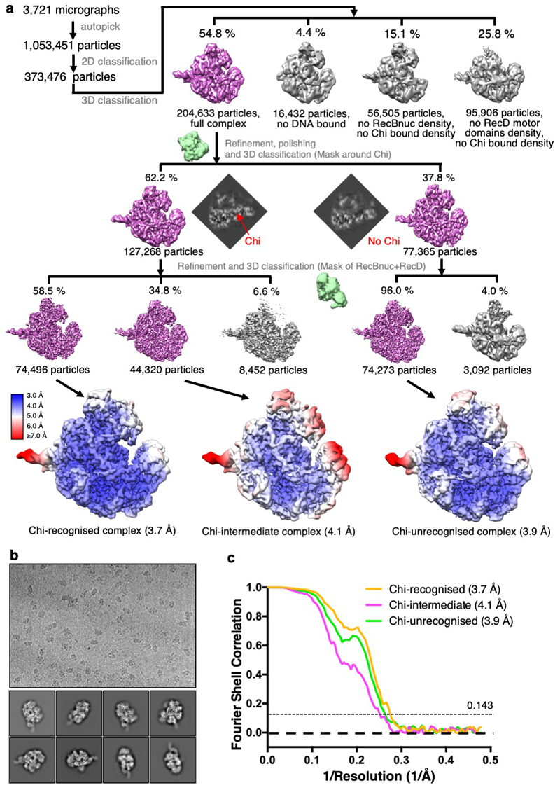
Extended Data Figure 1
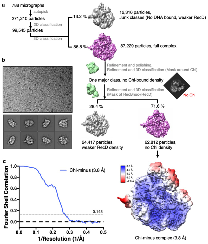
Extended Data Figure 2
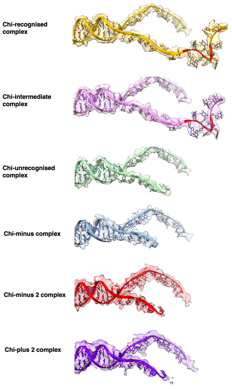
Extended Data Figure 3
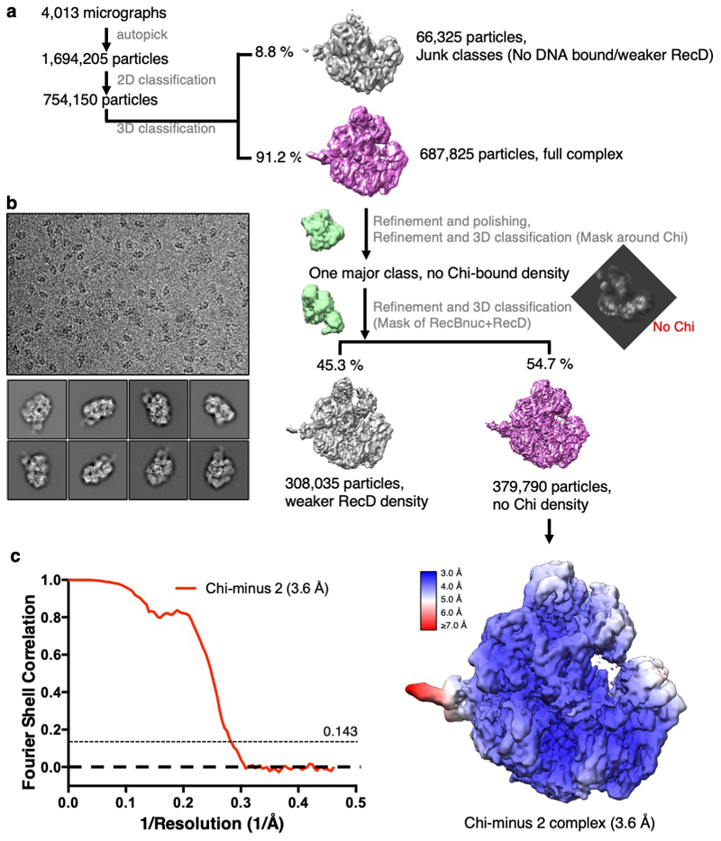
Extended Data Figure 4
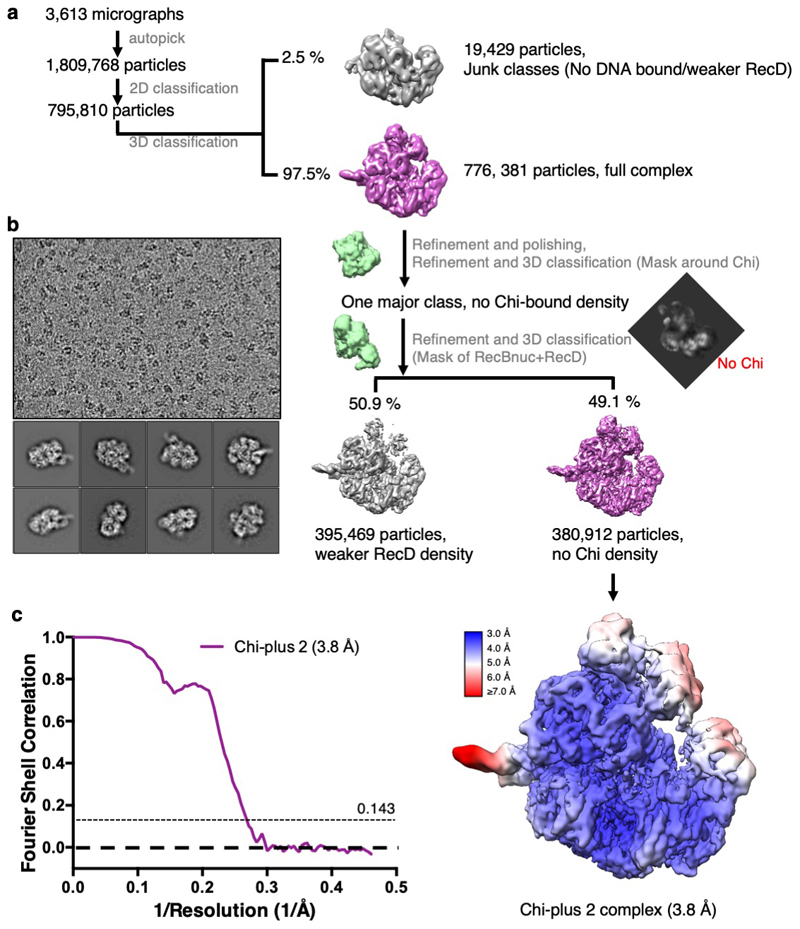
Extended Data Figure 5
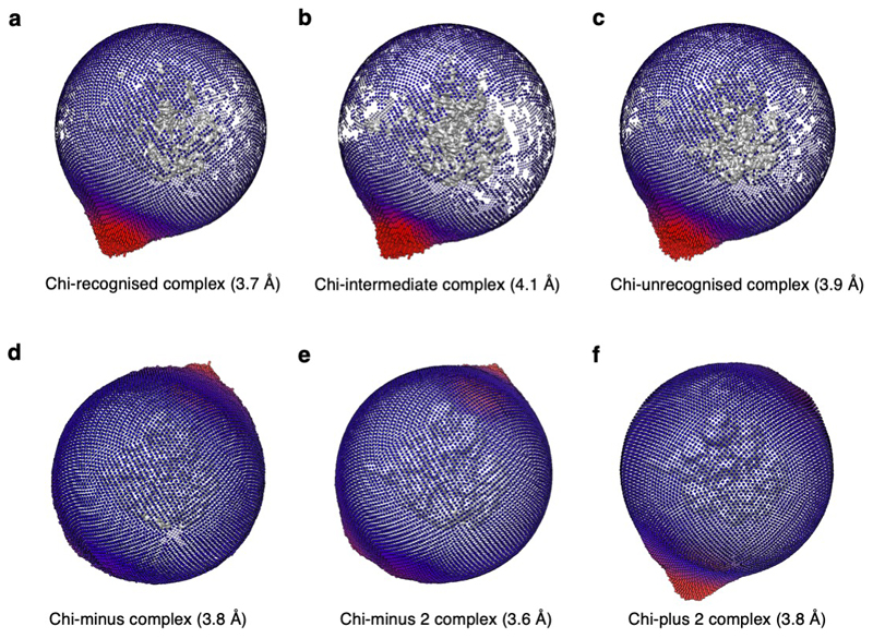
Extended Data Figure 6
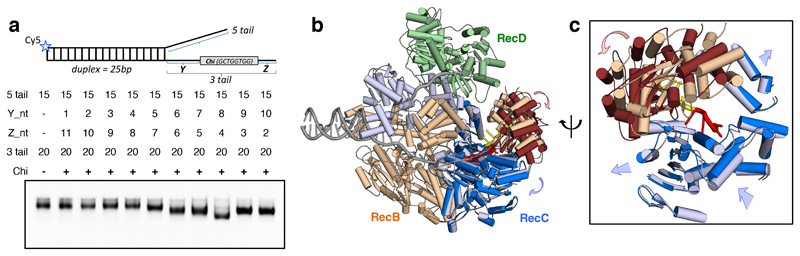
Fig. 1
Structural changes associated with Chi binding.
a, Native gel mobility shift assays (bottom) show an altered shift of the RecBCD-DNA complex band when the substrate contained eight bases between the fork junction and the Chi sequence (Y = 8), sugge...
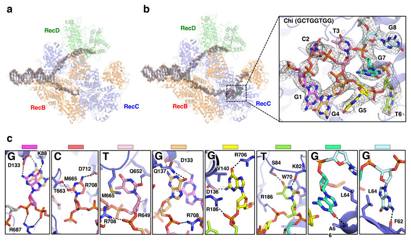
Fig. 2
Details of the Chi-binding site interactions.
a , Density corresponding to the bound DNA fork substrate in the Chi-unrecognised state with the protein complex shown as a faint cartoon. Strong density is observed for the duplex and 5u2019-tail reg...
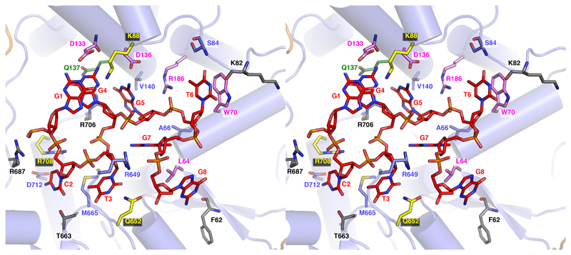
Fig. 3
Details of the full Chi-binding site.
A cross-eye stereo view of the Chi-binding site and interacting residues is shown. Interacting residues are shown as sticks and coloured according to previously published point mutation studies ( 19 ,...
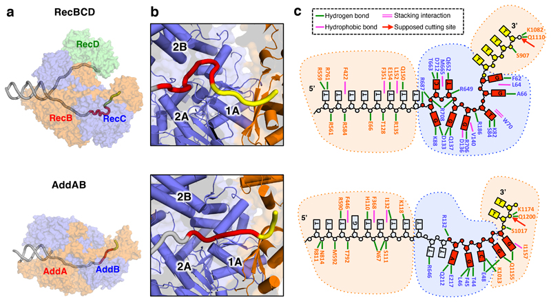
Fig. 4
Comparison of RecBCD and AddAB Chi-bound complexes.
a , Transparent surfaces of RecBCD (above) and AddAB (below) coloured by subunit with the DNA fork backbones overlaid as a cartoon in grey with their respective Chi sequences depicted in red and the b...
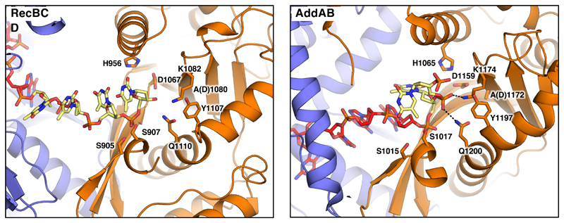
Fig. 5
The nuclease active site in the Chi-bound RecBCD and AddAB complexes.
The nuclease domains of RecBCD and AddAB are shown as cartoons. Key residues participating in DNA binding and catalysis are shown as sticks. Hydrogen bonds are shown as dotted lines. The DNA forks use...
Figure images are served from the NIH/NLM PubMed Central Open Access Subset or Europe PMC; copyright remains with the publishers and authors.
💬 Discussion
0 commentsNo comments yet. Be the first to start a discussion!
Leave a Comment