Abstract
Pancreatic islets are heavily vascularized in vivo with each insulin secreting beta-cell associated with at least one endothelial cell (EC). This structure is maintained immediately post-isolation; however, in culture the ECs slowly deteriorate, losing density and branched morphology. We postulate that this deterioration occurs in the absence of blood flow due to limited diffusion of media inside the tissue. To improve exchange of media inside the tissue, we created a microfluidic device to culture islets in a range of flow-rates. Culturing the islets from C57BL6 mice in this device with media flowing between 1 and 7 ml/24 hr resulted in twice the EC-density and -connected length compared to classically cultured islets. Media containing fluorescent dextran reached the center of islets in the device in a flow-rate-dependant manner consistent with improved penetration. We also observed deterioration of EC morphology using serum free media that was rescued by addition of bovine serum albumin, a known anti-apoptotic signal with limited diffusion in tissue. We further examined the effect of flow on beta-cells showing dampened glucose-stimulated Ca(2+)-response from cells at the periphery of the islet where fluid shear-stress is greatest. However, we observed normal two-photon NAD(P)H response and insulin secretion from the remainder of the islet. These data reveal the deterioration of islet EC-morphology is in part due to restricted diffusion of serum albumin within the tissue. These data further reveal microfluidic devices as unique platforms to optimize islet culture by introducing intercellular flow to overcome the restricted diffusion of media components.
🔬 Techniques
🔭 Microscopes
💻 Software
✨ Fluorophores
🧪 Sample Preparation
🔬 Cell Lines
🏭 Microscope Brands
🧪 Reagent Suppliers
🔎 Objectives
💻 Software Details
🏛️ Research Organizations (ROR)
Affiliated research institutions:
📋 Methods
Microfluidic Device Fabrication Devices were fabricated using elastomer polydimethylsiloxane (PDMS) (Dow-Corning) as described previously [16] , [17] . The cured mould was irreversibly bonded to a 24×50-mm coverslip (VWR Scientific) by oxygen plasma treatment (Harrick Scientific, Ossining, NY) and Tygon tubing was inserted directly into the cored-out port holes.
Microfluidic Device Design
The microfluidic device design and process are described in detail in the Supplemental Material ( Text S1 & Fig. S1 , S2 , and S3 ). Briefly, islets were brought into 125 µm tall ×300 µm wide microfluidic channels through inlet port tubes. Islets moved along these channels by media flow until they reached a dam wall. This dam structure was 25 µm tall, 1,800 µm wide, and 0.13 cm long, and allowed solution to flow past but blocked the movement of islets. Microfluidic devices provide only non-turbulent or laminar flow at the flow rates used in these studies [14] . Two main designs were used for these studies: a three-channel microfluidic device and an independent-channel microfluidic device ( Fig. S1 ). The three-channel device placed islets in three different flow-rates based on varied channel lengths and by using a single syringe pump. The independent-channel device provided a single flow-rate in parallel channels to facilitate subsequent live cell imaging or effluent collection.
Ethics Statement
Animal procedures were approved by the Animal Care Committee of the University Health Network, Toronto, Ontario, Canada in accordance with the policies and guidelines of the Canadian Council on Animal Care (Animal Use Protocol #1531).
Show full methods section
Microfluidic Device Fabrication Devices were fabricated using elastomer polydimethylsiloxane (PDMS) (Dow-Corning) as described previously [16] , [17] . The cured mould was irreversibly bonded to a 24×50-mm coverslip (VWR Scientific) by oxygen plasma treatment (Harrick Scientific, Ossining, NY) and Tygon tubing was inserted directly into the cored-out port holes.
Microfluidic Device Design
The microfluidic device design and process are described in detail in the Supplemental Material ( Text S1 & Fig. S1 , S2 , and S3 ). Briefly, islets were brought into 125 µm tall ×300 µm wide microfluidic channels through inlet port tubes. Islets moved along these channels by media flow until they reached a dam wall. This dam structure was 25 µm tall, 1,800 µm wide, and 0.13 cm long, and allowed solution to flow past but blocked the movement of islets. Microfluidic devices provide only non-turbulent or laminar flow at the flow rates used in these studies [14] . Two main designs were used for these studies: a three-channel microfluidic device and an independent-channel microfluidic device ( Fig. S1 ). The three-channel device placed islets in three different flow-rates based on varied channel lengths and by using a single syringe pump. The independent-channel device provided a single flow-rate in parallel channels to facilitate subsequent live cell imaging or effluent collection.
Ethics Statement
Animal procedures were approved by the Animal Care Committee of the University Health Network, Toronto, Ontario, Canada in accordance with the policies and guidelines of the Canadian Council on Animal Care (Animal Use Protocol #1531).
Pancreatic Islet Isolation and Treatment
Pancreatic islets were isolated from 8- to 12- week-old C57BL6 male mice by using collagenase digestion (Roche) [18] , [19] . Islets were cultured in RPMI medium 1640 containing 11 mM glucose, 10% FBS, 5 U/ml penicillin-streptomycin, and 20 mM HEPES. Control islets were incubated in a non-treated culture dishes in a humidified incubator at 37°C and 5% CO 2 . Device-treated islets were loaded into the microfluidic devices shortly after isolation and incubated in a custom-built desk-top incubator described in detail in the Supplemental Material ( Fig. S2 ). Briefly, the microfluidic device was submerged in a stirred water bath at 37°C with flow driven by a syringe pump. The reservoir media was also submerged in a separate water-bath maintained just above 37°C to reduce formation of air bubbles in the microfluidic channel. A cap of mineral oil was placed on top of the media in the reservoir to reduce evaporation and drift in pH.
Immunofluorescent Detection
Control and device-treated islets were labelled within the microfluidic device. Islets were fixed (2% PFA, PBS, 1 hr, 100 µL/hr), blocked (PBS, 0.1% TritonX-100, 4°C, 10% Normal Goat Serum, 4 hr, 15 µL/hr), incubated with primary antibody (PBS, 0.1% TritonX-100, 4°C, 1% Normal Goat Serum, 1∶100 dilution rat anti-mouse PECAM-1, 3 hr, 3 µL/hr), rinsed (PBS, 0.1% TritonX-100, 4°C, 10% Normal Goat Serum, 1 hr, 15 µL/hr) and incubated with 1∶500 goat anti-rat Alexa 633 (PBS, 0.1% TritonX-100, 4°C, 10% Normal Goat Serum, 3 hr, 15 µL/hr). Islets were imaged using a 20×0.50 NA objective lens in 3 µm steps for 16–20 slices (dependant on the size of the islet) on an Olympus FluoView 300 microscope. Images were analyzed using ImageJ 1.41o (Wayne Rasband, NIH, USA). EC fractional area was measured by calculating the area of PECAM-1 labelling divided by the total area of the islet (6 slices/islet). Total connected length was determined using Simple Neurite Tracer throughout the entire stack of images. PECAM-1 labelled regions were tracked through the stack and the resulting summation was normalized to the circumference of the islet. The average of individual islets was used for statistical analysis. A two tail, unpaired, two sample student's t-test was used and significance was determined with p
📊 Figures
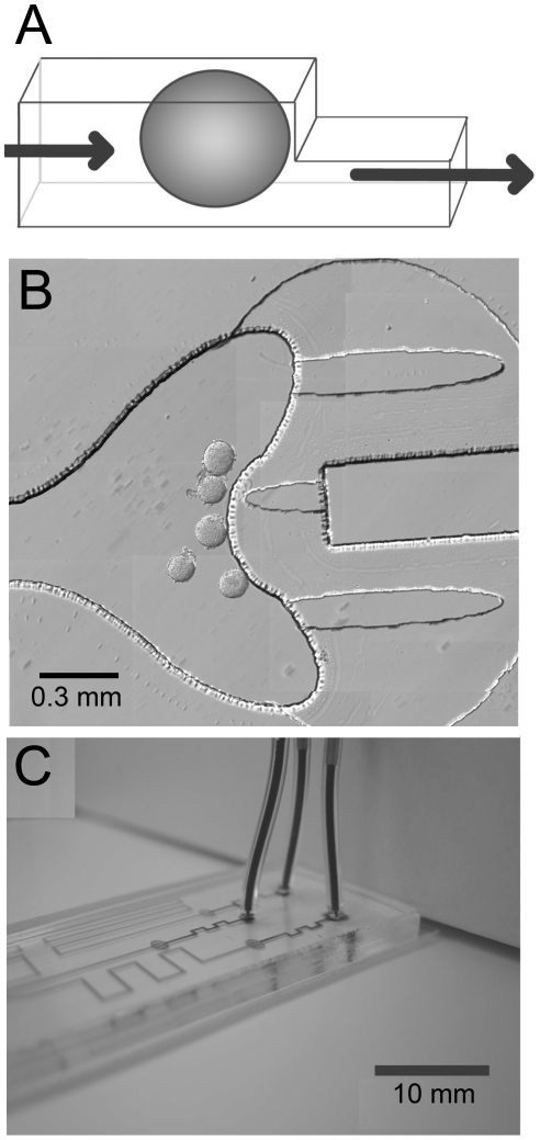
Figure 1
A microfluidic device to treat pancreatic islets with laminar flow.
(A) A schematic of a microfluidic channel is shown to demonstrate how pancreatic islets are held in laminar flow. Pancreatic islets ( oval ) are brought into microfluidic channels ( outlined ) and tra...
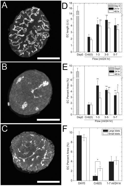
Figure 2
The effect of fluid-flow on EC area and connected length.
Pancreatic islets were cultured in either the microfluidic device with flow (device-treated) or in non-flowing media in an incubator ( control ) followed by anti-PECAM-1 immunolabelling. (A) A represe...
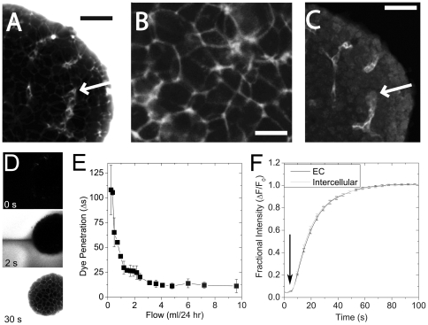
Figure 3
Media access in a microfluidic device measured using fluorescent dextran.
To explore the nature of the fluid flow inside treated islets, real-time fluorescent imaging was applied to 24 hr device-treated islets (3 ml/24 hr). (A) We flowed fluorescent dextran (3,000 MW) into ...
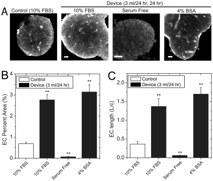
Figure 4
Effect of serum albumin on EC morphology.
Islets were cultured for 24 hr in normal culture (Control, 10% FBS) and the microfluidic device (3 ml/24 hr flow rate). The media used in the device was either normal media (10% FBS), serum free media...
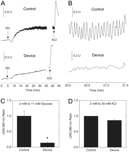
Figure 5
Measuring beta-cell [Ca 2+ ]-activity using Fura-2.
Islets subjected to 48 hr of fluid-flow (3 ml/hr) followed by labelling with Fura-2 to evaluate beta-cell [Ca 2+ ]-responses. (A) Typical control and device-treated islets at 2 mM glucose displayed a ...
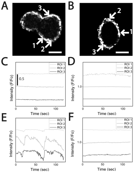
Figure 6
Monitoring synchronous beta-cell [Ca 2+ ]-response in pancreatic islets using Fluo-4.
Islets were cultured in control or microfluidic device flow (3 ml/24 hr) for 48 hr prior to labelling with Fluo-4. The dye labels cells as the periphery of the islet as observed using confocal microsc...
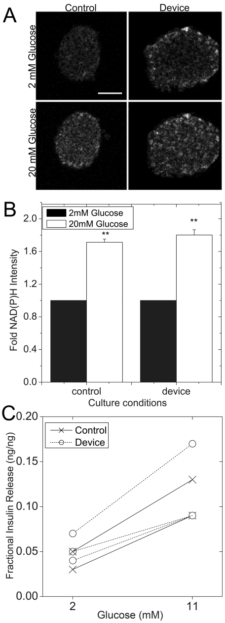
Figure 7
Glucose-stimulated NAD(P)H and insulin responses.
Islets were cultured in control conditions or in microfluidic device flow (3 ml/24 hr) for 48 hr. (A) Control and flow treated islets were subsequently treated with 2 and 20 mM glucose and imaged usin...
Figure images are served from the NIH/NLM PubMed Central Open Access Subset or Europe PMC; copyright remains with the publishers and authors.
💬 Discussion
0 commentsNo comments yet. Be the first to start a discussion!
Leave a Comment