Abstract
Ca(2+)-permeable type 2 two-pore channels (TPC2) are lysosomal proteins required for nicotinic acid adenine dinucleotide phosphate (NAADP)-evoked Ca(2+) release in many diverse cell types. Here, we investigate the importance of TPC2 proteins for the physiology and pathophysiology of the heart. NAADP-AM failed to enhance Ca(2+) responses in cardiac myocytes from Tpcn2(-/-) mice, unlike myocytes from wild-type (WT) mice. Ca(2+)/calmodulin-dependent protein kinase II inhibitors suppressed actions of NAADP in myocytes. Ca(2+) transients and contractions accompanying action potentials were increased by isoproterenol in myocytes from WT mice, but these effects of β-adrenoreceptor stimulation were reduced in myocytes from Tpcn2(-/-) mice. Increases in amplitude of L-type Ca(2+) currents evoked by isoproterenol remained unchanged in myocytes from Tpcn2(-/-) mice showing no loss of β-adrenoceptors or coupling mechanisms. Whole hearts from Tpcn2(-/-) mice also showed reduced inotropic effects of isoproterenol and a reduced tendency for arrhythmias following acute β-adrenoreceptor stimulation. Hearts from Tpcn2(-/-) mice chronically exposed to isoproterenol showed less cardiac hypertrophy and increased threshold for arrhythmogenesis compared with WT controls. Electron microscopy showed that lysosomes form close contacts with the sarcoplasmic reticulum (separation ∼ 25 nm). We propose that Ca(2+)-signaling nanodomains between lysosomes and sarcoplasmic reticulum dependent on NAADP and TPC2 comprise an important element in β-adrenoreceptor signal transduction in cardiac myocytes. In summary, our observations define a role for NAADP and TPC2 at lysosomal/sarcoplasmic reticulum junctions as unexpected but major contributors in the acute actions of β-adrenergic signaling in the heart and also in stress pathways linking chronic stimulation of β-adrenoceptors to hypertrophy and associated arrhythmias.
🔬 Techniques
🧪 Sample Preparation
🏭 Microscope Brands
🧪 Reagent Suppliers
📷 Detectors
💻 Software Details
🏛️ Research Organizations (ROR)
Affiliated research institutions:
📋 Methods
Cell Isolation, Guinea Pig Male guinea pigs (300–500 g) were killed in accordance with Schedule 1 of The Animals (Scientific Procedures) Act 1986 (HMSO). The heart was rapidly excised and washed in a modified Tyrode's solution containing EGTA and heparin to prevent clots from forming in the small coronary circulation. The heart was then mounted on a constant pressure Langendorff perfusion system for retrograde perfusion via the aorta. Following a 2-min initial perfusion with a modified Tyrode's solution (in m m : NaCl 136, KCl 5.4, NaHCO 3 12, Na + pyruvate 1, NaH 2 PO 4 1, MgCl 2 1, EGTA 0.04, glucose 5; gassed with 95% O 2 , 5% CO 2 to maintain a pH of 7.4), digestion was carried out with a Tyrode's solution (50 ml) of identical composition, but which lacked EGTA and contained 24–28 mg of collagenase type II (Worthington) and 0.1 m m CaCl 2 . Following digestion, the left atrium was separated and placed in high K + storage medium (in m m : KCl 70, MgCl 2 5, K + glutamine 5, taurine 20, EGTA 0.04, succinic acid 5, KH 2 PO 4 20, HEPES 5, glucose 10; pH 7.2 with KOH). Single cells were isolated by gentle trituration and stored at 4 °C in high K + storage medium until use. Ventricular tissue was dissected into several pieces and underwent mechanical agitation for a further 5 min in collagenase solution. Isolated myocytes were filtered through a 250-μm mesh, and collagenase was replaced with Dulbecco's modified Eagle's medium (DMEM, Invitrogen) warmed to 37 °C. Ventricular myocytes were stored in DMEM at room temperature and used within 9 h of isolation. Creation of TPC2 −/− Mice Mice lacking the TPC2 protein were created as described previously ( 9 , 11 ). Cell Isolation, Mouse Mice (13–18 weeks old) were killed in accordance with Schedule 1 of The Animals (Scientific Procedures) Act 1986 (HMSO). Hearts were excised, tied to a cannula via the aorta and perfused through a syringe containing a pre-gassed (with 95% O 2 , 5% CO 2 ) solution containing (m m ) NaCl 125, NaHCO 3 25, KCl 5.4, NaH 2 PO 4 1.2, MgCl 2 1, CaCl 2 1.0, glucose 5.5, pH 7.4, together with heparin (10 units/ml) and streptokinase (100 units/ml). The cannula was then removed from the syringe and mounted onto a Langendorff perfusion system. Once mounted, the hearts were perfused with a solution (36 °C) containing (m m ) NaCl 130, KCl 5.4, MgCl 2 3.5, glucose 10, HEPES 5, NaH 2 PO 4 0.4; pH 7.4, and gassed with 95% O 2 , 5% CO 2 . After 3 min, this isolation solution was replaced with a further 50 ml of a similar solution that also contained 0.3 mg/ml collagenase (type II, Worthington) and 0.1 m m CaCl 2 . Following enzymatic digestion (for a maximum time of 7 min), the heart was removed from the cannula. The ventricles were cut into pieces, and the myocytes were released during several gentle suspensions, then filtered through 250-μm mesh, and stored at room temperature in medium containing (in m m ) NaCl 130, KCl 5.4, MgCl 2 3.5, CaCl 2 0.1glucose 10, HEPES 5, NaH 2 PO 4 0.4, taurine 20; 0.1% bovine serum albumin; pH 7.4, and gassed with 95% O 2 , 5% CO 2 .
Show full methods section
Cell Isolation, Guinea Pig Male guinea pigs (300–500 g) were killed in accordance with Schedule 1 of The Animals (Scientific Procedures) Act 1986 (HMSO). The heart was rapidly excised and washed in a modified Tyrode's solution containing EGTA and heparin to prevent clots from forming in the small coronary circulation. The heart was then mounted on a constant pressure Langendorff perfusion system for retrograde perfusion via the aorta. Following a 2-min initial perfusion with a modified Tyrode's solution (in m m : NaCl 136, KCl 5.4, NaHCO 3 12, Na + pyruvate 1, NaH 2 PO 4 1, MgCl 2 1, EGTA 0.04, glucose 5; gassed with 95% O 2 , 5% CO 2 to maintain a pH of 7.4), digestion was carried out with a Tyrode's solution (50 ml) of identical composition, but which lacked EGTA and contained 24–28 mg of collagenase type II (Worthington) and 0.1 m m CaCl 2 . Following digestion, the left atrium was separated and placed in high K + storage medium (in m m : KCl 70, MgCl 2 5, K + glutamine 5, taurine 20, EGTA 0.04, succinic acid 5, KH 2 PO 4 20, HEPES 5, glucose 10; pH 7.2 with KOH). Single cells were isolated by gentle trituration and stored at 4 °C in high K + storage medium until use. Ventricular tissue was dissected into several pieces and underwent mechanical agitation for a further 5 min in collagenase solution. Isolated myocytes were filtered through a 250-μm mesh, and collagenase was replaced with Dulbecco's modified Eagle's medium (DMEM, Invitrogen) warmed to 37 °C. Ventricular myocytes were stored in DMEM at room temperature and used within 9 h of isolation. Creation of TPC2 −/− Mice Mice lacking the TPC2 protein were created as described previously ( 9 , 11 ). Cell Isolation, Mouse Mice (13–18 weeks old) were killed in accordance with Schedule 1 of The Animals (Scientific Procedures) Act 1986 (HMSO). Hearts were excised, tied to a cannula via the aorta and perfused through a syringe containing a pre-gassed (with 95% O 2 , 5% CO 2 ) solution containing (m m ) NaCl 125, NaHCO 3 25, KCl 5.4, NaH 2 PO 4 1.2, MgCl 2 1, CaCl 2 1.0, glucose 5.5, pH 7.4, together with heparin (10 units/ml) and streptokinase (100 units/ml). The cannula was then removed from the syringe and mounted onto a Langendorff perfusion system. Once mounted, the hearts were perfused with a solution (36 °C) containing (m m ) NaCl 130, KCl 5.4, MgCl 2 3.5, glucose 10, HEPES 5, NaH 2 PO 4 0.4; pH 7.4, and gassed with 95% O 2 , 5% CO 2 . After 3 min, this isolation solution was replaced with a further 50 ml of a similar solution that also contained 0.3 mg/ml collagenase (type II, Worthington) and 0.1 m m CaCl 2 . Following enzymatic digestion (for a maximum time of 7 min), the heart was removed from the cannula. The ventricles were cut into pieces, and the myocytes were released during several gentle suspensions, then filtered through 250-μm mesh, and stored at room temperature in medium containing (in m m ) NaCl 130, KCl 5.4, MgCl 2 3.5, CaCl 2 0.1glucose 10, HEPES 5, NaH 2 PO 4 0.4, taurine 20; 0.1% bovine serum albumin; pH 7.4, and gassed with 95% O 2 , 5% CO 2 .
Electrophysiology
Cardiac myocytes were mounted in a perfusion bath placed at the stage of an inverted microscope and perfused with a physiological salt solution (PSS) containing (in m m ) NaCl 125, NaHCO 3 25, KCl 5.4, NaH 2 PO 4 1.2, MgCl 2 1, glucose 5.5, CaCl 2 1.8 and oxygenated with 95% O 2 , 5% CO 2 to maintain a pH of 7.4. The PSS used for mouse experiments was identical except that CaCl 2 concentration was reduced to 1 m m . All electrophysiology was performed at 36 °C. Glass microelectrodes were manufactured from thin-walled, filamented borosilicate glass capillary tubing (GC150TF, Harvard Apparatus Ltd., Kent, UK). Electrode resistances of 3.0 ± 1.5 megohms were used for whole-cell experiments. During whole-cell recordings, a whole-cell patch solution was used containing(in m m ) K + aspartate 110, KCl 10, NaCl 5, MgCl 2 5.2, HEPES 5, K 2 ATP 5, pH 7.2, with KOH, to which Ca 2+ indicator dye, caged NAADP, and inhibitors were added at the stated concentrations. Perforated patch was carried out using whole-cell patch solution plus amphotericin B at 250 μg/ml. Current clamp recordings were made using an AxoClamp 2A amplifier system (Axon Instruments). Cells were stimulated to fire action potentials using a 2-ms current pulse applied via the microelectrode at a rate of 1 Hz. Voltage clamp recordings were made using an AxoPatch 200B amplifier system (Axon Instruments). To stimulate the L-type Ca 2+ current ( I CaL ), cells were maintained at a holding potential of −40 mV and stimulated by a 200-ms step depolarization to 0 mV at a rate of 0.2 Hz. I CaL amplitude was measured as maximum current minus current at the end of the voltage pulse. Ca 2+ Fluorescence, Dye Loading, and Cell Stimulation Protocol During whole-cell fluorescence experiments, Fluo-5F salt (100 μ m ) was added to the whole-cell patch solution. In mouse experiments, myocytes were incubated with Fluo-5F AM (20 μ m ) for 20 min. Ca 2+ transients were stimulated at 1 Hz either by a patch electrode (all guinea pig experiments) or by carbon fiber electrodes placed at the side of the superfusion bath (mouse experiments). Epifluorescence Guinea pig atrial myocytes were visualized using a Leica DMIRB inverted microscope. Excitation light was provided by 470 nm LED illumination, passed through a 475 ± 15 nm bandpass interference filter, and transmitted through the objective. Emitted light was passed through a 520-nm long pass filter and collected using a photomultiplier system (Cairn Research Ltd., Kent, UK). Photomultiplier signals were directed through an amplifier, followed by a low pass 100 Hz electronic filter, digitized (Axon Digidata 1200, Axon Instruments), and recorded with pClamp software at an acquisition rate of 10 kHz.
Spinning Disk Confocal Microscopy
Guinea pig and mouse ventricular myocytes were visualized using a Nikon Axiovert 200 inverted microscope with attached Nipkow spinning disk confocal unit (CSU-10, Andor Technology, UK). Excitation light was provided by a 488-nm diode laser (Cairn Research Ltd.) passed though the Nipkow unit and delivered to the sample through the objective. Emitted light passed back through the CSU-10 unit and was detected using an Andor iXON897 EM-CCD camera (Andor Technology) at a frame rate of 50 Hz. Images were recorded and analyzed using Andor iQ software (Andor Technology).
Line Scan Confocal Microscopy
Mouse ventricular myocytes were imaged with a confocal microscope system that consisted of a Leica TCS NT scanning head coupled to a Leica DMIRB inverted microscope with a 100× oil immersion objective lens (1.2 NA, Leica) in line scan mode (2.6 ms per line). Excitation light (488 nm) was provided by an air-cooled 488-nm argon ion laser system (Uniphase Ltd.), and emitted light was collected at wavelengths above 515 nm using a long- pass filter. Images were recorded using Leica TCS NT software and analyzed using ImageJ software. Analysis of Ca 2+ Transient Data For analysis, background fluorescence was subtracted, and multiple transients were averaged to collect data at a given time point. Data are presented as F / F 0 such that fluorescence data are presented relative to diastolic fluorescence. In the case of experiments in which NAADP was photoreleased in guinea pig ventricular myocytes, there was a small baseline change, and data are presented as F/F 0i where F 0i is equal to diastolic fluorescence before photorelease of NAADP.
Electron Microscopy
Rabbit ventricular tissue was fixed in Karnovsky fixative ( 39 ), resin-embedded, and sectioned at 80 nm (Reichart Ultracut). These were post-stained with 2% uranyl acetate and Reynolds lead citrate. Images were obtained using transmission electron microscopy (Jeol 1200EX II). Contraction Studies, Mouse Ventricular Myocytes Cells were stimulated to contract via a patch electrode (permeabilized configuration), and contractile properties were studied using the IonOptix system (IonOptix Corp.) to measure sarcomere length. Cells were visualized via a ×40 oil objective using an IonOptix MyoCam (IonOptix Corp.) that sampled images at a frequency of 240 Hz. Sinusoidal optical density traces arising from the alternating light and dark bands of the contractile machinery were then transformed into a signal of sarcomere length by application of a fast Fourier transform by the IonWizard sarcomere length acquisition software (IonOptix Corp.). Length measurements were calibrated using a stage micrometer with 2-μm graduations such that the number of pixels/μm recorded by the MyoCam could be entered into the software as a fixed value. Amplitude of sarcomere shortening was calculated by deduction of systolic from diastolic sarcomere length and expressed as a percentage of resting sarcomere length. Analysis was performed using IonWizard 5 software (IonOptix Corp.). All values represent an average of 10 contractions. Langendorff-perfused Whole Mouse Hearts Mice were killed in accordance with Schedule 1 of The Animals (Scientific Procedures) Act 1986 (HMSO). Hearts were excised, cannulated via the aorta under a microscope, and perfused for 30 s with PSS solution containing streptokinase (100 units/ml) via a syringe connected to the cannula. After initial perfusion, the syringe was removed and the cannula mounted on a Langendorff apparatus for retrograde perfusion with PSS containing 1.8 m m CaCl 2 (36 °C, gassed with 95% O 2 , 5% CO 2 ) at a constant flow rate (3.5 ml/min). Hearts were allowed to beat spontaneously, and contractile force was measured by means of a hook placed through the ventricles near the apex, attached to a tension transducer. Resting tension was set between 0.5 and 1.0 g initially. Heart rate was calculated from the force signal as the reciprocal of the time interval between contractions. Inflow pressure was measured using a pressure transducer positioned close to the aorta so that changes in coronary artery resistance could be measured as pressure changes at a constant perfusion flow rate. Data were acquired and analyzed using Chart software (ADInstruments, UK), with a data acquisition sampling rate of 100 samples/s. The tension signal was low pass-filtered with a cutoff frequency of 50 Hz. Photorelease of Caged NAADP NPE-caged NAADP, synthesized in-house by a method described previously ( 40 ), was included in whole-cell patch solution for use in both atrial and ventricular myocytes at the concentrations stated. NPE-caged NAADP is inactive until “uncaged” by exposure to UV light. UV photolysis was provided by rapid (∼1 ms) arc-lamp flash directed through a 355-nm filter and transmitted to the cell via the objective (atrial cells, Cairn Photolysis System, Cairn Research Ltd.; ventricular cells, Rapp Photolysis System, Rapp OptoElectronic, Vedel, Germany).
Cell Superfusion with NAADP-AM
NAADP-AM was synthesized in-house by a method described previously ( 41 ). Loading of NAADP into the cytosol was achieved by rapid switching the extracellular solution to one containing the membrane-permeant acetoxymethyl ester of NAADP (allowing rapid access of NAADP-AM to the cytosol and subsequent liberation of NAADP following action of intracellular esterases). Programmed Electrical Stimulation (PES) To assess propensity to arrhythmias, Langendorff-perfused hearts from WT and Tpcn2 −/− mice were subjected to PES. Monophasic action potentials were recorded from the left ventricular epicardial surface. Hearts from mice not previously exposed to chronic β-adrenergic stimulation were subjected to PES in the presence of 50 n m isoproterenol. Ventricular pacing first occurred with a progressively increasing pacing current (from pacing capture current to 35 mA). At each current amplitude, three bursts of 50 stimuli were delivered. The cycle length between each stimulus was held at 20 ms, and the bursts were separated by a 2-s pacing-free interval. Data were expressed as a cumulative percentage of pacing-free time during which ventricular arrhythmias were observed. A second set of experiments was then performed in which the cycle length was progressively reduced (from 90 to 10 ms), and the pacing current was held at 10 mA. These data were analyzed using the same method. Hearts from mice treated for 2 weeks with isoproterenol were subjected to a similar protocol in the absence of further β-adrenergic stimulation. A pacing train of eight stimuli (S1) was delivered with a cycle length of 100 ms, with a single (S2) premature extra stimulus introduced at progressively shorter intervals until arrhythmia was induced or the ventricular refractory period was reached. For the burst pacing protocol, ventricular pacing was carried out with a train of 50 S1 at a cycle length of 20 ms. Pacing current amplitude was progressively increased from the threshold of ventricular capture until ventricular tachycardia or fibrillation was induced or a current of 35 mA was reached. Ventricular tachycardia was defined as six or more consecutive premature ventricular waveforms (tachycardia with regular waveforms defined as ventricular tachycardia, whereas ventricular fibrillation was characterized by irregular fibrillating waveforms). Hypertrophy Studies Cardiac hypertrophy was induced by administration of isoproterenol (ISO, Sigma) at 10 mg/kg/day for 14 days via osmotic mini-pumps (Alzet) implanted subcutaneously in 8–10-week-old WT and Tpcn2 −/− mice. Echocardiography Mice were anesthetized with Avertin (Sigma, 200 mg/kg) via intraperitoneal injection. Transthoracic M-mode echocardiographic recordings were performed using an Acuson Sequoia C256 system (Siemens) following a protocol described previously ( 42 ). Measurements taken at end-systole and end-diastole were averaged to calculate parameters of end-diastolic left ventricular posterior wall thickness, left ventricular mass, and fractional shortening. Electrocardiography To monitor cardiac rhythms, we carried out in vivo electrocardiographic analysis on mice anesthetized with isoflurane (2.5%). RR interval, P wave duration, PR interval, QRS, JT, and QT durations were recorded.
Statistics
Statistical comparisons were made using paired or unpaired Student's t tests or one- or two-way analysis of variance (with repeated measures if appropriate) followed by either Tukey, Bonferroni, or Dunnett's post hoc test. Where a data set could not be deemed normally distributed, a Mann-Whitney test was used instead. A statistically significant difference was concluded when p was < 0.05. All data are expressed as mean values ± S.E.
📊 Figures
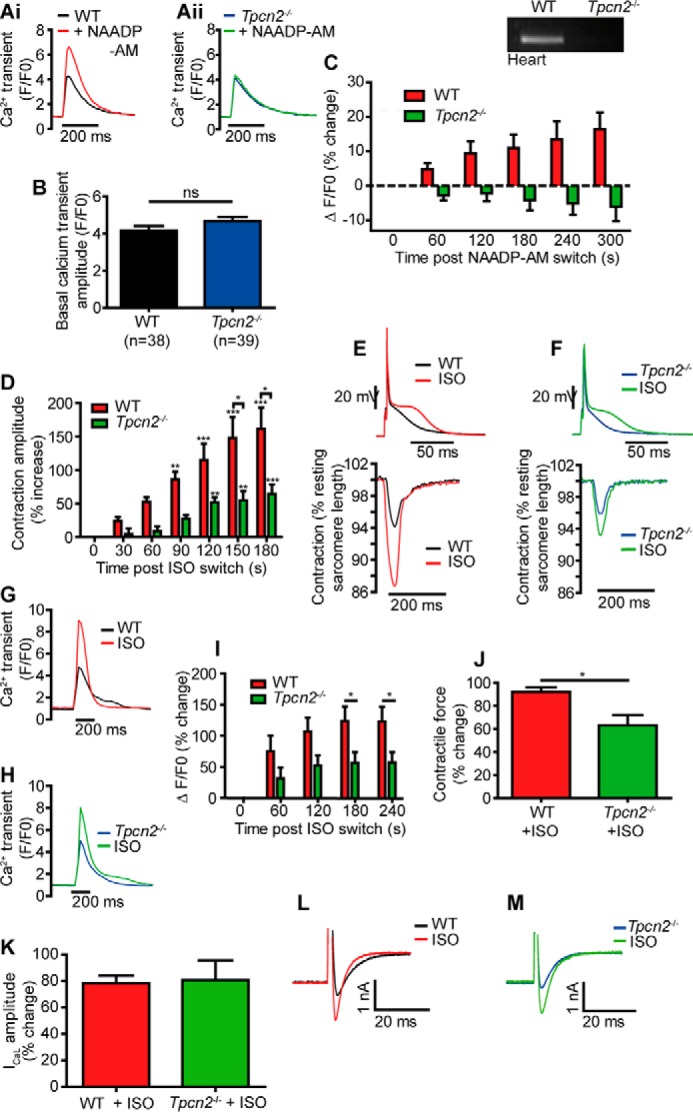
FIGURE 1.
NAADP actions were absent in cardiac ventricular myocytes from mice lacking TPC2 protein ( Tpcn2 u2212/u2212 ), and effects of u03b2-adrenoreceptor stimulation on contractions and Ca 2+ transients evo...
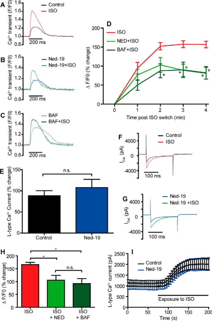
FIGURE 2.
Effects of u03b2-adrenoreceptor stimulation on Ca 2+ transients are reduced by suppressing lysosomal function or inhibiting NAADP actions in guinea pig myocytes. Au2013C show superimposed Ca 2+ transi...
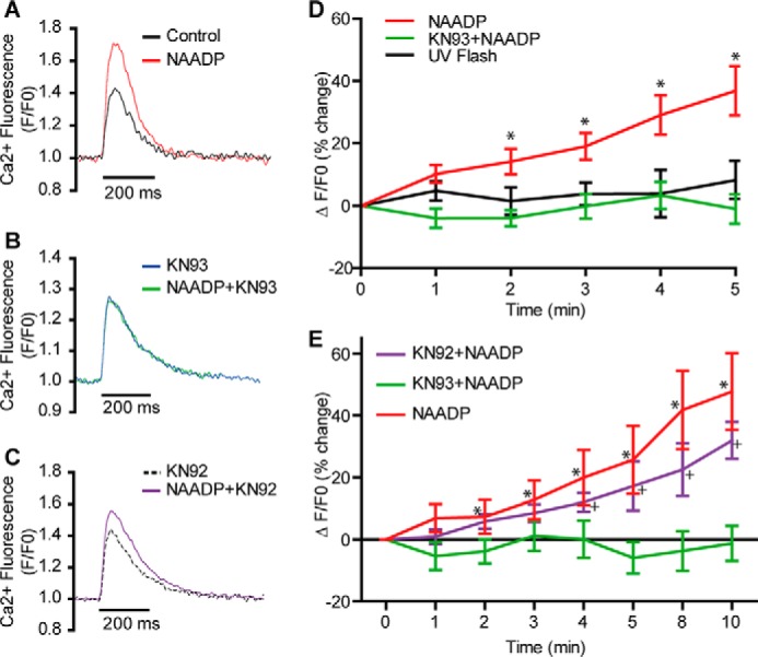
FIGURE 3.
CaMKII is required for atrial cardiomyocyte responses to NAADP from guinea pig. A and B show superimposed representative Ca 2+ transients (Fluo-5F, 1 Hz) before and after photorelease of NAADP from a ...
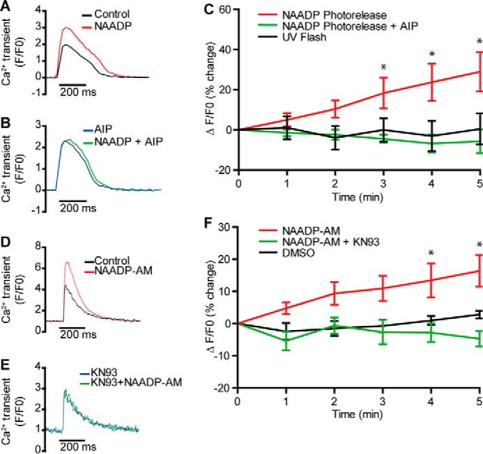
FIGURE 4.
CaMKII is required for ventricular cardiomyocyte responses to NAADP from guinea pig or WT mice. A and B show traces from similar experiments in guinea pig ventricular myocytes in the absence ( A ) and...
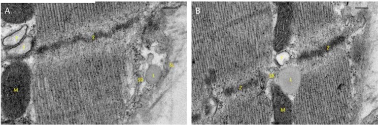
FIGURE 5.
Electron micrographs showing the subcellular relationships of cardiac lysosomes in rabbit. Lysosomes were identified according to their well established appearance and features ( i.e. a granular and a...
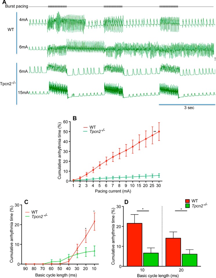
FIGURE 6.
Isolated whole hearts from Tpcn2 u2212/u2212 mice are less prone to arrhythmias induced by acute and high dose isoproterenol exposure (50 n m ). The susceptibility to arrhythmias was investigated usin...
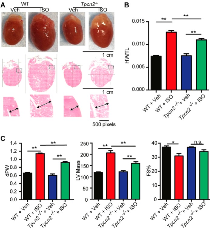
FIGURE 7.
Tpcn2 u2212/u2212 mice exhibit less hypertrophy and better cardiac function than wild-type mice after chronic u03b2-adrenergic stimulation for 14 days in vivo by osmotic mini-pump. The reduced hypertr...
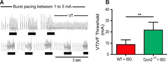
FIGURE 8.
Hearts from Tpcn2 u2212/u2212 mice exposed to chronic u03b2-adrenergic stimulation for 14 days in vivo by osmotic mini-pump show lower susceptibility to ventricular arrhythmias than hearts from wild-t...
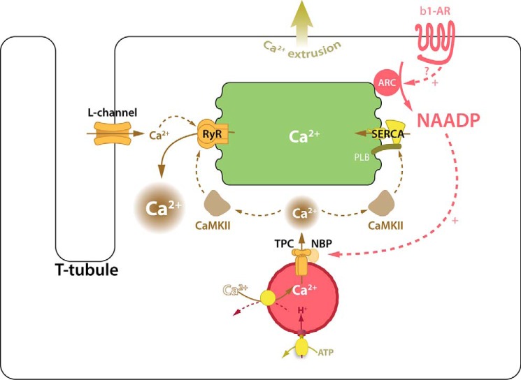
FIGURE 9.
Proposed scheme for actions of NAADP in cardiac myocytes. Scheme for action of NAADP in cardiac myocytes consistent with published observations with the addition of our present results. Activation of ...
Figure images are served from the NIH/NLM PubMed Central Open Access Subset or Europe PMC; copyright remains with the publishers and authors.
💬 Discussion
0 commentsNo comments yet. Be the first to start a discussion!
Leave a Comment