Abstract
Increased microvascular permeability to plasma proteins and neutrophil emigration are hallmarks of innate immunity and key features of numerous inflammatory disorders. Although neutrophils can promote microvascular leakage, the impact of vascular permeability on neutrophil trafficking is unknown. Here, through the application of confocal intravital microscopy, we report that vascular permeability-enhancing stimuli caused a significant frequency of neutrophil reverse transendothelial cell migration (rTEM). Furthermore, mice with a selective defect in microvascular permeability enhancement (VEC-Y685F-ki) showed reduced incidence of neutrophil rTEM. Mechanistically, elevated vascular leakage promoted movement of interstitial chemokines into the bloodstream, a response that supported abluminal-to-luminal neutrophil TEM. Through development of an in vivo cell labeling method we provide direct evidence for the systemic dissemination of rTEM neutrophils, and showed them to exhibit an activated phenotype and be capable of trafficking to the lungs where their presence was aligned with regions of vascular injury. Collectively, we demonstrate that increased microvascular leakage reverses the localization of directional cues across venular walls, thus causing neutrophils engaged in diapedesis to reenter the systemic circulation. This cascade of events offers a mechanism to explain how local tissue inflammation and vascular permeability can induce downstream pathological effects in remote organs, most notably in the lungs.
🔬 Techniques
🔭 Microscopes
✨ Fluorophores
🧪 Sample Preparation
🔬 Cell Lines
🏭 Microscope Brands
🧪 Reagent Suppliers
💻 Software Details
💾 Data Repositories
🏛️ Research Organizations (ROR)
Affiliated research institutions:
📋 Methods
Abs. The following Abs were obtained commercially: anti-CD31 (clone 390), PE–anti-CXCR4 (clone 2B11), anti–VE-cadherin (clone BV13), control anti-rabbit, anti–VE-cadherin (clone eBioBV14) mAbs from Thermo Fisher Scientific; PB–anti–Gr-1 (clone RB6-8C5), BV605–anti-CD62L (clone MEL-14), BV711–anti-CD11b (clone M1/70), AF488–anti-CD115 (clone AFS98), AF488–anti-CD102 (clone 3C4 MIC2/4), PE/Dazzle594–anti-CD54 (clone YN1/1.7.4), PE/Cy7–anti-CD29 (clone HMβ1-1), APC-Cy7–anti-CD115 (clone AFS98), biotin–anti–Ly-6G (clone 1A8) mAbs from BioLegend; blocking anti-CXCL1 (catalog 48415) from R&D Systems; rabbit anti–human CXCL8 (catalog NBP2-33819) from NOVUS; anti-NE (catalog ab68672) from Abcam; anti–α-SMA (clone 1A4), anti-CD31 (clone 2H8) from Sigma-Aldrich. Rabbit polyclonal anti–VE-PTP was generated as previously described ( 50 ). The following were gifts: rabbit polyclonal anti–JAM-C (clone H33; Michel Aurrand-Lions, INSERM, CRCM, France) and anti-MRP14 mAb (Nancy Hogg, Francis Crick Institute, London, United Kingdom). Animals. Male WT C57BL/6 (Charles River) and LysM-EGFP-ki ( 51 ) mice (8–12 weeks old) were used for all studies. VEC-Y685F-ki mutant mice that exhibit a single point mutation in VE-cadherin were generated as previously described ( 9 ). Generation of chimeric mice. VEC-WT and VEC-Y685F mice were lethally irradiated with 1 dose of 9 Gy over a time period of 11 minutes and were subsequently injected i.v. with 1.5 × 10 6 to 3 × 10 6 bone marrow cells from LysM-EGFP-ki mice. Level of engraftment was evaluated 4 weeks after reconstitution and all mice showed more than 99% of GFP + neutrophils with similar neutrophil counts. Confocal IVM of the mouse cremaster muscle. Anesthetized (3% isoflurane) male mice received an intrascrotal (i.s.) injection of fluorescently labeled anti-CD31 mAb (4 μg) and/or IL-1β (50 ng, R&D Systems), LTB 4 (300 ng, Cambridge Bioscience) to label vessels within the tissue and/or induce an inflammatory response, respectively. Control animals received PBS. The cremaster muscles were then prepared for intravital imaging 2 hours or 30 minutes after IL-1β or LTB 4 administration, respectively, as described previously ( 14 , 15 ). Topical application of histamine (30 μM, Sigma-Aldrich) onto exteriorized tissues, or i.v. injection of VEGF (4 μg/mouse, R&D Systems) was used to induce vascular leakage. IR injury was induced as described previously ( 14 , 15 ). In some experiments, anti-CXCL1 mAb or control IgG2a (1 mg/kg) and anti–VE-PTP Ab (100–200 μg) or rabbit IgG control (200 μg) was injected i.v. as indicated in relevant texts. In some experiments, recombinant mCXCL1 (50 ng, Peprotech) was injected i.v. 2 hours after IL-1β. To visualize vascular leakage, 75-kDa TRITC-dextran (40 mg/kg, Sigma-Aldrich) was injected i.v. (via tail vein cannula) 1 minute before the superfusion of histamine, in combination with i.v. injection of VEGF or during the reperfusion phase of IR-stimulated tissues. In some experiments, 10-kDa AF488-dextran (10 μg/mL, Thermo Fisher Scientific) was superfused 2 minutes after i.v. injection of 75-kDa TRITC-dextran for 10 minutes. The superfusate was then replaced with either Tyrode’s solution containing histamine (30 μM) or vehicle control for an additional 30 minutes. To label rTEM neutrophils, biotinylated anti-Ly6G mAb (2 μg) was injected i.v. 1.5 hours after IL-1β stimulation of tissues. Following exteriorization, the cremaster muscle was superfused with AF647-streptavidin (1 μg/mL, Thermo Fisher Scientific) with Tyrode’s solution with or without histamine (30 μM). Z -stack images of postcapillary venules (20–40 μm in diameter) were captured using a Leica SP5 or SP8 confocal microscope incorporating a ×20 water-dipping objective (NA 1.0), as detailed previously ( 6 , 14 ).
Show full methods section
Abs. The following Abs were obtained commercially: anti-CD31 (clone 390), PE–anti-CXCR4 (clone 2B11), anti–VE-cadherin (clone BV13), control anti-rabbit, anti–VE-cadherin (clone eBioBV14) mAbs from Thermo Fisher Scientific; PB–anti–Gr-1 (clone RB6-8C5), BV605–anti-CD62L (clone MEL-14), BV711–anti-CD11b (clone M1/70), AF488–anti-CD115 (clone AFS98), AF488–anti-CD102 (clone 3C4 MIC2/4), PE/Dazzle594–anti-CD54 (clone YN1/1.7.4), PE/Cy7–anti-CD29 (clone HMβ1-1), APC-Cy7–anti-CD115 (clone AFS98), biotin–anti–Ly-6G (clone 1A8) mAbs from BioLegend; blocking anti-CXCL1 (catalog 48415) from R&D Systems; rabbit anti–human CXCL8 (catalog NBP2-33819) from NOVUS; anti-NE (catalog ab68672) from Abcam; anti–α-SMA (clone 1A4), anti-CD31 (clone 2H8) from Sigma-Aldrich. Rabbit polyclonal anti–VE-PTP was generated as previously described ( 50 ). The following were gifts: rabbit polyclonal anti–JAM-C (clone H33; Michel Aurrand-Lions, INSERM, CRCM, France) and anti-MRP14 mAb (Nancy Hogg, Francis Crick Institute, London, United Kingdom). Animals. Male WT C57BL/6 (Charles River) and LysM-EGFP-ki ( 51 ) mice (8–12 weeks old) were used for all studies. VEC-Y685F-ki mutant mice that exhibit a single point mutation in VE-cadherin were generated as previously described ( 9 ). Generation of chimeric mice. VEC-WT and VEC-Y685F mice were lethally irradiated with 1 dose of 9 Gy over a time period of 11 minutes and were subsequently injected i.v. with 1.5 × 10 6 to 3 × 10 6 bone marrow cells from LysM-EGFP-ki mice. Level of engraftment was evaluated 4 weeks after reconstitution and all mice showed more than 99% of GFP + neutrophils with similar neutrophil counts. Confocal IVM of the mouse cremaster muscle. Anesthetized (3% isoflurane) male mice received an intrascrotal (i.s.) injection of fluorescently labeled anti-CD31 mAb (4 μg) and/or IL-1β (50 ng, R&D Systems), LTB 4 (300 ng, Cambridge Bioscience) to label vessels within the tissue and/or induce an inflammatory response, respectively. Control animals received PBS. The cremaster muscles were then prepared for intravital imaging 2 hours or 30 minutes after IL-1β or LTB 4 administration, respectively, as described previously ( 14 , 15 ). Topical application of histamine (30 μM, Sigma-Aldrich) onto exteriorized tissues, or i.v. injection of VEGF (4 μg/mouse, R&D Systems) was used to induce vascular leakage. IR injury was induced as described previously ( 14 , 15 ). In some experiments, anti-CXCL1 mAb or control IgG2a (1 mg/kg) and anti–VE-PTP Ab (100–200 μg) or rabbit IgG control (200 μg) was injected i.v. as indicated in relevant texts. In some experiments, recombinant mCXCL1 (50 ng, Peprotech) was injected i.v. 2 hours after IL-1β. To visualize vascular leakage, 75-kDa TRITC-dextran (40 mg/kg, Sigma-Aldrich) was injected i.v. (via tail vein cannula) 1 minute before the superfusion of histamine, in combination with i.v. injection of VEGF or during the reperfusion phase of IR-stimulated tissues. In some experiments, 10-kDa AF488-dextran (10 μg/mL, Thermo Fisher Scientific) was superfused 2 minutes after i.v. injection of 75-kDa TRITC-dextran for 10 minutes. The superfusate was then replaced with either Tyrode’s solution containing histamine (30 μM) or vehicle control for an additional 30 minutes. To label rTEM neutrophils, biotinylated anti-Ly6G mAb (2 μg) was injected i.v. 1.5 hours after IL-1β stimulation of tissues. Following exteriorization, the cremaster muscle was superfused with AF647-streptavidin (1 μg/mL, Thermo Fisher Scientific) with Tyrode’s solution with or without histamine (30 μM). Z -stack images of postcapillary venules (20–40 μm in diameter) were captured using a Leica SP5 or SP8 confocal microscope incorporating a ×20 water-dipping objective (NA 1.0), as detailed previously ( 6 , 14 ).
Quantification of neutrophil
TEM, microvascular leakage, and streptavidin labeling. Still images and 4D live recordings were analyzed using IMARIS software (Bitplane). Extravascular neutrophils were defined as those that had fully transmigrated and passed through the pericyte layer, recognizable via a change in their morphology, and expressed as number of cells/mm 3 of tissue. rTEM neutrophils were defined as cells that moved in an abluminal-to-luminal direction within EC junctions. Normal neutrophil TEM was classified as a response in which the cells migrated through EC junctions only in a luminal-to-abluminal direction, as previously described ( 14 , 15 ). Extravascular leakage was quantified by interstitial accumulation of i.v. 75-kDa TRITC-dextran and presented as mean fluorescence intensity (MFI) of indicated time points, measuring 6 to 8 regions of interest (ROI) in the interstitium (excluding areas exhibiting dextran-positive perivascular cells) and 30 μm away from the vessel wall. These readings were then normalized in relation to the first 2 MFI readings obtained after i.v. injection of the tracer and presented as normalized MFI. Similar analysis was conducted for the quantification of tissue levels of 10-kDa AF488-dextran (MFI normalized to the first 2 readings after superfusion of the tracer). Interstitial neutrophil speed and fluorescence intensity of AF647-streptavidin were analyzed using the spot or isosurface functions of IMARIS software, respectively. Bright-field IVM of the mouse cremaster muscle. Mice were injected i.s. with IL-1β (50 ng) or PBS alone for 2 hours before cremaster muscle exteriorization and before the superfusion of histamine (30 μM) or vehicle, as described above. Leukocyte rolling, firm arrest, and extravasation within 20- to 40-μm postcapillary venules were quantified by IVM over a 1.5-hour period using a bright-light microscope (Axioskop FS, Carl Zeiss), as detailed previously ( 44 ). Several vessel segments ( n = 3–5) from multiple vessels ( n = 3–5) were quantified for each animal. Quantification of chemokine and dextran levels in tissue and plasma. Anesthetized (3% isoflurane) mice were subjected to cremasteric ischemia (40 minutes) or injected i.s. with IL-1β (50 ng) or TNF (300 ng R&D Systems) in 200 μL PBS. Control animals received PBS only. Two hours later, animals were injected with i.s. histamine (30 μM solution) or PBS (both in 200 μL), or i.v. VEGF (4 μg/mouse) or PBS. Cremaster muscles and plasma samples (in 50 mM EDTA) were harvested 30 minutes later. In some experiments, hCXCL8 (500 ng, Peprotech) was coinjected with IL-1β (50 ng) for 1 hour before the end of the in vivo test period (i.e., 2.5 hours). In some experiments, mice were treated i.v. with a blocking anti–VE-PTP Ab (200 μg) ( 20 , 21 ) or rabbit IgG control (200 μg) 30 minutes after hCXCL8 and IL-1β injection. Other experiments involved treating the mice with a blocking anti–VE-cadherin mAb (BV13; 100 μg) ( 22 ) or a control mAb (anti-CD31 mAb, clone 390; 100 μg) i.s. for 3 hours followed by local injection of hCXCL8 for 1 hour before tissue and plasma collection. Tissues were homogenized in PBS containing 0.1% Triton X-100 and 1% Halt Protease and Phosphatase Inhibitor cocktail (Thermo Fisher Scientific) and mechanically dissociated using the Precellys 24 bead-beating system (Bertin Technologies). Levels of mCXCL1 and hCXCL8 were analyzed as per the manufacturer’s instructions by ELISA (R&D Systems and Thermo Fisher Scientific, respectively: sensitivity, 2 pg/mL). The quantity of chemokine detected in tissues was normalized to protein content as determined using a BCA assay (Thermo Fisher Scientific). Levels of 10-kDa AF488-dextran in plasma was quantified using a BMG NOVOstar microplate reader (BMG LABTECH). Immunofluorescence staining and confocal analysis of tissues. Whole-mount cremaster muscles were analyzed for expression of hCXCL8, JAM-C, red-(580/605)-microbeads (20 nm in diameter, 9.1 × 10 13 beads; Thermo Fisher Scientific), and AF647-streptavidin as previously described ( 6 , 15 ). Briefly, surgically removed tissues were fixed in ice-cold paraformaldehyde (PFA) (4% in PBS) for 45 minutes, blocked, and permeabilized at room temperature for 4 hours in PBS containing 25% FCS and 0.5% Triton X-100, and incubated overnight (anti-hCXCL8, anti-MRP14, anti–α-SMA, anti-CD31 staining) or 72 hours (anti–JAM-C staining) at 4°C with primary antibodies. Immunostained tissues were imaged with an inverted Zeiss 800 confocal laser-scanning microscope. JAM-C expression within the VE-cadherin channel was quantified as previously described ( 14 , 15 ). For analysis of hCXCL8 localization, ECs, neutrophils, and pericyte isosurfaces were created based on regions immunostained for CD31 (CD31 hi junctional and CD31 dim nonjunctional regions), MRP14, and α-SMA, respectively. EC body and junctional hCXCL8 expression was quantified as MFI within these isosurfaced regions. The MFI of fluorescent beads present in the subendothelial space (
📊 Figures
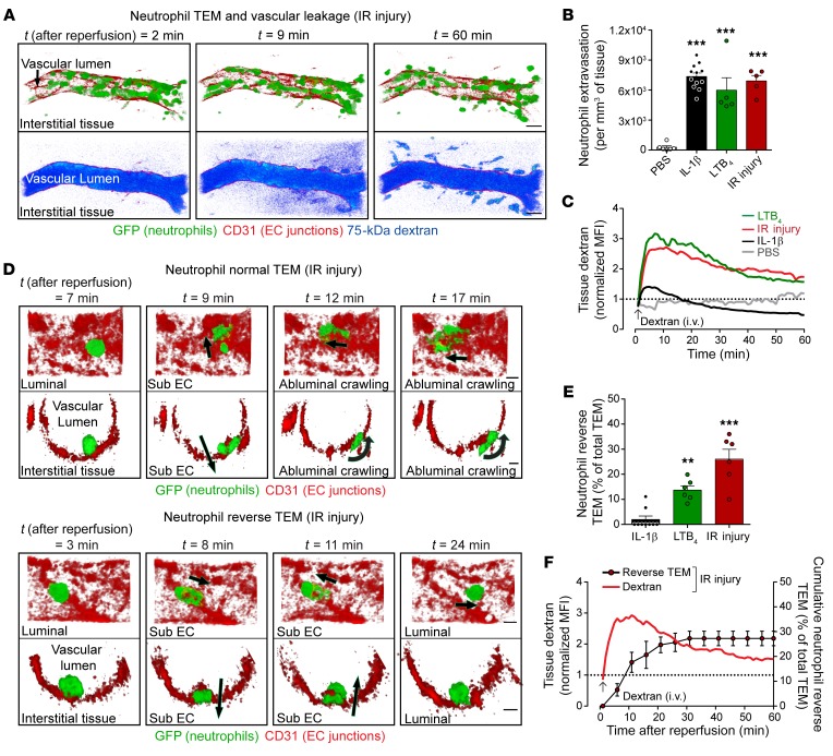
Figure 1
Hyperpermeability inflammatory reactions are associated with neutrophil reverse transendothelial migration.
Cremaster muscles of LysM-EGFP-ki mice were subjected to IL-1u03b2u2013 or LTB 4 -induced inflammation (120 and 30 minutes, respectively), or to IR injury (40 minutes ischemia + 1u20132 hours reperfus...
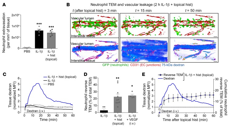
Figure 2
Induction of microvascular leakage promotes neutrophil reverse transendothelial migration.
Cremaster muscles of LysM-EGFP-ki mice were subjected to IL-1u03b2u2013induced inflammation for 120 minutes and analyzed by confocal IVM. AF647-labeled anti-CD31 mAb (i.s.) and 75-kDa TRITC-dextran (i...
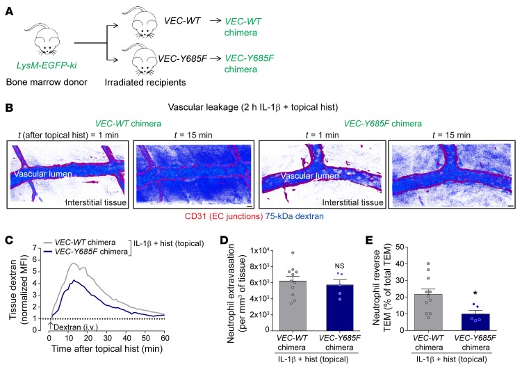
Figure 3
Chimeric VEC-Y658F mice exhibit reduced microvascular leakage induction and neutrophil reverse TEM.
( A ) Generation of chimeric mice exhibiting LysM-EGFP-ki hematopoietic cells within VEC-WT or VEC-Y658F recipients. ( B ) Representative confocal IVM images of postcapillary venular segments (stained...
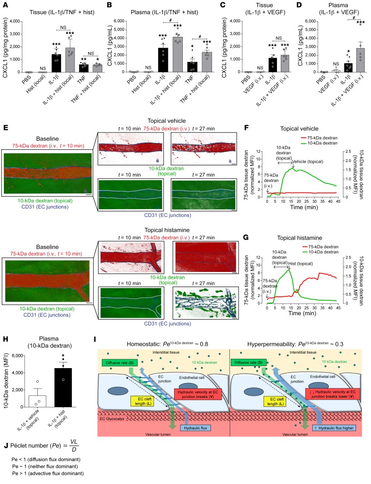
Figure 4
Stimulated microvascular leakage induces movement of small-molecular-weight proteins into the vascular lumen.
( A u2013 D ) Cremaster muscles of WT mice were stimulated with IL-1u03b2 or TNF for 2 hours followed by local injection of histamine or i.v. VEGF (or corresponding vehicle), for 30 minutes before col...
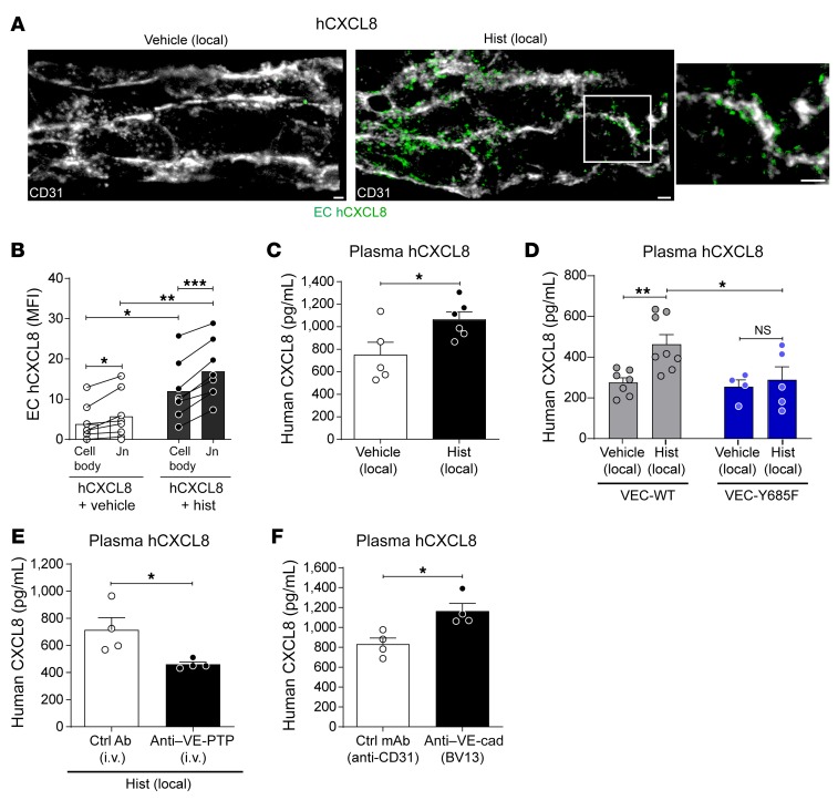
Figure 5
Vascular leakage induction promotes trafficking of tissue chemokine through venular walls.
Cremaster muscles were injected locally with IL-1u03b2 (50 ng) and human CXCL8 (hCXCL8, 500 ng) for 1 hour, followed by i.s. injection of vehicle (control) or histamine for an additional 1 hour. ( A )...
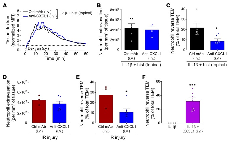
Figure 6
Systemic CXCL1 promotes neutrophil reverse TEM.
Cremaster muscles of LysM-EGFP-ki mice were stimulated with IL-1u03b2 (50 ng for 2 hours) followed by topical superfusion of histamine onto exteriorized tissues or subjected to IR injury. Blocking ant...
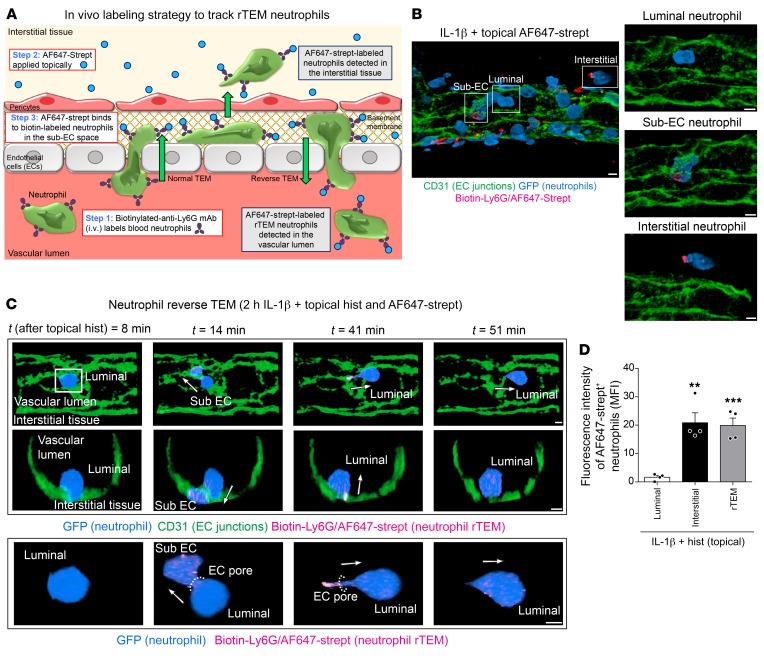
Figure 7
Development of an in vivo labeling strategy for tracking rTEM neutrophils.
( A ) Diagram detailing the labeling method. Cremaster muscles of LysM-EGFP-ki mice were stimulated with IL-1u03b2 (50 ng for 2 hours) followed by an i.v. injection of biotinylated anti-Ly6G (2 u03bcg...
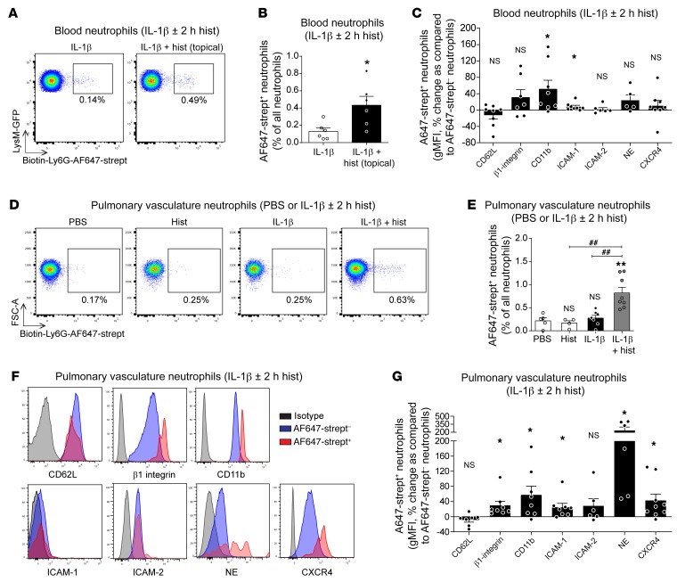
Figure 8
Labeled rTEM neutrophils are present in blood and pulmonary vasculature and show an activated phenotype.
( A and B ) LysM-EGFP-ki mice were subjected to local cremaster muscle stimulation with IL-1u03b2 (2 hours) and an i.v. injection of biotinylated anti-Ly6G (2 u03bcg) at t = 90 minutes. The tissues we...
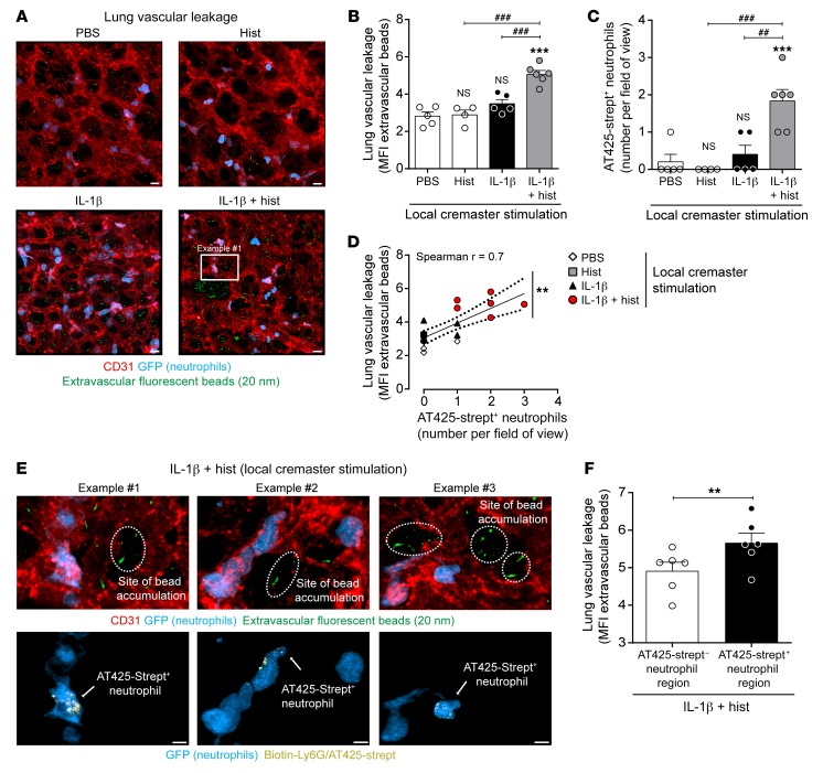
Figure 9
Accumulation of labeled rTEM neutrophils in lungs is linked with lung injury.
The cremaster muscles of LysM-EGFP-ki mice were locally stimulated with IL-1u03b2 or PBS (2 hours). Mice were injected i.v. with AF555u2013anti-CD31 mAb (10 u03bcg), biotinu2013anti-Ly6G mAb, and fluo...
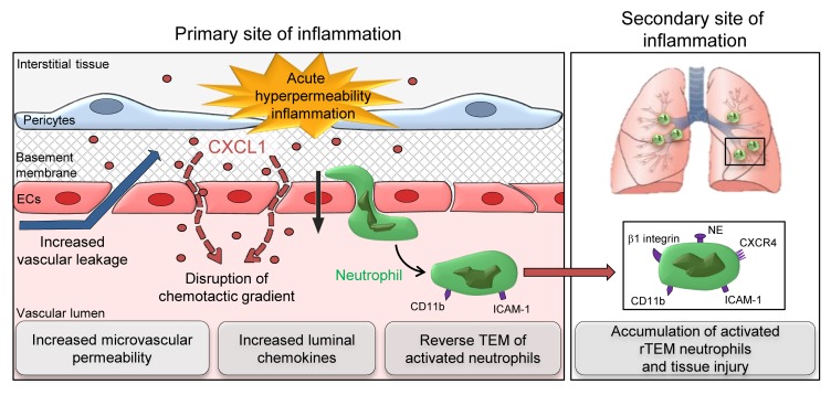
Figure 10
Schematic diagram of the cascade of events that link a local hyperpermeability inflammatory reaction to neutrophil rTEM and development of lung injury.
Figure images are served from the NIH/NLM PubMed Central Open Access Subset or Europe PMC; copyright remains with the publishers and authors.
💬 Discussion
0 commentsNo comments yet. Be the first to start a discussion!
Leave a Comment