Abstract
Abstract Introduction Breast cancer, the most common cause of cancer-related deaths worldwide among women, is a molecularly and clinically heterogeneous disease. Extensive genetic and epigenetic profiling of breast tumors has recently revealed novel putative driver genes, including p21-activated kinase (PAK)1. PAK1 is a serine/threonine kinase downstream of small GTP-binding proteins, Rac1 and Cdc42, and is an integral component of growth factor signaling networks and cellular functions fundamental to tumorigenesis. Methods PAK1 dysregulation (copy number gain, mRNA and protein expression) was evaluated in two cohorts of breast cancer tissues (n = 980 and 1,108). A novel small molecule inhibitor, FRAX1036, and RNA interference were used to examine PAK1 loss of function and combination with docetaxel in vitro. Mechanism of action for the therapeutic combination, both cellular and molecular, was assessed via time-lapse microscopy and immunoblotting. Results We demonstrate that focal genomic amplification and overexpression of PAK1 are associated with poor clinical outcome in the luminal subtype of breast cancer (P = 1.29 × 10⁻⁴ and P = 0.015, respectively). Given the role for PAK1 in regulating cytoskeletal organization, we hypothesized that combination of PAK1 inhibition with taxane treatment could be combined to further interfere with microtubule dynamics and cell survival. Consistent with this, administration of docetaxel with either a novel small molecule inhibitor of group I PAKs, FRAX1036, or PAK1 small interfering RNA oligonucleotides dramatically altered signaling to cytoskeletal-associated proteins, such as stathmin, and induced microtubule disorganization and cellular apoptosis. Live-cell imaging revealed that the duration of mitotic arrest mediated by docetaxel was significantly reduced in the presence of FRAX1036, and this was associated with increased kinetics of apoptosis. Conclusions Taken together, these findings further support PAK1 as a potential target in breast cancer and suggest combination with taxanes as a viable strategy to increase anti-tumor efficacy.
🔬 Techniques
🔭 Microscopes
💻 Software
✨ Fluorophores
🧪 Sample Preparation
🔬 Cell Lines
🏭 Microscope Brands
🧪 Reagent Suppliers
📷 Detectors
💻 Software Details
🏛️ Research Organizations (ROR)
Affiliated research institutions:
📋 Methods
PAK1 dysregulation (copy number gain, mRNA and protein expression) was evaluated in two cohorts of breast cancer tissues (n = 980 and 1,108). A novel small molecule inhibitor, FRAX1036, and RNA interference were used to examine PAK1 loss of function and combination with docetaxel in vitro . Mechanism of action for the therapeutic combination, both cellular and molecular, was assessed via time-lapse microscopy and immunoblotting.
Electronic supplementary material The online version of this article (doi:10.1186/s13058-015-0564-5) contains supplementary material, which is available to authorized users.
Materials and methods
Materials, cell culture and viability assays FRAX1036 was synthesized by Afraxis, Inc. (La Jolla, CA, USA) and docetaxel was purchased from Selleck Chemicals (Houston, TX, USA). Antibodies used for immunoblotting (p-MEK1-S298, p-CRAF-S338, Cleaved PARP, Cyclin D1, p-Stathmin-S16, p-β-catenin-S675, MCL-1, BCL-xL, p-Bad-S112 and PAK1) were purchased from Cell Signaling Technology (Danvers, MA, USA); anti-Actin was purchased from Sigma (St Louis, MO, USA). Cell lines were acquired from the American Type Culture Collection (ATCC; Manassas, VA, USA) and maintained at 37°C and 5% CO 2 in RPMI 1640 media with 10% fetal bovine serum and 2 mM L-glutamine. U2OS-red fluorescent protein (RFP)-Tubulin cells (Marinpharm, Luckenwalde, Germany) were stably transduced with a plasmid expressing green fluorescent protein (GFP)-histone H2B. Cell transfections and treatments were performed using short interfering RNA oligonucleotides for PAK1 from Dharmacon RNAi Technologies (Chicago, IL, USA). Cellular viability was assessed via ATP content using the CellTiter-Glo Luminescent Assay (Promega, Madison, WI, USA) and results represent mean ± standard deviation from three experiments. PAK1/CCND1 survival analysis Breast tumors from the Molecular Taxonomy of Breast Cancer International Consortium (METABRIC) dataset [ 15 ] with survival and DNA copy number data were selected, yielding 980 patients. DNA copy number was calculated using Affymetrix SNP6.0 arrays and a modified version of the PICNIC algorithm [ 19 ], published recently [ 20 ]. Samples were identified as having amplification of either PAK1 or CCND1 if the absolute copy number of the respective gene was >5 copies. The Kaplan-Meier plot and log-rank test were performed using the censored survival values (days since diagnosis) provided with the METABRIC dataset and our calculated PAK1 amplification status using the R language [ 21 ], version 3.1, and the R package “survival”, version 2.37-7. A Cox proportional hazard model was constructed using the METABRIC censored survival data, Nottingham prognostic index (NPI), patient age, and patient PAM50 breast cancer subtype classification in addition to the interaction of CCND1 and PAK1 amplification statuses. More specifically, the model “survival ~ NPI + age + PAM50 + CCND1 * PAK1 ” was fit using the “coxph” R package, where ccnd1 and pak1 are binary variables, as discussed above. The forest plot was produced using the coefficients from this model and their P -values. The whiskers on this plot represent ±1.96 × the standard error for each coefficient. The coefficient for amplification of both CCND1 and PAK1 (dual amplification) in the same sample was calculated as the sum of the coefficients “pak1Amplified”, “ccnd1Amplified”, and the coefficient for the interaction term for these two terms.
Show full methods section
PAK1 dysregulation (copy number gain, mRNA and protein expression) was evaluated in two cohorts of breast cancer tissues (n = 980 and 1,108). A novel small molecule inhibitor, FRAX1036, and RNA interference were used to examine PAK1 loss of function and combination with docetaxel in vitro . Mechanism of action for the therapeutic combination, both cellular and molecular, was assessed via time-lapse microscopy and immunoblotting.
Electronic supplementary material The online version of this article (doi:10.1186/s13058-015-0564-5) contains supplementary material, which is available to authorized users.
Materials and methods
Materials, cell culture and viability assays FRAX1036 was synthesized by Afraxis, Inc. (La Jolla, CA, USA) and docetaxel was purchased from Selleck Chemicals (Houston, TX, USA). Antibodies used for immunoblotting (p-MEK1-S298, p-CRAF-S338, Cleaved PARP, Cyclin D1, p-Stathmin-S16, p-β-catenin-S675, MCL-1, BCL-xL, p-Bad-S112 and PAK1) were purchased from Cell Signaling Technology (Danvers, MA, USA); anti-Actin was purchased from Sigma (St Louis, MO, USA). Cell lines were acquired from the American Type Culture Collection (ATCC; Manassas, VA, USA) and maintained at 37°C and 5% CO 2 in RPMI 1640 media with 10% fetal bovine serum and 2 mM L-glutamine. U2OS-red fluorescent protein (RFP)-Tubulin cells (Marinpharm, Luckenwalde, Germany) were stably transduced with a plasmid expressing green fluorescent protein (GFP)-histone H2B. Cell transfections and treatments were performed using short interfering RNA oligonucleotides for PAK1 from Dharmacon RNAi Technologies (Chicago, IL, USA). Cellular viability was assessed via ATP content using the CellTiter-Glo Luminescent Assay (Promega, Madison, WI, USA) and results represent mean ± standard deviation from three experiments. PAK1/CCND1 survival analysis Breast tumors from the Molecular Taxonomy of Breast Cancer International Consortium (METABRIC) dataset [ 15 ] with survival and DNA copy number data were selected, yielding 980 patients. DNA copy number was calculated using Affymetrix SNP6.0 arrays and a modified version of the PICNIC algorithm [ 19 ], published recently [ 20 ]. Samples were identified as having amplification of either PAK1 or CCND1 if the absolute copy number of the respective gene was >5 copies. The Kaplan-Meier plot and log-rank test were performed using the censored survival values (days since diagnosis) provided with the METABRIC dataset and our calculated PAK1 amplification status using the R language [ 21 ], version 3.1, and the R package “survival”, version 2.37-7. A Cox proportional hazard model was constructed using the METABRIC censored survival data, Nottingham prognostic index (NPI), patient age, and patient PAM50 breast cancer subtype classification in addition to the interaction of CCND1 and PAK1 amplification statuses. More specifically, the model “survival ~ NPI + age + PAM50 + CCND1 * PAK1 ” was fit using the “coxph” R package, where ccnd1 and pak1 are binary variables, as discussed above. The forest plot was produced using the coefficients from this model and their P -values. The whiskers on this plot represent ±1.96 × the standard error for each coefficient. The coefficient for amplification of both CCND1 and PAK1 (dual amplification) in the same sample was calculated as the sum of the coefficients “pak1Amplified”, “ccnd1Amplified”, and the coefficient for the interaction term for these two terms.
Bliss analysis
Cellular viability was assessed via ATP content using the CellTiter-Glo Luminescent Assay (Promega, Fitchburg, WI, USA) after a 4-day incubation period, and results represent mean ± standard deviation from three experiments. Total luminescence was measured on a Wallac Multilabel Reader (Perkin-Elmer, Waltham, MA, USA). Cells were treated simultaneously with FRAX1036 (dose range = 0 to 5 μM) or docetaxel (dose range = 0 to 0.4 nM) in an 8 × 10 matrix of concentrations. Combination synergy of FRAX1036 and docetaxel was determined by Bliss independence analyses. A Bliss expectation for a combined response C was calculated by the equation: C = (A + B) - (A × B) where A and B are the fractional growth inhibitions of given doses of drug A and B. ΔBliss scores were summed across the dose matrix to generate a Bliss sum. Bliss sum = 0 indicates that the combination effect is additive while Bliss sum >0 indicates synergy effect and Bliss sum
📊 Figures
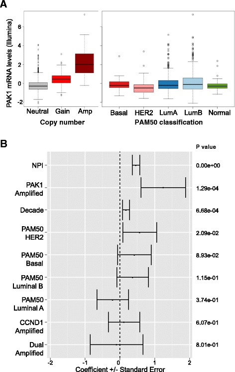
Figure 1
p21-Activated kinase (PAK)1 copy number and expression is elevated and associated with poor clinical outcome in breast tumors analyzed by the Molecular Taxonomy of Breast Cancer International Consorti...
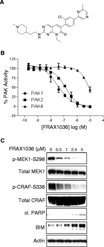
Figure 2
FRAX1036 inhibition of group I p21-activated kinase (PAK) isoforms. (A) Chemical structure of the group I PAK inhibitor, FRAX1036. (B) Concentration-response analysis of FRAX1036 against PAK1, PAK2 or...
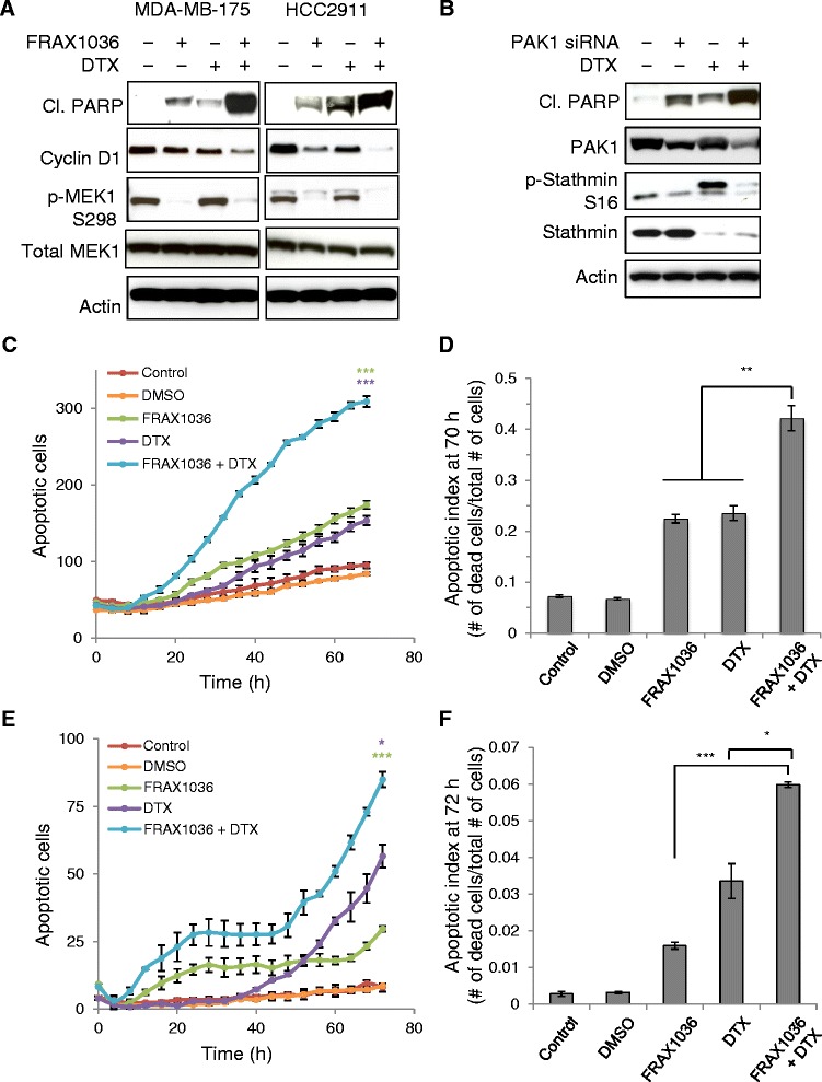
Figure 3
FRAX1036 and docetaxel (DTX) combine to alter stathmin phosphorylation, induce the apoptotic marker cleaved PARP and increase kinetics of apoptosis. (A) MDA-MB-175 and HCC2911 cells were treated with ...
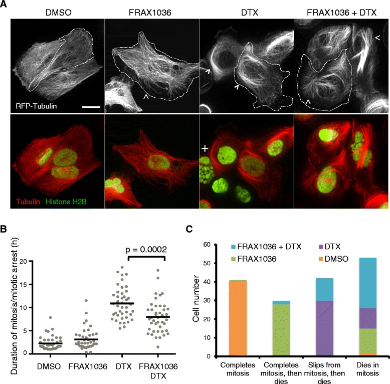
Figure 4
FRAX1036, docetaxel (DTX) and their combination affects microtubule organization, mitosis and cell fate. (A) Spinning-disk confocal images of live U2OS cells expressing red fluorescent protein (RFP)-T...
Figure images are served from the NIH/NLM PubMed Central Open Access Subset or Europe PMC; copyright remains with the publishers and authors.
💬 Discussion
0 commentsNo comments yet. Be the first to start a discussion!
Leave a Comment