Abstract
AbstractThroughout the genome, nucleosomes often form regular arrays that differ in nucleosome repeat length (NRL), occupancy of linker histone H1 and transcriptional activity. Here, we report cryo-EM structures of human H1-containing tetranucleosome arrays with four physiologically relevant NRLs. The structures show a zig-zag arrangement of nucleosomes, with nucleosomes 1 and 3 forming a stack. H1 binding to stacked nucleosomes depends on the NRL, whereas H1 always binds to the non-stacked nucleosomes 2 and 4. Short NRLs lead to altered trajectories of linker DNA, and these altered trajectories sterically impair H1 binding to the stacked nucleosomes in our structures. As the NRL increases, linker DNA trajectories relax, enabling H1 contacts and binding. Our results provide an explanation for why arrays with short NRLs are depleted of H1 and suited for transcription, whereas arrays with long NRLs show full H1 occupancy and can form transcriptionally silent heterochromatin regions.
🔬 Techniques
🧬 Organisms
🧪 Sample Preparation
🔬 Cell Lines
🏭 Microscope Brands
🧪 Reagent Suppliers
📷 Detectors
💻 Software Details
💾 Data Repositories
🏛️ Research Organizations (ROR)
Affiliated research institutions:
📋 Methods
Plasmids and DNA preparation
Plasmids contained human core histones, H2B1K, H3.2 and H4 (ref. 57 ). Full-length human linker histone H1.4 (UniProt ID P10412 ) was codon-optimized for Escherichia coli and synthesized by IDT as a gBlock. The DNA sequence for the GyrA intein was as described 58 and was synthesized by IDT as a gBlock. The DNA construct coding for Smt3-H1.4-GyrA was generated by overlap PCR to include a carboxy-terminal 6×His tag and cloned into LIC1B to include an N-terminal 6×His tag. Plasmids containing EcoRV-flanked repeats of the Widom-601 sequence 56 with DNA linker lengths of 30 bp, 40 bp, 50 bp and 60 bp were synthesized by GeneArt (Thermo Fisher). Linker sequences were based on the design of the 12×177 array 25 . Full DNA sequences are provided in the supplementary information. For DNA preparation, large cultures of E. coli XL1 blue transfected with plasmids containing the Widom-601 repeats were grown and prepared using the NucleoBond PC 10000 kit (Macherey-Nagel) according to the manufacturer’s instructions. Purified plasmids were digested with EcoRV (New England Biolabs) overnight, and the DNA templates containing the tandem Widom-601 repeats were purified by precipitation with PEG-6000 (ref. 59 ).
Protein purification
Human core histones H2A.1, H2B1K, H3.2 and H4 were purified as previously described 57 , 60 . Purified proteins were flash-frozen in liquid nitrogen and lyophilized. Histone octamer was reconstituted as described 57 , 60 . In brief, core histones were resuspended in unfolding buffer (6 M guanidinium hydrochloride, 20 mM HEPES pH 7.5, 10 mM dithiothreitol (DTT)), core histones were mixed at molar ratio 1.2:1.2:1:1, dialyzed 3 times against gel filtration buffer (20 mM HEPES pH 7.5, 1 mM EDTA, 2 M NaCl, 2 mM DTT) and loaded onto a Superdex 200 increase 10/300 GL (GE Healthcare) gel filtration column. Peak fractions containing core histone octamer were collected and directly used for nucleosome reconstitution or were flash-frozen in liquid nitrogen and stored at −80 °C. Full-length human linker histone H1.4 was purified as described 58 , with minor modifications. Briefly, Smt3-H1.4-GyrA was expressed in E. coli Rosetta 2 (DE3) cells and purified by His-Trap 5 ml HP (GE Healthcare). Peak fractions containing full-length Smt3-H1.4-GyrA were cleaved by Ulp1 for 1 hour at room temperature, followed by incubation with 500 mM β-mercaptoethanol for 4 hours at room temperature. The sample was adjusted to 8 M urea by weighing in solid urea, added to 1 L of buffer A (50 mM Tris-HCl pH 9.0, 200 mM NaCl, 8 M urea) and purified using a HiTrap SP 1 ml (GE Healthcare) column. The sample was adjusted to 200 mM HEPES pH 7.5 and run over a His-Trap 1 ml HP (GE Healthcare) column. The flowthrough was dialyzed 2 times against buffer B (20 mM HEPES pH 7.0, 600 mM NaCl), concentrated using Amicon Ultra-4 10 kDa MWCO centrifugal filters (Merck Millipore) and directly used for nucleosome reconstitution or flash-frozen in liquid nitrogen and stored at −80 °C. Nucleosome array reconstitution Nucleosome arrays containing H1.4 were reconstituted by salt-gradient dialysis as described 25 . Briefly, histone octamer and DNA were mixed at a molar ratio of 1:1 with respect to Widom-601 sequences in nucleosome reconstitution buffer A (20 mM HEPES pH 7.0, 2 M NaCl, 1 mM EDTA, 1 mM DTT), transferred into Slide-A-Lyzer MINI Dialysis Units 3,500 MWCO (Thermo Fisher) dialysis cups and gradually dialyzed over 16 hours from nucleosome reconstitution buffer A to nucleosome reconstitution buffer B (20 mM HEPES pH 7.0, 600 mM NaCl, 1 mM EDTA, 1 mM DTT). The sample was recovered and reconstituted with H1.4 in 1.2-fold molar excess over the number of Widom-601 sequences and dialyzed for 6 h from nucleosome reconstitution buffer B to nucleosome reconstitution buffer C (20 mM HEPES pH 7.0, 1 mM EDTA, 1 mM DTT). The sample was recovered and cleared from aggregation by spinning down in a table-top centrifuge at the top speed for 10 min at 4 °C. To probe stoichiometric binding of histone octamer to the Widom-601 nucleosome positioning sequence, nucleosome arrays were reconstituted without H1.4 and analyzed by BanI restriction enzyme digestion. For EMSAs of H1-containing arrays, 300 ng of sample was run on a 1.2% agarose gel in 0.5× TBE buffer for 1.5 hours at 110 V at 4 °C. To test differential binding of H1.4 to arrays of different NRLs, nucleosome arrays were reconstituted in the absence of H1.4 and adjusted to 100 nM DNA and 150 mM NaCl. H1.4 was then added to different molar ratios of H1 to Widom-601 sequence and incubated on ice for 30 min, and binding was probed by EMSA as described above. For sample in buffer with salt, the sample was adjusted to 60 mM NaCl and incubated for 30 min on ice prior to cryo-EM grid preparation. Cryo-EM sample preparation and data collection Quantifoil Cu 300 R 1.2/1.3 holey carbon grids were glow-discharged using a PELCO easiGlow (Ted Pella) for 100 s at 15 mA and 0.4 bar. In a Vitrobot Mark IV (FEI) chamber set to 100% humidity at 16 °C, 2 μl of sample was applied to each side of the grid. Excess liquid was blotted away using blot force 5 for 3 seconds, and the grid was vitrified by plunging into liquid ethane. Data were collected on a Titan Krios 300 kV transmission electron microscope (FEI) equipped with a Gatan Imaging Filter set to 20 eV and a K3 direct electron detector (Gatan). Movies containing 60 frames with a total fluence of 60 e – /Å 2 were collected using SerialEM 61 at a nominal magnification of ×81,000 and a pixel size of 1.05 Å/pixel with 40° stage tilt.
Show full methods section
Plasmids and DNA preparation
Plasmids contained human core histones, H2B1K, H3.2 and H4 (ref. 57 ). Full-length human linker histone H1.4 (UniProt ID P10412 ) was codon-optimized for Escherichia coli and synthesized by IDT as a gBlock. The DNA sequence for the GyrA intein was as described 58 and was synthesized by IDT as a gBlock. The DNA construct coding for Smt3-H1.4-GyrA was generated by overlap PCR to include a carboxy-terminal 6×His tag and cloned into LIC1B to include an N-terminal 6×His tag. Plasmids containing EcoRV-flanked repeats of the Widom-601 sequence 56 with DNA linker lengths of 30 bp, 40 bp, 50 bp and 60 bp were synthesized by GeneArt (Thermo Fisher). Linker sequences were based on the design of the 12×177 array 25 . Full DNA sequences are provided in the supplementary information. For DNA preparation, large cultures of E. coli XL1 blue transfected with plasmids containing the Widom-601 repeats were grown and prepared using the NucleoBond PC 10000 kit (Macherey-Nagel) according to the manufacturer’s instructions. Purified plasmids were digested with EcoRV (New England Biolabs) overnight, and the DNA templates containing the tandem Widom-601 repeats were purified by precipitation with PEG-6000 (ref. 59 ).
Protein purification
Human core histones H2A.1, H2B1K, H3.2 and H4 were purified as previously described 57 , 60 . Purified proteins were flash-frozen in liquid nitrogen and lyophilized. Histone octamer was reconstituted as described 57 , 60 . In brief, core histones were resuspended in unfolding buffer (6 M guanidinium hydrochloride, 20 mM HEPES pH 7.5, 10 mM dithiothreitol (DTT)), core histones were mixed at molar ratio 1.2:1.2:1:1, dialyzed 3 times against gel filtration buffer (20 mM HEPES pH 7.5, 1 mM EDTA, 2 M NaCl, 2 mM DTT) and loaded onto a Superdex 200 increase 10/300 GL (GE Healthcare) gel filtration column. Peak fractions containing core histone octamer were collected and directly used for nucleosome reconstitution or were flash-frozen in liquid nitrogen and stored at −80 °C. Full-length human linker histone H1.4 was purified as described 58 , with minor modifications. Briefly, Smt3-H1.4-GyrA was expressed in E. coli Rosetta 2 (DE3) cells and purified by His-Trap 5 ml HP (GE Healthcare). Peak fractions containing full-length Smt3-H1.4-GyrA were cleaved by Ulp1 for 1 hour at room temperature, followed by incubation with 500 mM β-mercaptoethanol for 4 hours at room temperature. The sample was adjusted to 8 M urea by weighing in solid urea, added to 1 L of buffer A (50 mM Tris-HCl pH 9.0, 200 mM NaCl, 8 M urea) and purified using a HiTrap SP 1 ml (GE Healthcare) column. The sample was adjusted to 200 mM HEPES pH 7.5 and run over a His-Trap 1 ml HP (GE Healthcare) column. The flowthrough was dialyzed 2 times against buffer B (20 mM HEPES pH 7.0, 600 mM NaCl), concentrated using Amicon Ultra-4 10 kDa MWCO centrifugal filters (Merck Millipore) and directly used for nucleosome reconstitution or flash-frozen in liquid nitrogen and stored at −80 °C. Nucleosome array reconstitution Nucleosome arrays containing H1.4 were reconstituted by salt-gradient dialysis as described 25 . Briefly, histone octamer and DNA were mixed at a molar ratio of 1:1 with respect to Widom-601 sequences in nucleosome reconstitution buffer A (20 mM HEPES pH 7.0, 2 M NaCl, 1 mM EDTA, 1 mM DTT), transferred into Slide-A-Lyzer MINI Dialysis Units 3,500 MWCO (Thermo Fisher) dialysis cups and gradually dialyzed over 16 hours from nucleosome reconstitution buffer A to nucleosome reconstitution buffer B (20 mM HEPES pH 7.0, 600 mM NaCl, 1 mM EDTA, 1 mM DTT). The sample was recovered and reconstituted with H1.4 in 1.2-fold molar excess over the number of Widom-601 sequences and dialyzed for 6 h from nucleosome reconstitution buffer B to nucleosome reconstitution buffer C (20 mM HEPES pH 7.0, 1 mM EDTA, 1 mM DTT). The sample was recovered and cleared from aggregation by spinning down in a table-top centrifuge at the top speed for 10 min at 4 °C. To probe stoichiometric binding of histone octamer to the Widom-601 nucleosome positioning sequence, nucleosome arrays were reconstituted without H1.4 and analyzed by BanI restriction enzyme digestion. For EMSAs of H1-containing arrays, 300 ng of sample was run on a 1.2% agarose gel in 0.5× TBE buffer for 1.5 hours at 110 V at 4 °C. To test differential binding of H1.4 to arrays of different NRLs, nucleosome arrays were reconstituted in the absence of H1.4 and adjusted to 100 nM DNA and 150 mM NaCl. H1.4 was then added to different molar ratios of H1 to Widom-601 sequence and incubated on ice for 30 min, and binding was probed by EMSA as described above. For sample in buffer with salt, the sample was adjusted to 60 mM NaCl and incubated for 30 min on ice prior to cryo-EM grid preparation. Cryo-EM sample preparation and data collection Quantifoil Cu 300 R 1.2/1.3 holey carbon grids were glow-discharged using a PELCO easiGlow (Ted Pella) for 100 s at 15 mA and 0.4 bar. In a Vitrobot Mark IV (FEI) chamber set to 100% humidity at 16 °C, 2 μl of sample was applied to each side of the grid. Excess liquid was blotted away using blot force 5 for 3 seconds, and the grid was vitrified by plunging into liquid ethane. Data were collected on a Titan Krios 300 kV transmission electron microscope (FEI) equipped with a Gatan Imaging Filter set to 20 eV and a K3 direct electron detector (Gatan). Movies containing 60 frames with a total fluence of 60 e – /Å 2 were collected using SerialEM 61 at a nominal magnification of ×81,000 and a pixel size of 1.05 Å/pixel with 40° stage tilt.
Data processing and analysis
Gain normalization, motion correction and CTF estimation of cryo-EM movies were performed using Warp 62 , and particles were picked using an instance of Warp’s neural network retrained on the 4×177 data set. Particles were extracted at 8.4 Å/pixel in RELION 3.1 (refs. 63 , 64 ) and sorted by 2–3 rounds of two-dimensional classification in cryoSPARC 65 . Particles belonging to classes showing 2 or more nucleosomes were reextracted at 3.15 Å/pixel, and all subsequent processing was done in RELION 3.1. For the 4×177+H1.4 data set (Supplementary Fig. 2 ), several rounds of 3D classification yielded particles that were refined to a 7.2-Å resolution map of a 4×177 trinucleosome. From this, 3D classification with a mask around the presumed location of the nucleosome 4 yielded particles that were refined to a 9.5-Å resolution map of the 4×177 tetranucleosome. The signal of the trinucleosome was subtracted from these particles, and the output was refined to the 7.9-Å resolution map of the fourth nucleosome. From the 4×177 trinucleosome map, masked refinements on the nucleosome stack or the connecting nucleosome were signal subtracted for the other nucleosomes and refined to yield the focused-refined maps of nucleosomes 1, 2 and 3. Similarly, the 4×187 (Supplementary Fig. 4 ), 4×197 (Supplementary Fig. 6 ) and 4×207 (Supplementary Fig. 8 ) cryo-EM data were subjected to several rounds of 3D classification and 3D refinement to yield maps with a defined nucleosome stack and blurred density for the connecting nucleosome. From this map, several more rounds of 3D classification were performed, and the selected particles were refined to the 4×187, 4×197 and 4×207 trinucleosome at 11 Å, 9.7 Å and 9.8 Å resolution, respectively. Particles from the 3D refinement of the stack with less defined connecting nucleosome were extracted, unbinned and further processed using signal subtraction, 3D classifications and masked refinements to yield maps for nucleosomes 1, 2 and 3. For the 4×187 data set, the same strategy was applied to obtain the map for nucleosome 4 but proved unsuccessful for the 4×197+H1.4 and 4×207+H1.4 data sets. The angular distribution of views for each map was plotted using Warp, local resolution and global FSC was determined using RELION, and the directional FSCs were calculated using the 3D FSC server 66 .
Model building and refinement
The local-resolution-filtered maps were used for model building, except for the 4×177 trinucleosome, 4×177 nucleosome 1, 4×177 nucleosome 2 and 4×177 nucleosome 4, for which the post-processed maps were used. For each data set, the structure of the H1-bound mononucleosome (PDB 7K5Y (ref. 19 )), with protein and DNA sequences mutated to the ones used in this study, was rigid-body fitted into the density of nucleosomal unit in UCSF Chimera 67 . Protein termini, entry DNA and exit DNA were manually adjusted in COOT 68 , and the resulting structures were real-space refined in PHENIX 69 . The refined nucleosomal units were then rigid-body fitted into corresponding densities of the nucleosome stack, trinucleosome and tetranucleosome, respectively, using UCSF Chimera. In case of the trinucleosome and tetranucleosome structures, the linker DNA was manually built in COOT. The models were real-space refined in PHENIX and were validated using Molprobity 70 (Tables 1 and 2 ). Figures were generated using PyMOL (Schrödinger), UCSF Chimera and UCSF ChimeraX 70 .
Analysis of linker DNA trajectories
The models for the nucleosome stacks were used to measure linker DNA trajectories for nucleosomes 1 and 3, and the models of the focused-refined maps of nucleosomes 2 and 4 were used to measure linker DNA deviation for nucleosomes 2 and 4. The corresponding maps were used to rigid-body fit the structure of the H1-bound 197 bp mononucleosome (PDB 7K5Y (ref. 19 )). The plane of the nucleosome disc needs to be defined to determine the angle α , and a plane normal to the nucleosome disc along the dyad axis needs to be defined to determine the angle β . For definition of these planes, we defined 3 points for each nucleosomal unit: (1) the centroid of the coordinates of the central base pair of the 147-bp Widom-601 sequence, (2) the centroid of the coordinates of the base pair 38 bp upstream of point 1 and (3) the centroid of the coordinates of the base pair 39 bp downstream of point 1. Points 2 and 3 are on two different DNA gyres and on the opposite side of the nucleosome dyad. We defined vectors v using points 2 and 3 to approximate the normal to the nucleosome disc, and u using point 1 and the centroid of points 2 and 3 to approximate the dyad axis. We used u and v to describe the plane perpendicular to the nucleosome disc. We determined the normal w to this plane by taking the normalized cross product of u and v , and we use u and w to describe the plane of the nucleosome disc. Linker DNA vectors were defined by using (4) the centroid of coordinates of the base pair 5 bp into the Widom-601 sequence and (5) the centroid of the coordinates of the base pair 10 bp outside of the Widom-601 sequence. For measurement of the angle β , as shown in Fig. 6b , we projected linker DNA vectors onto the plane generated by u and v and calculated the angle between the projected vectors. For the angle α , linker DNA vectors were projected onto plane the plane generated by u and w and we calculated the angle between the projected vectors. Calculations were done in MATLAB R2017a. Reporting Summary Further information on research design is available in the Nature Research Reporting Summary linked to this article.
Online content Any methods, additional references, Nature Research reporting summaries, source data, extended data, supplementary information, acknowledgements, peer review information; details of author contributions and competing interests; and statements of data and code availability are available at 10.1038/s41594-022-00768-w.
Supplementary information Supplementary Information Supplementary Figures 1–12, Supplementary Table 1, Supplementary Videos 1–4 descriptions, DNA sequences, Supplementary References Reporting Summary Supplementary Video 1 Nucleosomes 1 and 3 form a stack, while nucleosome 2 loops out between them and nucleosome 4 extends separately from the stack. H1 binds to non-stacking nucleosome 2 and 4 but not to stacking nucleosomes 1 and 3. Supplementary Video 2 Nucleosomes 1 and 3 form a stack, while nucleosome 2 loops out between them and nucleosome 4 extends separately from the stack. H1 binds to non-stacking nucleosome 2 and 4 and to stacking nucleosome 1 but not to stacking nucleosome 3. Supplementary Video 3 Nucleosomes 1 and 3 form a stack, while nucleosome 2 loops out between them. H1 binds to non-stacking nucleosome 2 and to stacking nucleosome 1 but not to stacking nucleosome 3. Supplementary Video 4 Nucleosomes 1 and 3 form a stack, while nucleosome 2 loops out between them. H1 binds to non-stacking nucleosome 2 and to stacking nucleosome 1 and 3. Supplementary Data 1 Uncropped EMSAs for Supplementary Fig. 1: a, 4×177; b, 4×187; c, 4×197; d, 4×207 Supplementary Data 2 Uncropped EMSA for Supplementary Fig. 11 Supplementary Data 3 Uncropped EMSAs Supplementary Fig. 12: a, 4×177; b, 4×187; c, 4×197; d, 4×207
📊 Figures
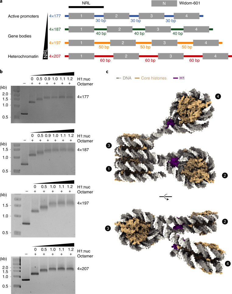
Fig. 1
Reconstitution of tetranucleosome arrays for structural studies.
a , DNA templates contain four Widom-601 (ref. 56 ) nucleosome positioning sequences and variable linker DNA: 4u00d7177 with 30-bp linker, 4u00d7187 with 40-bp linker, 4u00d7197 with 50-bp linker, and...
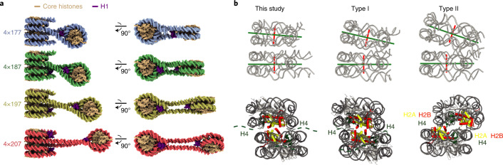
Fig. 2
Structure of trinucleosome cores of tetranucleosome arrays.
a . The trinucleosome cores of the 4u00d7177, 4u00d7187, 4u00d7197 and 4u00d7207 structures. Nucleosome 2 is rotated relative to the stack in all structures and is located at a greater distance from t...
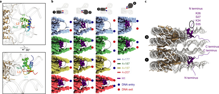
Fig. 3
NRL determines H1 binding to arrays.
a , H1 binds to nucleosomes of the array near the nucleosome dyad. The N-terminal part of the u03b12-helix (Nu03b12) and the L3 loop contact the DNA around the dyad, whereas the u03b13-helix and the L...
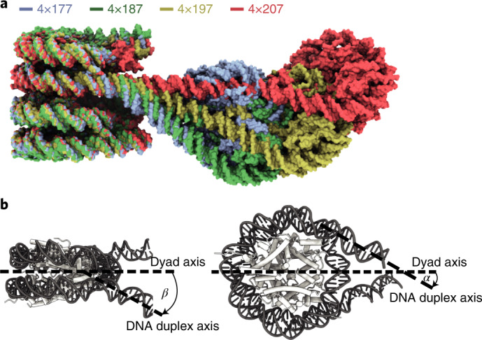
Fig. 4
NRL alters linker DNA trajectory at stacked nucleosomes.
a , Overlay of all four trinucleosome structures shown in Fig. 2 . With increasing NRL, linker DNA trajectories at the stacked nucleosomes are altered. b , u03b2 is defined as the angle between the nu...
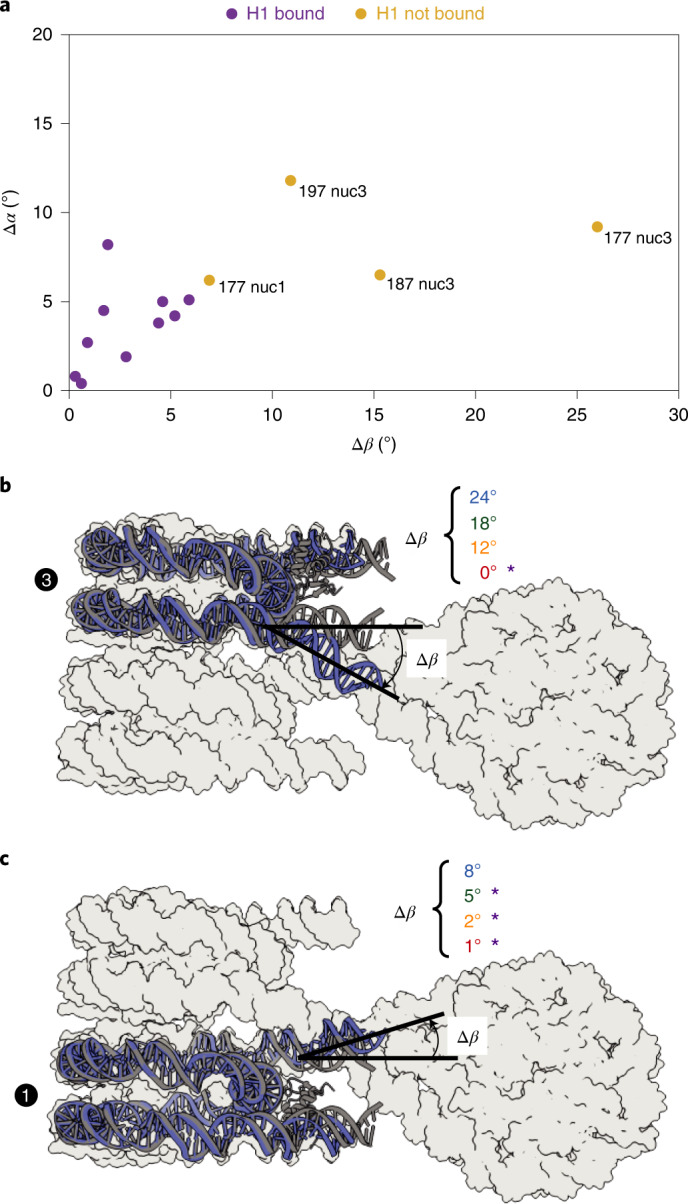
Fig. 5
Linker DNA trajectory determines H1 binding.
For each nucleosome, u0394 u03b1 and u0394 u03b2 describe the difference in u03b1 and u03b2 , respectively, between isolated H1-bound mononucleosomal linker DNA (PDB 7K5Y (ref. 19 )) and the linker DN...
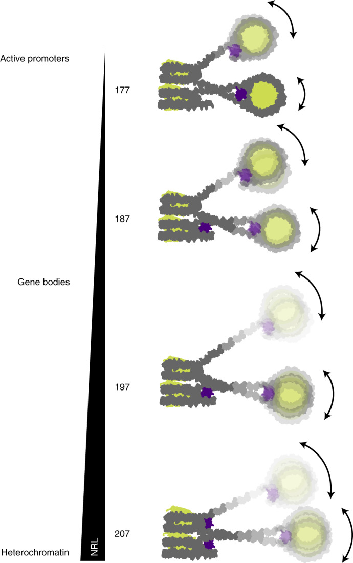
Fig. 6
Overview of H1 binding to tetranucleosome arrays.
Note that H1 binding to stacked nucleosomes depends on linker DNA trajectory that in turn depends on the NRL. For details, compare text.
Figure images are served from the NIH/NLM PubMed Central Open Access Subset or Europe PMC; copyright remains with the publishers and authors.
💬 Discussion
0 commentsNo comments yet. Be the first to start a discussion!
Leave a Comment