Abstract
G-protein-coupled receptors (GPCRs), which constitute the largest family of cell surface receptors, were originally thought to function as monomers, but are now recognized as being able to act in a wide range of oligomeric states and indeed, it is known that the oligomerization state of a GPCR can modulate its pharmacology and function. A number of experimental techniques have been devised to study GPCR oligomerization including those based upon traditional biochemistry such as blue-native PAGE (BN-PAGE), co-immunoprecipitation (Co-IP) and protein-fragment complementation assays (PCAs), those based upon resonance energy transfer, FRET, time-resolved FRET (TR-FRET), FRET spectrometry and bioluminescence resonance energy transfer (BRET). Those based upon microscopy such as FRAP, total internal reflection fluorescence microscopy (TIRFM), spatial intensity distribution analysis (SpIDA) and various single molecule imaging techniques. Finally with the solution of a growing number of crystal structures, X-ray crystallography must be acknowledged as an important source of discovery in this field. A different, but in many ways complementary approach to the use of more traditional experimental techniques, are those involving computational methods that possess obvious merit in the study of the dynamics of oligomer formation and function. Here, we summarize the latest developments that have been made in the methods used to study GPCR oligomerization and give an overview of their application.
🔬 Techniques
🧬 Organisms
✨ Fluorophores
🧪 Sample Preparation
💾 Data Repositories
🏛️ Research Organizations (ROR)
Affiliated research institutions:
📋 Methods
Methods to study oligomers of GPCRs Biochemical methods Co-IP and Western blotting Co-IP and Western blotting are probably one of the first techniques that provided direct evidence for β 2 -AR that it can form homodimers [ 12 ]. It has been used to detect oligomeric complexes of a number of GPCRs, such as β 2 -AR [ 12 ], DOR [ 31 ] and mGluR 5 [ 15 ] receptors. Using epitope tags and appropriate antibodies, the β 2 -AR was found to form homodimers and furthermore, these could be stabilized by agonists, whereas inverse agonists treatment was found to stabilize monomeric species. This suggests the possibility that interconversion between monomeric and dimeric forms may be important for biological activity [ 12 ]. Co-IP studies have also revealed that the adenosine receptor A 2 AR, dopamine receptor D 2 R and mGluR 5 receptors can form higher order oligomers [ 32 ], whereas the OX 1 receptor can form homodimers and even higher order oligomers [ 23 ]. When this approach is used to study GPCR quaternary structure, the cells containing the putative oligomers must first be lysed and solubilized. An antibody raised against one of the receptors or against epitope sequences such as V5, His or Myc fused to one of the receptors is used to separate the oligomer from the mixture of other proteins present in the lysate by, for instance the addition of protein G or A sepharose. This binds the antibody and allows separation by centrifugation. The oligomer may then be resolved by SDS/PAGE and the second receptor detected by Western blotting with a second antibody raised against the second receptor or an epitope tag to which it is fused [ 33 ]. Co-IP can be effective for detecting GPCR dimers and oligomers, both homodimers/oligomers and heterodimers/oligomers. However, a significant restriction to the use of Co-IP in the study of oligmerization in native systems is the paucity of specific and high-affinity antibodies to GPCRs themselves. Therefore, the ability to differentially epitope-tag GPCRs has been central to the use of Co-IP, though this does tend to limit the use of the technique to heterologously expressed receptors [ 22 ]. A further drawback of this methodology is that the sample lysis and solubilization required for releasing the protein of interest from its insoluble membrane environment, is a step that may, by itself, lead to artificial protein–protein associations being formed or possibly destroying existing associations. However, as an early and classic method, the Co-IP plus Western blotting approach is still used for detecting receptor–receptor interactions in vivo [ 33 ].
Show full methods section
Methods to study oligomers of GPCRs Biochemical methods Co-IP and Western blotting Co-IP and Western blotting are probably one of the first techniques that provided direct evidence for β 2 -AR that it can form homodimers [ 12 ]. It has been used to detect oligomeric complexes of a number of GPCRs, such as β 2 -AR [ 12 ], DOR [ 31 ] and mGluR 5 [ 15 ] receptors. Using epitope tags and appropriate antibodies, the β 2 -AR was found to form homodimers and furthermore, these could be stabilized by agonists, whereas inverse agonists treatment was found to stabilize monomeric species. This suggests the possibility that interconversion between monomeric and dimeric forms may be important for biological activity [ 12 ]. Co-IP studies have also revealed that the adenosine receptor A 2 AR, dopamine receptor D 2 R and mGluR 5 receptors can form higher order oligomers [ 32 ], whereas the OX 1 receptor can form homodimers and even higher order oligomers [ 23 ]. When this approach is used to study GPCR quaternary structure, the cells containing the putative oligomers must first be lysed and solubilized. An antibody raised against one of the receptors or against epitope sequences such as V5, His or Myc fused to one of the receptors is used to separate the oligomer from the mixture of other proteins present in the lysate by, for instance the addition of protein G or A sepharose. This binds the antibody and allows separation by centrifugation. The oligomer may then be resolved by SDS/PAGE and the second receptor detected by Western blotting with a second antibody raised against the second receptor or an epitope tag to which it is fused [ 33 ]. Co-IP can be effective for detecting GPCR dimers and oligomers, both homodimers/oligomers and heterodimers/oligomers. However, a significant restriction to the use of Co-IP in the study of oligmerization in native systems is the paucity of specific and high-affinity antibodies to GPCRs themselves. Therefore, the ability to differentially epitope-tag GPCRs has been central to the use of Co-IP, though this does tend to limit the use of the technique to heterologously expressed receptors [ 22 ]. A further drawback of this methodology is that the sample lysis and solubilization required for releasing the protein of interest from its insoluble membrane environment, is a step that may, by itself, lead to artificial protein–protein associations being formed or possibly destroying existing associations. However, as an early and classic method, the Co-IP plus Western blotting approach is still used for detecting receptor–receptor interactions in vivo [ 33 ].
Biochemical methods Co-IP and Western blotting
Co-IP and Western blotting are probably one of the first techniques that provided direct evidence for β 2 -AR that it can form homodimers [ 12 ]. It has been used to detect oligomeric complexes of a number of GPCRs, such as β 2 -AR [ 12 ], DOR [ 31 ] and mGluR 5 [ 15 ] receptors. Using epitope tags and appropriate antibodies, the β 2 -AR was found to form homodimers and furthermore, these could be stabilized by agonists, whereas inverse agonists treatment was found to stabilize monomeric species. This suggests the possibility that interconversion between monomeric and dimeric forms may be important for biological activity [ 12 ]. Co-IP studies have also revealed that the adenosine receptor A 2 AR, dopamine receptor D 2 R and mGluR 5 receptors can form higher order oligomers [ 32 ], whereas the OX 1 receptor can form homodimers and even higher order oligomers [ 23 ]. When this approach is used to study GPCR quaternary structure, the cells containing the putative oligomers must first be lysed and solubilized. An antibody raised against one of the receptors or against epitope sequences such as V5, His or Myc fused to one of the receptors is used to separate the oligomer from the mixture of other proteins present in the lysate by, for instance the addition of protein G or A sepharose. This binds the antibody and allows separation by centrifugation. The oligomer may then be resolved by SDS/PAGE and the second receptor detected by Western blotting with a second antibody raised against the second receptor or an epitope tag to which it is fused [ 33 ]. Co-IP can be effective for detecting GPCR dimers and oligomers, both homodimers/oligomers and heterodimers/oligomers. However, a significant restriction to the use of Co-IP in the study of oligmerization in native systems is the paucity of specific and high-affinity antibodies to GPCRs themselves. Therefore, the ability to differentially epitope-tag GPCRs has been central to the use of Co-IP, though this does tend to limit the use of the technique to heterologously expressed receptors [ 22 ]. A further drawback of this methodology is that the sample lysis and solubilization required for releasing the protein of interest from its insoluble membrane environment, is a step that may, by itself, lead to artificial protein–protein associations being formed or possibly destroying existing associations. However, as an early and classic method, the Co-IP plus Western blotting approach is still used for detecting receptor–receptor interactions in vivo [ 33 ].
Biophysical methods FRET
FRET is a non-invasive and non-destructive method for studying protein–protein interactions. The principle of resonance energy transfer was first illustrated in the late 1940s by Förster [ 40 ]. FRET is a physical phenomenon in which energy transfers from one excited fluorescent protein (the donor) to another (the acceptor) in a non-radiative (dipole–dipole) manner [ 41 ], assuming certain conditions that are required for FRET to occur are met [ 42 , 43 ]. One key parameter is that there must be an overlap between the emission spectrum of the donor molecule and the excitation spectrum of the acceptor molecule, whereas another is that the donor and the acceptor must be in close proximity, generally less than 100-Å apart [ 44 ]. GFP from the crystal jelly Aequorea victoria and its variants such as CFP/YFP were the fluorophores most commonly used in early analyses for labelling potential ‘partner’ proteins of interest and we now refer to the use of these as classical FRET analyses ( Figure 3 A). Previously, in some applications, YFP has been replaced with a fluorescein arsenical hairpin binder (FlAsH) sequence, the binding of which by an inherently non-fluorescent ligand generates a fluorescent species that can function as an energy acceptor for the donor CFP [ 45 , 46 ]. Figure 3 Principles of FRET, BRET and time-resolved FRET (TR-FRET) ( A ) The principle of FRET using CFP and YFP as a donor-acceptor pair. FRET occurs when the donor (CFP, shown in cyan) and the acceptor (YFP, shown in yellow) are in close proximity (
📊 Figures
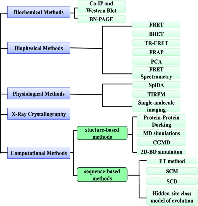
Figure 1
Techniques for studying oligomerization of GPCRs
The techniques used to study oligomerization of GPCRs may be divided into five categories, which are biochemical methods, biophysical methods, physiological methods, X-ray crystallography and computat...
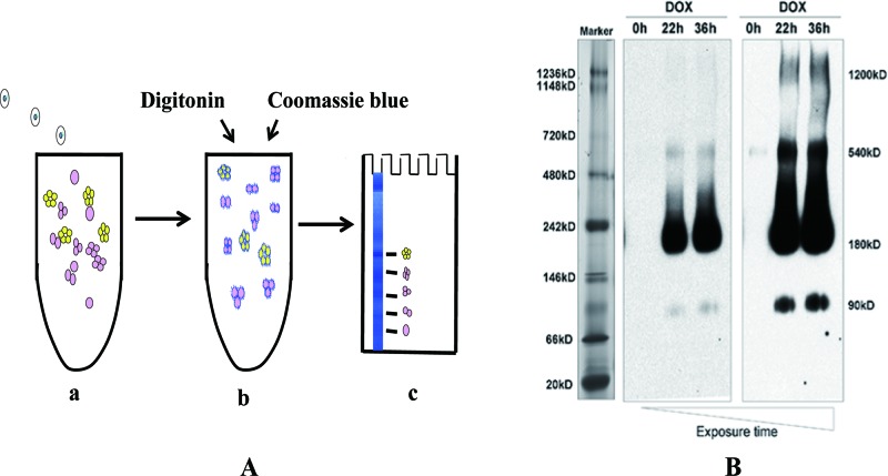
Figure 2
Principles of BN-PAGE
( A ) Cartoon representation of the major steps involved in BN-PAGE. ( i ) A mixture of proteins and protein complexes. ( ii ) Protein complexes are solubilized by mild non-ionic detergents (such as d...
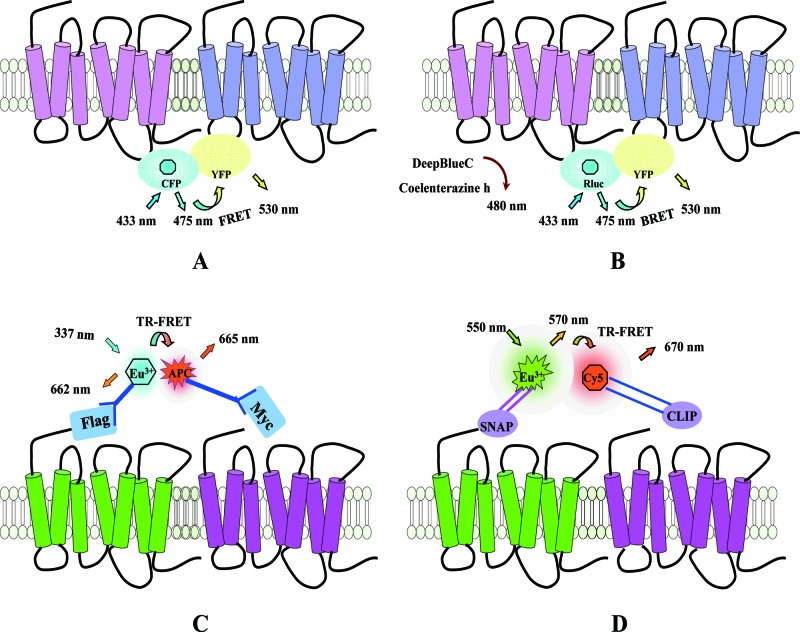
Figure 3
Principles of FRET, BRET and time-resolved FRET (TR-FRET)
( A ) The principle of FRET using CFP and YFP as a donor-acceptor pair. FRET occurs when the donor (CFP, shown in cyan) and the acceptor (YFP, shown in yellow) are in close proximity (<10 nm), so t...
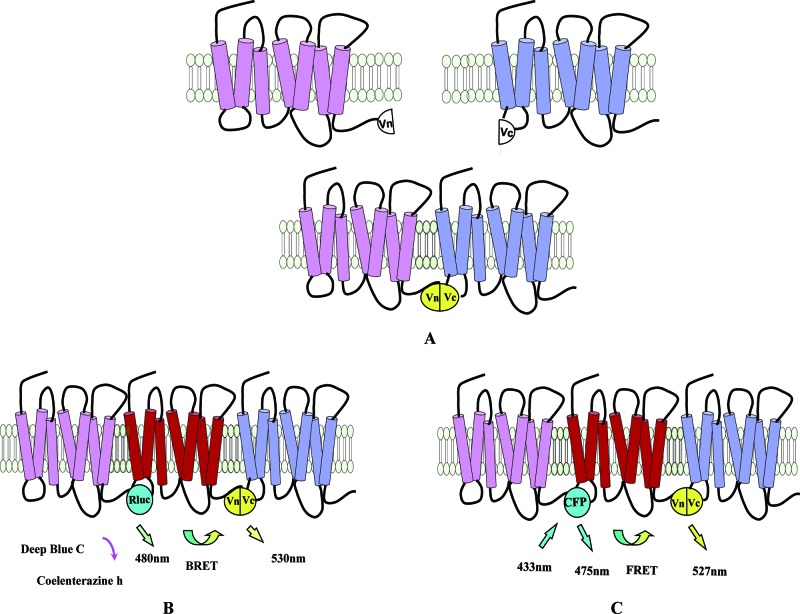
Figure 4
Schematic representation of PCA technology
( A ) Cartoon representation of PCA. When the two receptors fused to two complementary fluorescent protein fragments (Vn and Vc) are in close proximity, fluorescence can be detected. ( B ) When three ...
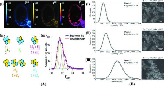
Figure 5
The application of FRET spectrometry to the study of GPCR oligomerization
( A ) FRET spectrometry analysis of Flp-In TM T-REx TM-293 cells expressing Mycu2013Y149C/A239GM 3 Ru2013Cerulean (inducible) and FLAGu2013M 3 Ru2013Citrine (constitutive). ( i ) Photomicrograph of a ...
Figure images are served from the NIH/NLM PubMed Central Open Access Subset or Europe PMC; copyright remains with the publishers and authors.
💬 Discussion
0 commentsNo comments yet. Be the first to start a discussion!
Leave a Comment