Abstract
Protein secretion, a key intercellular event for transducing cellular signals, is thought to be strictly regulated. However, secretion dynamics at the single-cell level have not yet been clarified because intercellular heterogeneity results in an averaging response from the bulk cell population. To address this issue, we developed a novel assay platform for real-time imaging of protein secretion at single-cell resolution by a sandwich immunoassay monitored by total internal reflection microscopy in sub-nanolitre-sized microwell arrays. Real-time secretion imaging on the platform at 1-min time intervals allowed successful detection of the heterogeneous onset time of nonclassical IL-1β secretion from monocytes after external stimulation. The platform also helped in elucidating the chronological relationship between loss of membrane integrity and IL-1β secretion. The study results indicate that this unique monitoring platform will serve as a new and powerful tool for analysing protein secretion dynamics with simultaneous monitoring of intracellular events by live-cell imaging.
🔬 Techniques
🧬 Organisms
💻 Software
✨ Fluorophores
🧪 Sample Preparation
🔬 Cell Lines
🏭 Microscope Brands
🧪 Reagent Suppliers
📷 Detectors
🎨 Filters
💻 Software Details
🏛️ Research Organizations (ROR)
Affiliated research institutions:
📋 Methods
Reagents The DuoSet ELISA Development
System was purchased from R&D Systems (Minneapolis, MN, USA), and the set of capture and detection antibodies was used for sandwich immunoassays for human IL-1β (DY201). Human IL-6 monoclonal antibody (clone 6708; MAB206) and human IL-6 biotinylated affinity-purified polyclonal antibody (BAF206) were also purchased from R&D Systems. Adenosine 5′-triphosphate disodium salt hydrate (ATP, A7699) and lipopolysaccharides from Escherichia coli 055:B5 (LPS, L4524) were purchased from Sigma-Aldrich (St. Louis, MO, USA). Lipidure BL802, a water-soluble polymer of 2-methacryloyloxy ethyl phosphorylcholine, was purchased from NOF Corporation (Tokyo, Japan). Calcein AM (C3099) and SYTOX Blue nucleic acid stain (S11348) were purchased from Life Technologies (Carlsbad, CA, USA). CF660R streptavidin was purchased from Biotium (29040; Hayward, CA, USA). Dimethyl pimelimidate-2HCl (DMP, 21666) was purchased from Thermo Fisher Scientific (Rockford, IL, USA). Foetal bovine serum (FBS, s1560) was purchased from Biowest (Nuaillé, France).
Cell culture
The incubation of cells was performed in a CO 2 incubator at 37°C in a humidified atmosphere with 5% CO 2 , unless otherwise indicated. For the isolation of human peripheral blood monocytes, 20 mL of venous blood was drawn from a healthy donor after obtaining institutional approval of the ethical committee of the Kyoto University Hospital, in accordance with Declaration of Helsinki. Monocytes from different donors were used for different experiments. The cell fraction was separated with Lymphoprep (Axis-Shield, Dundee, UK) according to the manufacturer's instructions. CD14 + monocytes were sequentially purified by MACS (Miltenyi Biotec, Bergisch Gladbach, Germany) with a negative selection reagent (i.e. monocyte isolation kit II) and a positive selection reagent (i.e. CD14 microbeads). The isolated monocytes were incubated overnight in RPMI medium containing 10% FBS. Optical arrangement All measurements were performed with a completely automated inverted microscope (ECLIPSE Ti-E; Nikon, Tokyo, Japan) equipped with a high NA 60× objective lens (TIRF 60 × H; NA, 1.49; Nikon). The microscope was customized to introduce external laser beams (635 nm; Radius 635-25; Coherent, Santa Clara, CA, USA) at the indicated incident angle to actualize TIRF illumination. The following sets of excitation (Ex) and emission (Em) filters and a dichroic mirror (DM) were used with a high-pressure xenon lamp: for SYTOX, Ex = FF01-448/20-25, Em = FF01-482/25-25, and DM = Di02-R442-25x36; and for calcein, Ex = FF02-472/30-25, Em = FF01-520/35-25, and DM = FF495-Di03-25x36. The following Ex and Em filters and DM were used with the 632 nm laser for the CF660R dye: Ex = FF02-628/40-25, Em = FF01-692/40-25, and DM = FF660-Di02-25x36. These optical filters were purchased from Semrock (Rochester, NY, USA). Each image was projected on an EM-CCD camera (ImagEM C9100-13; Hamamatsu Photonics K.K., Sizuoka, Japan) through a 0.7× lens (C-0.7x DXM Relay Lens; Nikon). A stage-top incubator (ONICS; Tokai Hit Co., Shizuoka, Japan) was used to control temperature, humidity, and gas concentration. MWA chips MWA chips were prepared from PDMS and microscopic-grade coverslips. The PDMS MWA chip was prepared as follows: A thin PDMS sheet, consisting of a 50 × 50 array of 50-μm or 70-μm through-holes with 100-μm centre-to-centre spacing and 80-μm thickness, was purchased from Fluidware Technologies (Saitama, Japan). The PDMS sheet and a glass slide were permanently bonded together after the contact surfaces between them were plasma-treated (SEDE-PFA; Meiwafosis, Tokyo, Japan). The bare glass surfaces that now functioned as the bottom of the microwells after bonding were aminated with Vecatabond Reagent (SP-1800; Vector Laboratories, Burlingame, CA, USA). The chip was mounted on a coverslip holder (Attofluor cell chamber, A7819; Life Technologies) adjacent to another PDMS block (Sylgard184; Dow Corning Toray, Tokyo, Japan) with an 8-mm diameter through-hole that served as a reservoir well. The interstices between the PDMS-glass chip and the PDMS block were filled with uncured PDMS. The integrated chip was baked at 130°C for 3 h. A 100-μL mixture of capture antibodies (100 μg/mL) and dimethyl pimelimidate-2HCl (DMP; 7 mg/mL) was loaded onto the aminated glass surface to fix the capture antibody. The surface was blocked with monoethanolamine (0.1 M, pH 8.2) and Lipidure reagent (0.2% [w/v]), and stored at 4°C until use.
Show full methods section
Reagents The DuoSet ELISA Development
System was purchased from R&D Systems (Minneapolis, MN, USA), and the set of capture and detection antibodies was used for sandwich immunoassays for human IL-1β (DY201). Human IL-6 monoclonal antibody (clone 6708; MAB206) and human IL-6 biotinylated affinity-purified polyclonal antibody (BAF206) were also purchased from R&D Systems. Adenosine 5′-triphosphate disodium salt hydrate (ATP, A7699) and lipopolysaccharides from Escherichia coli 055:B5 (LPS, L4524) were purchased from Sigma-Aldrich (St. Louis, MO, USA). Lipidure BL802, a water-soluble polymer of 2-methacryloyloxy ethyl phosphorylcholine, was purchased from NOF Corporation (Tokyo, Japan). Calcein AM (C3099) and SYTOX Blue nucleic acid stain (S11348) were purchased from Life Technologies (Carlsbad, CA, USA). CF660R streptavidin was purchased from Biotium (29040; Hayward, CA, USA). Dimethyl pimelimidate-2HCl (DMP, 21666) was purchased from Thermo Fisher Scientific (Rockford, IL, USA). Foetal bovine serum (FBS, s1560) was purchased from Biowest (Nuaillé, France).
Cell culture
The incubation of cells was performed in a CO 2 incubator at 37°C in a humidified atmosphere with 5% CO 2 , unless otherwise indicated. For the isolation of human peripheral blood monocytes, 20 mL of venous blood was drawn from a healthy donor after obtaining institutional approval of the ethical committee of the Kyoto University Hospital, in accordance with Declaration of Helsinki. Monocytes from different donors were used for different experiments. The cell fraction was separated with Lymphoprep (Axis-Shield, Dundee, UK) according to the manufacturer's instructions. CD14 + monocytes were sequentially purified by MACS (Miltenyi Biotec, Bergisch Gladbach, Germany) with a negative selection reagent (i.e. monocyte isolation kit II) and a positive selection reagent (i.e. CD14 microbeads). The isolated monocytes were incubated overnight in RPMI medium containing 10% FBS. Optical arrangement All measurements were performed with a completely automated inverted microscope (ECLIPSE Ti-E; Nikon, Tokyo, Japan) equipped with a high NA 60× objective lens (TIRF 60 × H; NA, 1.49; Nikon). The microscope was customized to introduce external laser beams (635 nm; Radius 635-25; Coherent, Santa Clara, CA, USA) at the indicated incident angle to actualize TIRF illumination. The following sets of excitation (Ex) and emission (Em) filters and a dichroic mirror (DM) were used with a high-pressure xenon lamp: for SYTOX, Ex = FF01-448/20-25, Em = FF01-482/25-25, and DM = Di02-R442-25x36; and for calcein, Ex = FF02-472/30-25, Em = FF01-520/35-25, and DM = FF495-Di03-25x36. The following Ex and Em filters and DM were used with the 632 nm laser for the CF660R dye: Ex = FF02-628/40-25, Em = FF01-692/40-25, and DM = FF660-Di02-25x36. These optical filters were purchased from Semrock (Rochester, NY, USA). Each image was projected on an EM-CCD camera (ImagEM C9100-13; Hamamatsu Photonics K.K., Sizuoka, Japan) through a 0.7× lens (C-0.7x DXM Relay Lens; Nikon). A stage-top incubator (ONICS; Tokai Hit Co., Shizuoka, Japan) was used to control temperature, humidity, and gas concentration. MWA chips MWA chips were prepared from PDMS and microscopic-grade coverslips. The PDMS MWA chip was prepared as follows: A thin PDMS sheet, consisting of a 50 × 50 array of 50-μm or 70-μm through-holes with 100-μm centre-to-centre spacing and 80-μm thickness, was purchased from Fluidware Technologies (Saitama, Japan). The PDMS sheet and a glass slide were permanently bonded together after the contact surfaces between them were plasma-treated (SEDE-PFA; Meiwafosis, Tokyo, Japan). The bare glass surfaces that now functioned as the bottom of the microwells after bonding were aminated with Vecatabond Reagent (SP-1800; Vector Laboratories, Burlingame, CA, USA). The chip was mounted on a coverslip holder (Attofluor cell chamber, A7819; Life Technologies) adjacent to another PDMS block (Sylgard184; Dow Corning Toray, Tokyo, Japan) with an 8-mm diameter through-hole that served as a reservoir well. The interstices between the PDMS-glass chip and the PDMS block were filled with uncured PDMS. The integrated chip was baked at 130°C for 3 h. A 100-μL mixture of capture antibodies (100 μg/mL) and dimethyl pimelimidate-2HCl (DMP; 7 mg/mL) was loaded onto the aminated glass surface to fix the capture antibody. The surface was blocked with monoethanolamine (0.1 M, pH 8.2) and Lipidure reagent (0.2% [w/v]), and stored at 4°C until use.
Preparation of detection medium
Detection antibody labelled with biotin was coupled with CF-labelled streptavidin at 1:10 molar ratios at room temperature in the dark for 3 h. Unoccupied sites on streptavidin were blocked with excess dPEG4-biotin acid (10199; Quanta BioDesign, Ltd., Powell, OH, USA). Unconjugated streptavidin and dPEG4-biotin were removed by ultrafiltration (Amicon Ultra-0.5, 100 kDa; Merck Millipore, Billerica, MA, USA). The detection media contained prepared CF-labelled detection antibody (30 nM), BSA (2.5% [w/v]), and the indicated combination of the following additives (with the final concentrations): SYTOX (0.8 μM), glycine (5 mM), LPS (1 μg/mL), and/or ATP (5 mM).
Handling and treatment of cells
Monocytes were stimulated with LPS for 3 h and incubated with 0.4 μM Calcein AM for 15 min before IL-1β secretion analysis on a PDMS MWA chip. The cells were washed with fresh medium and recovered from the bottom of 96-well plates by gentle pipetting. Approximately 2,000 cells were introduced into an MWA chip and allowed to settle by gravity for 10 min in a CO 2 incubator. Next, the medium was replaced with the detection medium containing SYTOX, glycine, and LPS. The cells were first monitored for 20 min before ATP stimulation; after ATP administration, monitoring was resumed during the 10-min incubation period. The medium was then replaced with fresh detection medium containing SYTOX, glycine, and LPS. Measurements were then resumed and continued for 4 h. Time-resolved monitoring of cytokine secretion from stimulated monocytes Ten positions were selected in order to include as many single cell-containing microwells as possible. Monocytes in the selected position of microwells were monitored at 1-min intervals by multichannel microscopy, i.e. DIA images, epifluorescence images for calcein and SYTOX, and TIRF images for CF660R for cytokine secretion at controlled temperature and gas concentrations. The MFIs of each microwell at each time point were measured with the NIS Elements imaging software. Transition time estimation of calcein, SYTOX and IL-1β signals obtained from LPS/ATP stimulated monocytes The transition time of calcein disappearance, SYTOX influx, and IL-1β release were detected by data analysis software (Origin 8.6; OriginLab Co., Northampton, MA, USA) by using the following equations: for calcein disappearance, for SYTOX influx, and for IL-1β release, where I(t) represents intensity, I 0 represents the background, m represents intensity drift, I 1 represents amplitude, t 0 represents transition time, and τ represents the time constant of exponential decay for each fluorescence signal. The subscripts c , s , and i of each parameter indicate calcein, SYTOX, and IL-1β labelling, respectively. The time constant of IL-1β ( τ i ) was determined from a supplementary experiment with recombinant IL-1β ( Fig. 2 ). The IL-1β secretion dataset was fitted by equation (3) .
Supplementary Material Supplementary Information movie 1 Supplementary Information movie 2 Supplementary Information Supplementary information
📊 Figures
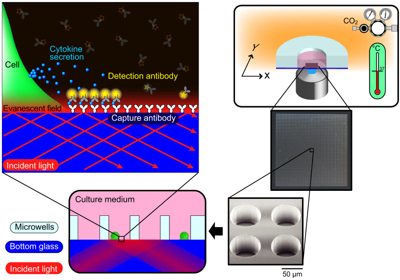
Figure 1
Concept of the real-time single cell secretion assay platform.
The schematic illustrates the concept of the platform for the real-time single cell secretion assay. The platform works with micro-fabricated well-array chip on a fully automated fluorescence microsco...
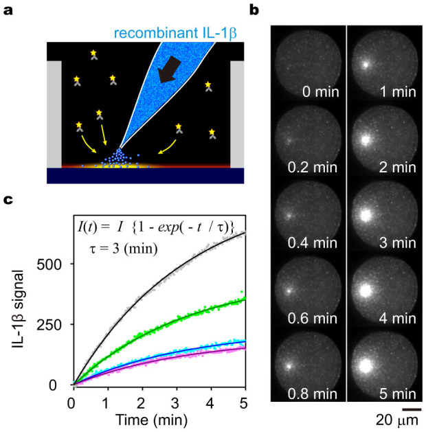
Figure 2
Performance evaluation of time-resolved FIA in the MWA on model experiments using a microinjector.
(a) Schema of the model experiment using pulsed injection of IL-1u03b2 via a microinjector. An increase in fluorescence signal was observed after pulse injecting 100u2005ng/mL of recombinant IL-1u03b2...
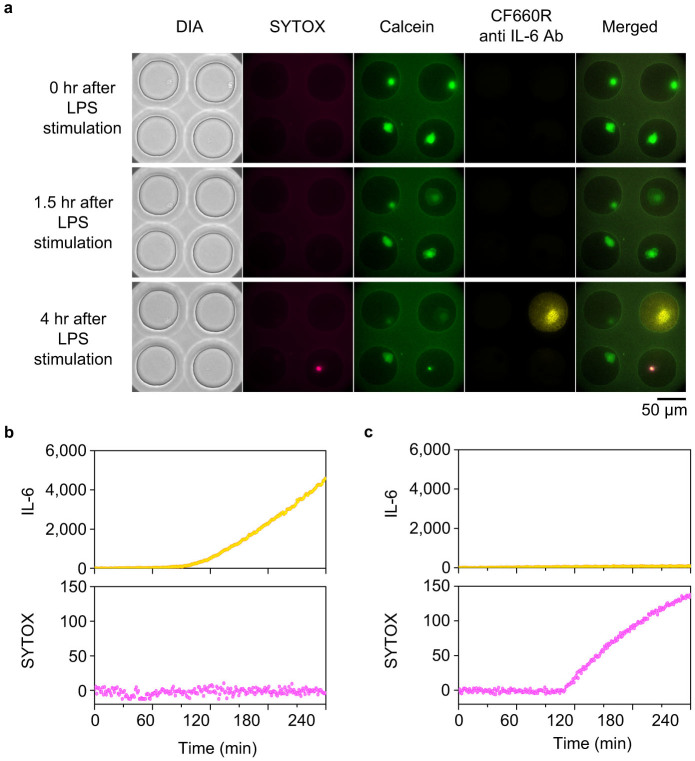
Figure 3
Time-resolved monitoring of IL-6 secretion after a classical secretion pathway.
Calcein-charged human peripheral blood monocytes were introduced into the MWA chip. Signals of SYTOX, calcein and IL-6 secretion were observed with 1-min time intervals after administration of 1u2005u...
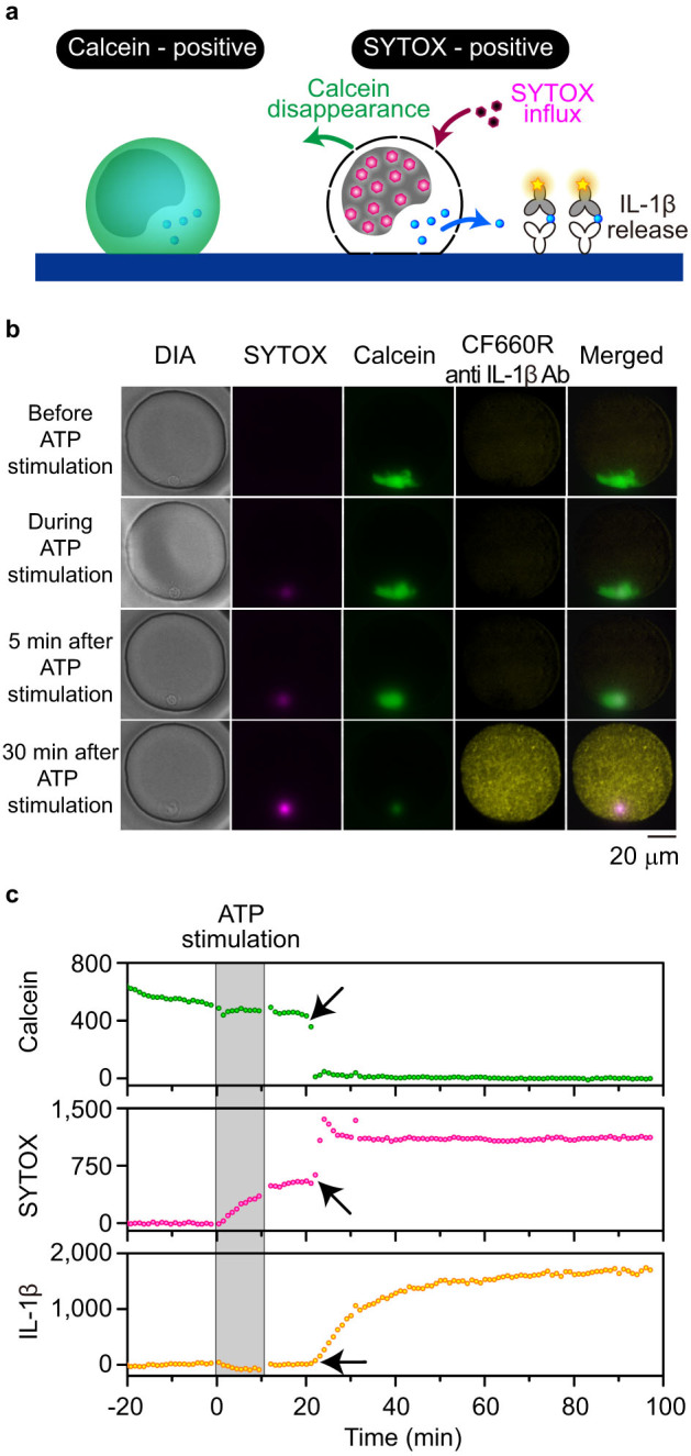
Figure 4
Time-resolved monitoring of IL-1u03b2 secretion on the PDMS MWA chip.
(a) Schematic of simultaneous monitoring of IL-1u03b2 secretion and cell membrane integrity using calcein and SYTOX staining. SYTOX influx and fluorescent calcein disappearance was observed due to com...
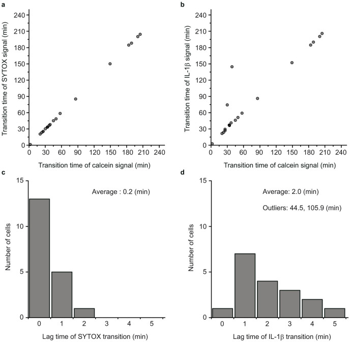
Figure 5
High concordance of the three transition times observed in individual single cells.
(a), (b) Scatter plots of the signal transition times for SYTOX or IL-1u03b2 versus calcein. Signal transition occurred virtually simultaneously between calcein, SYTOX, and IL-1u03b2. Each circle repr...
Figure images are served from the NIH/NLM PubMed Central Open Access Subset or Europe PMC; copyright remains with the publishers and authors.
💬 Discussion
0 commentsNo comments yet. Be the first to start a discussion!
Leave a Comment