Abstract
Leukocyte transendothelial migration involves the active participation of the endothelium through the formation of apical membrane protrusions that embrace adherent leukocytes, termed docking structures. Using live-cell imaging, we find that prior to transmigration, endothelial docking structures form around 80% of all neutrophils. Previously we showed that endothelial RhoG and SGEF control leukocyte transmigration. In this study, our data reveal that both full-length Trio and the first DH-PH (TrioD1) domain of Trio, which can activate Rac1 and RhoG, interact with ICAM-1 and are recruited to leukocyte adhesion sites. Moreover, upon clustering of ICAM-1, the Rho-guanine nucleotide exchange factor Trio activates Rac1, prior to activating RhoG, in a filamin-dependent manner. We further show that docking structure formation is initiated by ICAM-1 clustering into ring-like structures, which is followed by apical membrane protrusion. Interestingly, we find that Rac1 is required for ICAM-1 clustering, whereas RhoG controls membrane protrusion formation. Finally, silencing endothelial Trio expression or reducing TrioD1 activity without affecting SGEF impairs both docking structure formation and leukocyte transmigration. We conclude that Trio promotes leukocyte transendothelial migration by inducing endothelial docking structure formation in a filamin-dependent manner through the activation of Rac1 and RhoG.
🔬 Techniques
🔭 Microscopes
🧬 Organisms
💻 Software
✨ Fluorophores
🧪 Sample Preparation
🔬 Cell Lines
🏭 Microscope Brands
🧪 Reagent Suppliers
🎨 Filters
💻 Software Details
🏛️ Research Organizations (ROR)
Affiliated research institutions:
📋 Methods
Antibodies and reagents
Trio (clone D-20), Vav2 (clone H-200), ICAM-1 (clone H-108), and VE-cadherin (F8) antibodies were from Tebu-Bio (Heerhugowaard, Netherlands). ICAM-1 mAb (clone BBIG-I1/11C81) was purchased from R&D Systems (Abingdon, United Kingdom). Actin (clone AC-40) mAb antibody was purchased from Sigma-Aldrich (Zwijndrecht, Netherlands). Filamin A mAb (clone PM6/317) antibody was purchased from Serotec (Oxford, United Kingdom). Polyclonal filamin B (A301-726A) antibody was from Bethyl Laboratories (Montgomery, AL). Mouse polyclonal Trio antibody was from Abnova (Heidelberg, Germany). Polyclonal filamin C antibody was from Kinasource (Dundee, United Kingdom). Polyclonal α-actinin-4 antibody was purchased from Enzo Life Sciences BVBA (Zandhoven, Belgium). Cortactin (4F11) mAb was from Millipore (Leiden, Netherlands). c-Myc (clone 9E10) antibody was purchased from Invitrogen (Breda, Netherlands). Rac1 (clone 102) mAb antibody was purchased from BD Biosciences (Breda, Netherlands). RhoG monoclonal antibody was a kind gift of J. Meller and M. A. Schwartz (University of Virginia, Charlottesville, VA). Secondary horseradish peroxidase (HRP)-conjugated goat anti-mouse, swine anti-rabbit, and rabbit anti-goat antibodies were purchased from Dako (Heverlee, Belgium). Monoclonal GFP (JL-8) antibody, secondary goat anti-rabbit IR 680, goat anti-mouse IR 800, and donkey anti-goat IR 800 antibodies were purchased from Licor Westburg (Leusden, Netherlands). Texas Red- and Alexa Fluor 633–conjugated phalloidin were from Invitrogen (Breda, Netherlands). TNF-α was purchased from Peprotech EC (London, United Kingdom). NSC23766 was purchased from Merck (Nottingham, United Kingdom). ITX3 was from Chembridge (San Diego, CA). Cell culture and transfection HEK-293, COS-7, and HeLa were maintained in Iscove's modified Dulbecco's medium (IMDM; BioWhittaker, Verviers, Belgium) containing 10% (vol/vol) heat-inactivated fetal calf serum (Invitrogen, Breda, Netherlands), 300 μg/ml glutamine, 100 U/ml penicillin and streptomycin, at 37°C and 5% CO 2 . Cells were transiently transfected with the expression vectors indicated in each experiment according to the manufacturer's protocol with Trans IT - LT1 reagent (Mirus, Madison, WI). Primary HUVEC were purchased from Lonza (Baltimore, MD) and cultured during regular passaging following fibronectin (10 μg/ml) coating of the tissue culture flasks (TPP, Switzerland) or glass slides in EGM2-containing SingleQuots (Lonza). Endothelial cells were cultured until passage 9.
Show full methods section
Antibodies and reagents
Trio (clone D-20), Vav2 (clone H-200), ICAM-1 (clone H-108), and VE-cadherin (F8) antibodies were from Tebu-Bio (Heerhugowaard, Netherlands). ICAM-1 mAb (clone BBIG-I1/11C81) was purchased from R&D Systems (Abingdon, United Kingdom). Actin (clone AC-40) mAb antibody was purchased from Sigma-Aldrich (Zwijndrecht, Netherlands). Filamin A mAb (clone PM6/317) antibody was purchased from Serotec (Oxford, United Kingdom). Polyclonal filamin B (A301-726A) antibody was from Bethyl Laboratories (Montgomery, AL). Mouse polyclonal Trio antibody was from Abnova (Heidelberg, Germany). Polyclonal filamin C antibody was from Kinasource (Dundee, United Kingdom). Polyclonal α-actinin-4 antibody was purchased from Enzo Life Sciences BVBA (Zandhoven, Belgium). Cortactin (4F11) mAb was from Millipore (Leiden, Netherlands). c-Myc (clone 9E10) antibody was purchased from Invitrogen (Breda, Netherlands). Rac1 (clone 102) mAb antibody was purchased from BD Biosciences (Breda, Netherlands). RhoG monoclonal antibody was a kind gift of J. Meller and M. A. Schwartz (University of Virginia, Charlottesville, VA). Secondary horseradish peroxidase (HRP)-conjugated goat anti-mouse, swine anti-rabbit, and rabbit anti-goat antibodies were purchased from Dako (Heverlee, Belgium). Monoclonal GFP (JL-8) antibody, secondary goat anti-rabbit IR 680, goat anti-mouse IR 800, and donkey anti-goat IR 800 antibodies were purchased from Licor Westburg (Leusden, Netherlands). Texas Red- and Alexa Fluor 633–conjugated phalloidin were from Invitrogen (Breda, Netherlands). TNF-α was purchased from Peprotech EC (London, United Kingdom). NSC23766 was purchased from Merck (Nottingham, United Kingdom). ITX3 was from Chembridge (San Diego, CA). Cell culture and transfection HEK-293, COS-7, and HeLa were maintained in Iscove's modified Dulbecco's medium (IMDM; BioWhittaker, Verviers, Belgium) containing 10% (vol/vol) heat-inactivated fetal calf serum (Invitrogen, Breda, Netherlands), 300 μg/ml glutamine, 100 U/ml penicillin and streptomycin, at 37°C and 5% CO 2 . Cells were transiently transfected with the expression vectors indicated in each experiment according to the manufacturer's protocol with Trans IT - LT1 reagent (Mirus, Madison, WI). Primary HUVEC were purchased from Lonza (Baltimore, MD) and cultured during regular passaging following fibronectin (10 μg/ml) coating of the tissue culture flasks (TPP, Switzerland) or glass slides in EGM2-containing SingleQuots (Lonza). Endothelial cells were cultured until passage 9.
Human microvascular endothelial cells
(HMVEC) were purchased from Lonza. HCMEC/D3 human brain endothelial cells were a kind gift from P. O. Couraud, CNRS, Paris, France. Knockdown with siRNA siRNA duplexes against filamin A and B have been described previously ( Kanters et al ., 2008 ). As control siRNA, the following sequence targeting the firefly luciferase GL2 gene ( Elbashir et al ., 2001 ) was used: CGUACGCGGAAUACUUCGA. Oligos were purchased from Eurogentec (Liege, Belgium). Filamin C siRNA SMARTpool was purchased from Dharmacon (Lafayette, CO). Cells were transfected with 1 nM siRNA by means of interferin transfection reagent (Polyplus/Westburg, Leusden, Netherlands) according to the manufacturer's instructions. After 72 h, cells were assayed as described below. Note that the oligos efficiently reduced protein expression in human (HUVEC) as well as monkey (Cos7) cells. Cloning of GFP-Trio FL Trio fragment (1091–8593) was digested out of Myc-Trio FL ( McPherson et al ., 2005 ) into peGFP(C1)-TrioN with Nru I and Kpn I, resulting in peGFP(C1)-Trio FL pt1. The C-terminal part (8594–9117) was obtained by PCR amplification with Myc-Trio as template and JR43F (GAGATCGGTACCTGCACAACTGCAGGA) and JR44R (GAGATC GGTACCTCAAACTCTAGGCAGAAGC) as 5′ and 3′ primers, respectively. The PCR product was cloned as a Kpn I-digested fragment into peGFP(C1)-Trio FL pt1, resulting in peGFP(C1)-Trio FL.
Western blotting
SDS–PAGE samples were analyzed on 7.5, 10, or 15% (wt/vol) polyacrylamide gels, depending on the size of the proteins of interest, and transferred onto nitrocellulose (Whatman, Piscataway, NJ) or PVDF membrane (Bio-Rad, Hercules, CA). Following blocking in 5% (wt/vol) low-fat milk in TBST Tris-buffered saline–Tween-20 (TBST), the blots were incubated with the primary antibody for 1 h at room temperature, washed 3 times for 20 min in TBST, and subsequently incubated with HRP-coupled secondary antibodies (dilution: 1:7000) in TBST for 1 h at room temperature; this was followed by three washes with TBST for 20 min each and development of the blot by enhanced chemiluminescence (ECL) or SuperSignal West nano-ECL (ThermoScientific, Etten-Leur, The Netherlands). Alternatively, blots were incubated with IR 680 or IR 800 dye-conjugated secondary antibodies (dilution 1:5000) in TBST for 1 h at room temperature; this was followed by three washes with TBST for 20 min each. Infrared signal was detected and analyzed with the Odyssey infrared detection system (LI-COR Biosciences, Lincoln, NE). For Trio protein expression, 3–8% (wt/vol) Tris-acetate precast gels (Invitrogen) were used according to the manufacturer's instructions, and samples were transferred onto nitrocellulose membrane by blotting for 18 h at 20 mA.
Confocal laser-scanning microscopy
For immunofluorescence, cells were grown on fibronectin-coated 14-mm coverslips. After treatment, cells were washed in cold PBSA (phosphate-buffered saline [PBS], 0.5% [wt/vol] bovine serum albumin [BSA], 1 mM CaCl 2 , 0.5 mM MgCl 2 ) and fixed in 4% (vol/vol) formaldehyde in PBS (+1 mM CaCl 2 , 0.5 mM MgCl 2 ) for 10 min. After fixation, cells were permeabilized in PBS-T (PBS + 1% Triton X-100 and 10% glycerol) for 4 min; this was followed by a blocking step in PBS supplemented with 2% (wt/vol) BSA. Cells were incubated with primary and secondary antibodies, and after each step were washed three times in PBSA. Coverslips were mounted with Vectashield with 4′,6-diamidino-2-phenylindole (Vector Laboratories, Peterborough, UK) on microscope slides. For live-cell imaging, cells were seeded on 30-mm coverslips and transfected or transduced, as indicated. After 24–72 h, cells were placed in a heating chamber at 37°C and 5% CO 2 and imaged with a confocal microscope (LSM510 META; Carl Zeiss MicroImaging, Jena, Germany). Adenovirus production The ICAM-1-GFP fragment was obtained by PCR amplification with peICAM-1-GFP ( Barreiro et al ., 2002 ) as template and JR11F (GAGATCGTCGACATG GCTCCCAGCAGCCC) and JR18R (GAGATCGCGGCCGCTTTACTTGTAC) as 5′ and 3′ primers, respectively. The PCR product was cloned as a Sal I/ Not I fragment into pENTR1A (Invitrogen). GFP-TrioD1 was obtained by PCR amplification with peGFP-TrioD1 as template and primer pairs JR22F (GAGATCGTCGACCTG GTTTAGTGAACCGTCAGA) and JR4R (GAGATCGAATTCCTAGGCGTCATTG CTGGAGAC). The PCR product was cloned as a Sal I/ Eco RI fragment into pENTR1A (Invitrogen). pENTR1A-constructs were recombined into the pAd/CMV/V5-DEST vector (Invitrogen) with Clonase II enzyme mix according to the manufacturer's instructions (Invitrogen). Adenovirus expressing ICAM-1-GFP or GFP-TrioD1 was produced by transfecting Pac I-digested (Westburg, Leusden) constructs into HEK293T cells. shRNA lentivirus production shRNA constructs (Sigma Mission library) targeting Trio (TRC_10561), Rac1 (TRC_4873), and RhoG (TRC_48019), or nontargeting shCtrl construct were packaged into lentivirus in HEK293T cells with third-generation lentiviral packaging plasmids ( Dull et al ., 1998 ). Lentivirus-containing supernatant was harvested on days 2 and 3 after transfection. Lentivirus was concentrated by centrifugation at 20,000 × g for 2 h. Fusion proteins The fusion proteins GST-ELMO, GST-Rac1 G15A, and GST-RhoG G15A were purified from BL21 Escherichia coli cells (Agilent Technologies, Amstelveen, Netherlands) with glutathione–Sepharose 4B, as previously described ( Ellerbroek et al ., 2004 ). GST-fusion proteins were stored in 30% (vol/vol) glycerol at −80°C.
Pulldown assay
A synthetic, biotinylated peptide encoding the intracellular domains of human ICAM-1 or VCAM-1, or empty beads, was used in pulldown assays, as previously described ( Kanters et al ., 2008 ). In short, a confluent monolayer of cells in a 100-mm Petri dish was washed with cold PBS (+ 1 mM CaCl 2 ; 0.5 mM MgCl 2 ) and lysed in cold NP-40 lysis buffer (50 mM Tris-HCl, pH 7.4, 100 mM NaCl, 10 mM MgCl 2 , 10% [vol/vol] glycerol, and 1% [vol/vol] Nonidet P40) supplemented with protease inhibitors (complete mini EDTA, Roche, Almere, The Netherlands) for 5 min. After lysis, cell debris was removed by centrifugation (10,000 × g , 5 min at 4°C). Supernatant was incubated with 30 μg of biotinylated peptide comprising the intracellular tail of ICAM-1 or indicated controls and 30 μl (concentration 2.4 mg/ml packed gel) of streptavidin agarose (Sigma-Aldrich, Zwijndrecht, Netherlands) for 2–3 h under constant head-over-head rotation at 4°C. Beads were subsequently washed five times with NP40 lysis buffer and resuspended in SDS–PAGE sample buffer.
RhoG and Rac1 activation assay
A confluent monolayer of HUVEC in a 100-mm Petri dish was washed with cold PBS (+ 1 mM CaCl 2 ; 0.5 mM MgCl 2 ) and lysed in 50 mM Tris, pH 7.4, 0.5 mM MgCl 2 , 500 mM NaCl, 1% (vol/vol) Triton X-100, 0.5% (wt/vol) deoxycholic acid, and 0.1% (wt/vol) SDS supplemented with protease inhibitors. Lysates were cleared at 14,000 × g for 5 min. GTP-bound RhoG was isolated by rotating supernatants for 30 min with 60–90 μg of GST-ELMO (GST-fusion protein containing the full-length RhoG effector ELMO) conjugated to glutathione–Sepharose beads (GE Healthcare, Zeist, Netherlands; van Buul et al. , 2007 ; Wittchen and Burridge, 2008 ). GTP-bound Rac1 was isolated with biotinylated Pak1-Crib peptide coupled to streptavidin agarose ( ten Klooster et al ., 2006 ). Beads were washed four times in 50 mM Tris, pH 7.4, 0.5 mM MgCl 2 , 150 mM NaCl, 1% (vol/vol) Triton X-100, and protease inhibitors. Pulldowns and lysates were immunoblotted with monoclonal RhoG and Rac1 antibodies. Antibody-coated beads Polystyrene beads (3-μm beads for biochemical assays and 3- or 10-μm beads for immunofluorescence; Polysciences, Eppelheim, Germany) were pretreated with 8% (vol/vol) glutaraldehyde overnight, washed five times with PBS, and incubated with 300 μg/ml ICAM-1 mAb according to the manufacturer's protocol. Free glutaraldehyde groups were blocked with 0.5 M ethanolamine and 2 mg/ml BSA. Electric cell-substrate impedance sensing Monolayer permeability was determined by measuring the electrical resistance with electric cell–substrate impedance sensing (ECIS). Electrode arrays (10WE; Applied BioPhysics) were precoated with 10 mM l -cysteine (Sigma-Aldrich, Steinheim, Germany) for 15 min at 37°C, after which they were washed twice with 0.9% (wt/vol) NaCl and subsequently coated with fibronectin (Sigma-Aldrich) in 0.9% (wt/vol) NaCl for 1 h at 37°C. A measure of 1 × 10 5 cells per well (0.8 cm 2 ) were seeded, grown to confluency, and treated with TNF-α for 20 h. Electrical resistance was continuously measured at 37°C and 5% CO 2 with ECIS-Model-9600 Controller (Applied BioPhysics, Troy, NY).
Neutrophil isolation
Polymorphonuclear neutrophils were isolated from whole blood derived from healthy donors by means of a Ficoll-Paque plus (Pharmacia Biotech, Uppsala, Sweden) density gradient. After erythrocyte lysis in a ice-cold isotonic lysis buffer (155 mM NH 4 Cl, 10 mM KHCO 3 , 0.1 mM EDTA pH 7.4), neutrophils were washed once with lysis buffer, once with 10% (vol/vol) trisodium citrate/PBS, resuspended in HEPES medium (20 mM HEPES, 132 mM NaCl, 6 mM KCl, 1 mM CaCl 2 , 1 mM MgSO 4 , 1.2 mM K 2 HPO 4 , 5 mM glucose [all from Sigma-Aldrich], and 0.4% [wt/vol] human serum albumin [Sanquin Reagents], pH 7.4), and kept at room temperature for not longer than 4 h until use. Neutrophil TEM across Transwell Transmigration of neutrophils over endothelial monolayers was analyzed with Falcon FluoroBlok Transwell inserts with 3.0-μm pore size (BD Biosciences, Breda, Netherlands). At 24 h after transduction with shCtrl or shTrio-producing lentivirus, 2 × 10 5 HUVEC per well were seeded on fibronectin-coated filters and grown for another 48 h. At 20 h prior to the experiment, HUVEC were stimulated with 10 ng/ml TNF-α. Freshly isolated, CellTrace calcein green–labeled (Invitrogen) neutrophils were added to the upper compartment, and cells were allowed to migrate toward 10 nM formyl-Met-Leu-Phe (fMLP) in the lower compartment for 30 min. During this time, fluorescence was measured with a Tecan GENios Plus microplate reader with a 530/30 band-pass filter. The percentage of TEM was calculated as (fluorescence of calcein-labeled neutrophils that transmigrated across a Transwell filter coated with HUVEC)/(fluorescence of calcein-labeled neutrophils input) × 100. Neutrophil TEM under flow HUVEC were cultured in a fibronectin-coated Ibidi μ-slide VI 0.4 (Ibidi, München, Germany) for 2–3 d until confluency and were stimulated overnight with TNF-α (10 ng/ml). Freshly isolated neutrophils were resuspended at 0.4 × 10 6 cells/ml in HEPES medium and were incubated for 30 min at 37ºC. Neutrophils were perfused over HUVEC monolayers at 0.5 ml/min (shear stress of 1.0 dyne/cm 2 ). After 3 min, the neutrophil-containing HEPES solution was replaced by HEPES medium for 30 min. During this time leukocyte–endothelial interactions were recorded in six random fields with a Zeiss Axiovert 200 microscope (10× objective) equipped with a motorized stage. Images were recorded with Zeiss Axiovision 4.7 software. Live imaging was performed at 37ºC and 5% CO 2 . Transmigrated neutrophils were distinguished from those adhering to the apical surface of the endothelium by their transition from bright to phase-dark morphology. HUVEC that were pretreated with ITX3 were carefully washed before being exposed to flow. Owing to the washing and flow conditions, no ITX3 was present in the medium at times of neutrophil TEM. ITX3 was incubated overnight but was also active after only 1 h of incubation on HUVEC. In addition, ITX3 did not need to be present in the medium to inhibit Trio activity, as wash-out experiments showed that ITX3 was able to inhibit Trio activity for at least 60 min after wash-out.
Statistical analysis
Statistical comparisons between experimental groups were performed by the Student's t test. A two-sided p value of ≤0.05 was considered significant. Unless otherwise stated, a representative experiment out of at least three independent experiments is shown.
📊 Figures
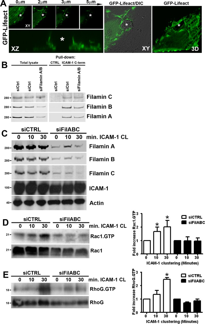
FIGURE 1:
Rac1 and RhoG activation upon ICAM-1 clustering requires filamin. (A) Formation of F-actinu2013positive docking structures around transmigrating neutrophils was visualized by GFP-LifeAct in TNF-u03b1u...
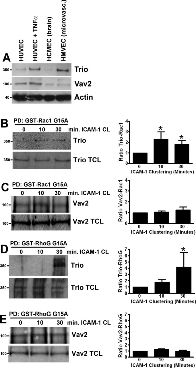
FIGURE 2:
ICAM-1 clustering induces activation of the GEF Trio. (A) Trio (350 kDa) and Vav2 (100 kDa) protein expression in HUVEC (HUVEC, lane 1; HUVEC treated for 20 h with TNF-u03b1, lane 2), human brain endo...
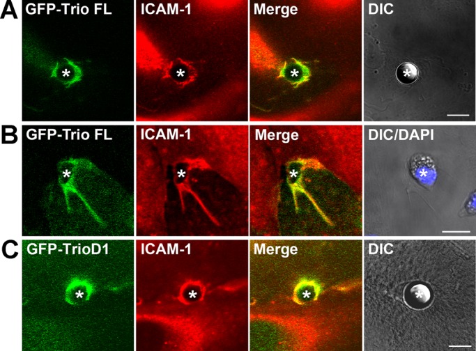
FIGURE 3:
Trio and TrioD1 colocalize with ICAM-1 upon clustering. Endogenous ICAM-1 (red) in TNF-u03b1u2013stimulated endothelial cells colocalizes with GFP-Trio full length to 10-u03bcm u03b1-ICAM-1u2013antibo...
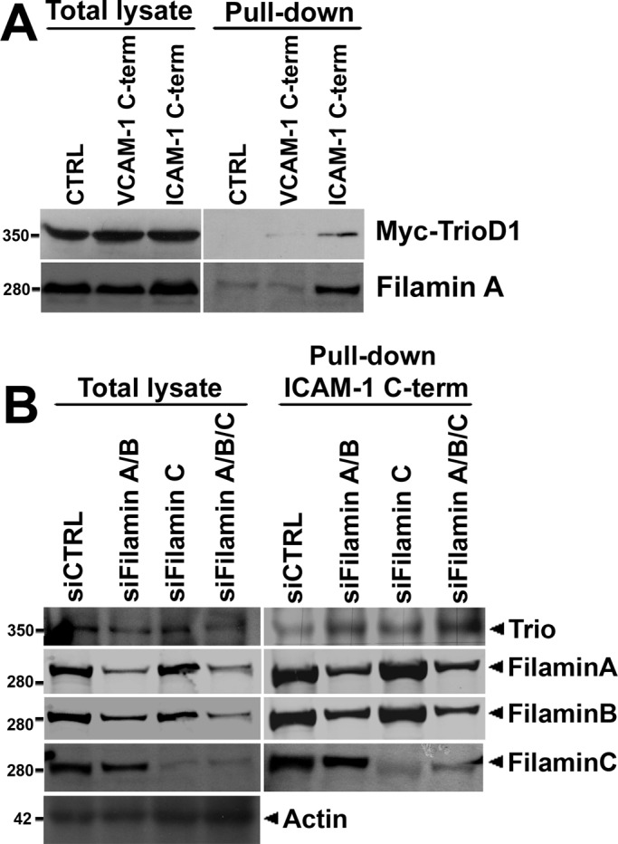
FIGURE 4:
Trio interacts with ICAM-1 independently of filamin. (A) Cos7 cells were transiently transfected with Myc-TrioD1 (B), and pulldown assays were performed with the ICAM-1 C-terminal peptide, streptavidi...
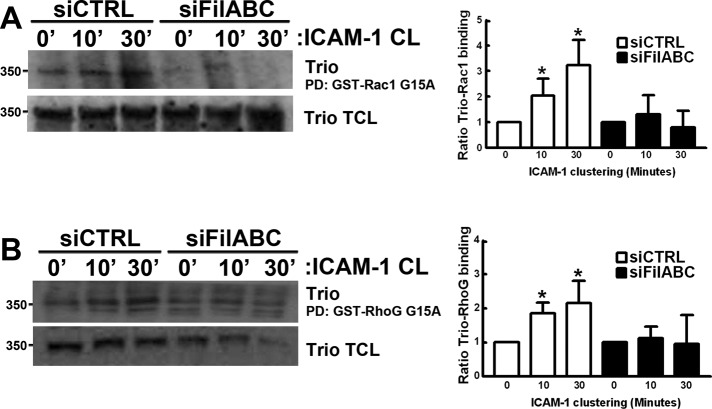
FIGURE 5:
Trio activation upon ICAM-1 clustering requires filamin. ICAM-1 was clustered (CL) for 10 and 30 min on TNF-u03b1u2013stimulated HUVEC transfected with siRNA targeting filamin A/B/C. Trio activation w...
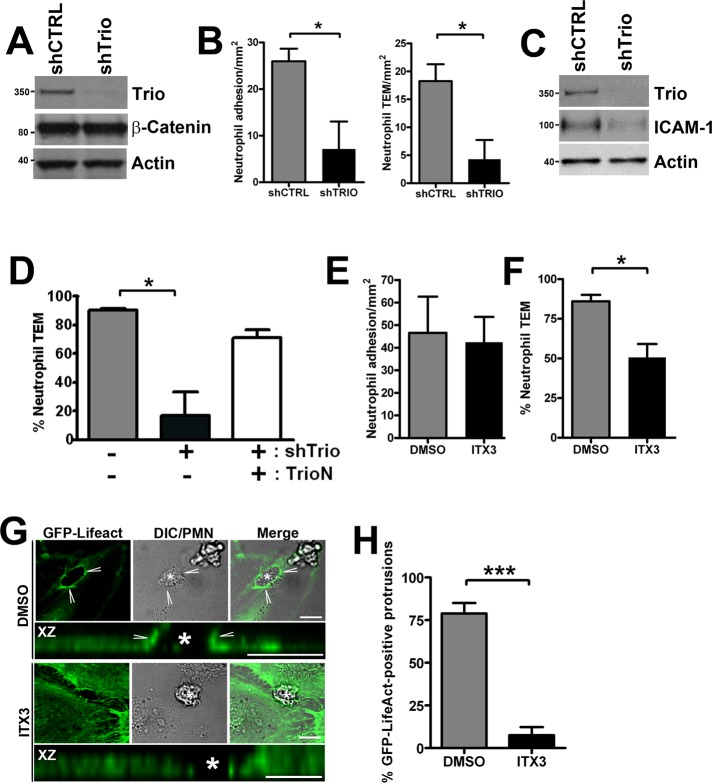
FIGURE 6:
Trio knockdown or inhibition reduces neutrophil adhesion and TEM. HUVEC were transduced with shTrio-expressing lentivirus to silence Trio expression. (A) Knockdown of Trio expression was confirmed by ...
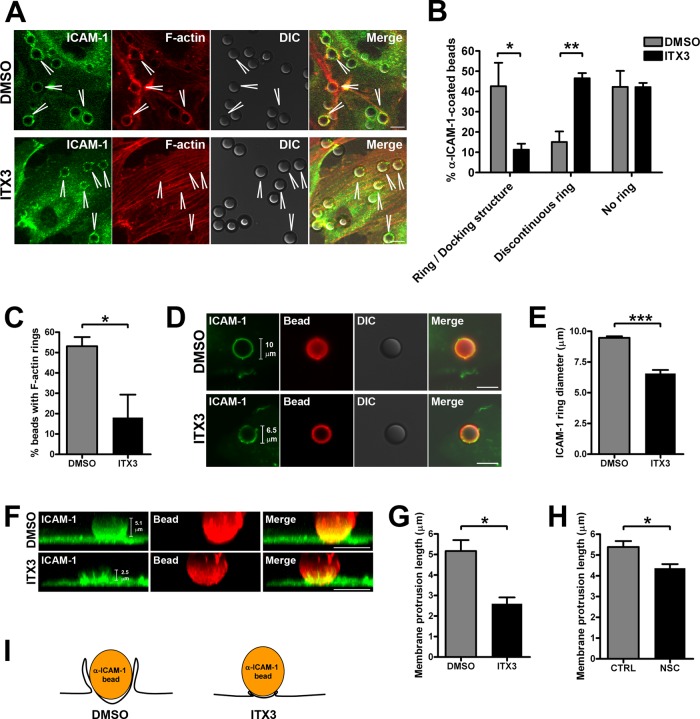
FIGURE 7:
Trio inhibition with ITX3 impairs endothelial docking structure formation. (A) ICAM-1 was clustered with 10-u03bcm u03b1-ICAM-1u2013antibody beads on TNF-u03b1u2013stimulated HUVEC were treated with 1...
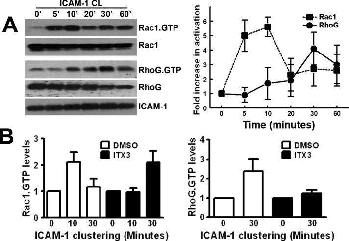
FIGURE 8:
Sequential Rac1 and RhoG activation upon ICAM-1 clustering. (A) ICAM-1 was clustered (CL) with u03b1-ICAM-1u2013antibody beads for indicated time points, and Rac1-GTP and RhoG-GTP were precipitated as...
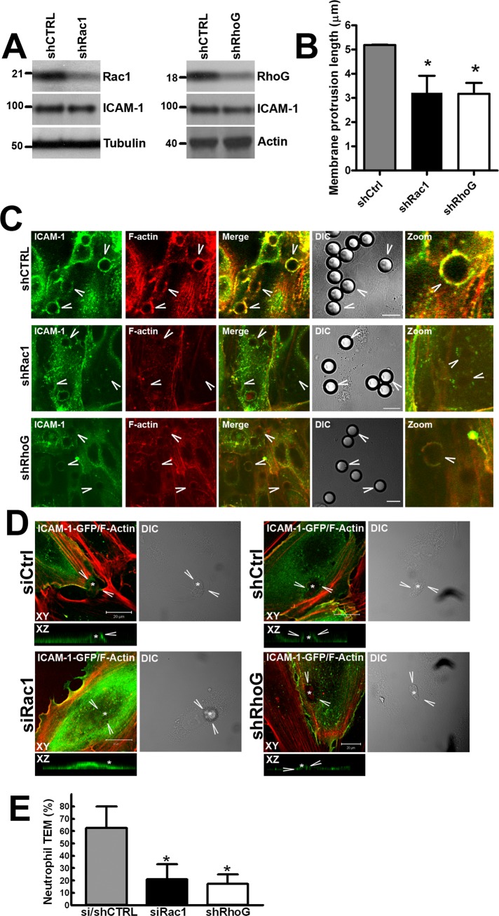
FIGURE 9:
Trio mediates endothelial docking structure formation through Rac1 and RhoG. (A) To silence Rac1 and RhoG expression, we used lentivirally delivered constructs targeting Rac1 and RhoG mRNA in TNF-u03b...
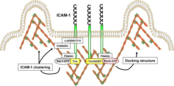
FIGURE 10:
Model for ICAM-1u2013induced endothelial docking structure formation. Clustering of ICAM-1 induces the recruitment of the F-actin cross-linker protein filamin. Filamin functions as a scaffold for subs...
Figure images are served from the NIH/NLM PubMed Central Open Access Subset or Europe PMC; copyright remains with the publishers and authors.
💬 Discussion
0 commentsNo comments yet. Be the first to start a discussion!
Leave a Comment