Abstract
Chromosome segregation requires coordinated separation of sister chromatids following biorientation of all chromosomes on the mitotic spindle. Chromatid separation at the metaphase-to-anaphase transition is accomplished by cleavage of the cohesin complex that holds chromatids together. Here we show using live-cell imaging that extending the metaphase bioriented state using five independent perturbations (expression of non-degradable Cyclin B, expression of a Spindly point mutant that prevents spindle checkpoint silencing, depletion of the anaphase inducer Cdc20, treatment with a proteasome inhibitor, or treatment with an inhibitor of the mitotic kinesin CENP-E) leads to eventual scattering of chromosomes on the spindle. This scattering phenotype is characterized by uncoordinated loss of cohesion between some, but not all sister chromatids and subsequent spindle defects that include centriole separation. Cells with scattered chromosomes persist long-term in a mitotic state and eventually die or exit. Partial cohesion loss-associated scattering is observed in both transformed cells and in karyotypically normal human cells, albeit at lower penetrance. Suppressing microtubule dynamics reduces scattering, suggesting that cohesion at centromeres is unable to resist dynamic microtubule-dependent pulling forces on the kinetochores. Consistent with this view, strengthening cohesion by inhibiting the two pathways responsible for its removal significantly inhibits scattering. These results establish that chromosome scattering due to uncoordinated partial loss of chromatid cohesion is a common outcome following extended arrest with bioriented chromosomes in human cells. These findings have important implications for analysis of mitotic phenotypes in human cells and for development of anti-mitotic chemotherapeutic approaches in the treatment of cancer.
🔬 Techniques
💻 Software
✨ Fluorophores
🧪 Sample Preparation
🔬 Cell Lines
🏭 Microscope Brands
🧪 Reagent Suppliers
📷 Detectors
💻 Software Details
🏛️ Research Organizations (ROR)
Affiliated research institutions:
📋 Methods
Cell lines and drugs
Stable isogenic HeLa Flp-In T-Rex (HeLa FRT) cell lines expressing the nondegradable Cyclin B1 (lacking the N-terminal 86 amino acids) and the Spindly point mutant, both stably expressing H2b:mRFP, were previously generated [16] , [40] . Stable expression of H2b:mRFP, and mRFP:H2b and YFP:α-tubulin, in hTERT-RPE1 and HeLa cells respectively, was done by retroviral delivery as previously described [41] . hTERT-RPE1 and HeLa cells were obtained from the ATCC (American Type Culture Collection, www.atcc.org ). The CENP-E inhibitor used was a close analog of GSK923295 [19] , [20] and was a gift from D. Cleveland and A. Shiau. For all experiments it was used at a final concentration of 1 µM. Reversine [23] was used at a final concentration of 0.5 µM. MG132 (Sigma) was used at a final concentration of 20 µM.
Cell culture and RNAi
Cells were maintained at 37°C in a 5% CO 2 atmosphere. HeLa cells were maintained in Dulbecco's modified Eagle's medium (Gibco), hTERT-RPE1 cells were maintained in DMEM/F12+Glutamax (Gibco) and both were supplemented with 10% tetracycline-free fetal bovine serum (Clontech), 100 U/mL penicillin and 100 U/mL streptomycin. For immunofluorescence, cells were seeded on 12-mm poly-L-lysine coated coverslips in 12-well plates 24 h prior to treatment. For live-cell imaging experiments, cells were seeded in a 35-mm glass-bottom dish coated with poly-D-lysine (MatTek). Cells were transfected using Oligofectamine and reduced-serum Opti-MEM (Invitrogen) according to manufacturer's instructions. A predesigned (Dharmacon/Thermo Scientific) siRNA for Spindly ( GAAAGGGUCUCAAACUGAA ), ESPL1/separase ( GCUUGUGAUGCCAUCCUGAUU ) or a nontargeting control ( UGGUUUACAUGUCGACUAA ) was used at a final concentration of 100 nM. ON-TARGETplus SMARTpool siRNA (Dharmacon/Thermo Scientific) was used for knockdown of Cdc20 (L-003225-00) and Wapl (L-026287-01) at a final concentration of 100 nM. After incubation for 5–6 hrs, 1 vol of medium and fetal bovine serum (10% final) was added. After 24 hrs, the transfection mixture was replaced with fresh medium. For live imaging of HeLa Flp-In T-Rex cells, transgene expression was induced with tetracycline (0.2 mg/mL) 24 hrs after transfection, and the filming session was initiated 8 hrs later.
Show full methods section
Cell lines and drugs
Stable isogenic HeLa Flp-In T-Rex (HeLa FRT) cell lines expressing the nondegradable Cyclin B1 (lacking the N-terminal 86 amino acids) and the Spindly point mutant, both stably expressing H2b:mRFP, were previously generated [16] , [40] . Stable expression of H2b:mRFP, and mRFP:H2b and YFP:α-tubulin, in hTERT-RPE1 and HeLa cells respectively, was done by retroviral delivery as previously described [41] . hTERT-RPE1 and HeLa cells were obtained from the ATCC (American Type Culture Collection, www.atcc.org ). The CENP-E inhibitor used was a close analog of GSK923295 [19] , [20] and was a gift from D. Cleveland and A. Shiau. For all experiments it was used at a final concentration of 1 µM. Reversine [23] was used at a final concentration of 0.5 µM. MG132 (Sigma) was used at a final concentration of 20 µM.
Cell culture and RNAi
Cells were maintained at 37°C in a 5% CO 2 atmosphere. HeLa cells were maintained in Dulbecco's modified Eagle's medium (Gibco), hTERT-RPE1 cells were maintained in DMEM/F12+Glutamax (Gibco) and both were supplemented with 10% tetracycline-free fetal bovine serum (Clontech), 100 U/mL penicillin and 100 U/mL streptomycin. For immunofluorescence, cells were seeded on 12-mm poly-L-lysine coated coverslips in 12-well plates 24 h prior to treatment. For live-cell imaging experiments, cells were seeded in a 35-mm glass-bottom dish coated with poly-D-lysine (MatTek). Cells were transfected using Oligofectamine and reduced-serum Opti-MEM (Invitrogen) according to manufacturer's instructions. A predesigned (Dharmacon/Thermo Scientific) siRNA for Spindly ( GAAAGGGUCUCAAACUGAA ), ESPL1/separase ( GCUUGUGAUGCCAUCCUGAUU ) or a nontargeting control ( UGGUUUACAUGUCGACUAA ) was used at a final concentration of 100 nM. ON-TARGETplus SMARTpool siRNA (Dharmacon/Thermo Scientific) was used for knockdown of Cdc20 (L-003225-00) and Wapl (L-026287-01) at a final concentration of 100 nM. After incubation for 5–6 hrs, 1 vol of medium and fetal bovine serum (10% final) was added. After 24 hrs, the transfection mixture was replaced with fresh medium. For live imaging of HeLa Flp-In T-Rex cells, transgene expression was induced with tetracycline (0.2 mg/mL) 24 hrs after transfection, and the filming session was initiated 8 hrs later.
Live-cell imaging
For live-cell imaging, medium was replaced with CO 2 -independent medium (Gibco) supplemented with 10% tetracycline-free fetal bovine serum, 100 U/mL penicillin and 100 U/mL streptomycin. Tetracycline (0.2 mg/mL) was added to maintain transgene expression, and the medium was covered with mineral oil immediately before filming. Time-lapse images were recorded (every 4 minutes for the 10 hr filming, every 12 minutes for the 48 hr filming and every 30 sec for the 3 hr imaging) on a Deltavision microscope (Applied Precision) equipped with an environmental chamber heated to 37°C. Images were acquired with a CoolSnap charge-coupled device camera (Roper Scientific) and a 40× NA 1.35 U-planApo objective (Olympus) at 2×2 binning. Images were viewed and analyzed with MetaMorph software (Molecular Dynamics).
Indirect Immunofluorescence
Cells were washed once with PBS and fixed with 4% paraformaldehyde in PBS for 10–15 min at room temperature, then permeabilized for 3–5 min with 0.1% Triton X-100 in PBS. Primary antibodies and dilutions used: ACA 1∶500 (Antibodies Incorporated), α-tubulin 1∶500 (DM1α – Sigma), Sas4 1∶500 (A. Dammermann, K. Oegema Lab), Mad1 1∶40 (a gift from A. Musacchio), Cyclin B 1∶100 (Santa Cruz) and securin 1∶100 (Abcam). Images were recorded on a Deltavision microscope at 1×1 binning with a 100× NA 1.3 U-planApo objective. Z-stacks (0.2-µm sections) were deconvolved using softWorRx (Applied Precision) and maximum intensity projections were imported into Adobe Photoshop CS4 (Adobe) for further processing. Determination of interkinetochore stretched was done as previously described [16] .
Supporting Information Figure S1 Cells with scattered chromosomes maintain the hallmarks of mitosis. ( A, B, C ) Immunofluorescence images of control or scattered cells for (A and C) securin and (B) Cyclin B. For (A and B) the arrowhead indicates cells in metaphase, the arrow indicates cells in anaphase. In (C), the arrowhead indicates a scattered cell, the arrow indicates a cell that has entered an anaphase-like state. Scale bar, 5 µm. (TIF) Click here for additional data file. Figure S2 Immunoblot of protein inhibition via RNAi. Whole cell lysates were probed for either separase or Wapl for the various conditions. (TIF) Click here for additional data file. Movie S1 Chromosome scattering results from perturbations that arrest cells in a metaphase-like state. Movies start at NEBD. Time lapse is 4 min and playback speed is 1440×real time. (MOV) Click here for additional data file. Movie S2 Chromosome scattering results from perturbations that arrest cells in a metaphase-like state. Movies start at NEBD. Time lapse is 4 min and playback speed is 1440×real time. (MOV) Click here for additional data file. Movie S3 Uncoordinated loss of cohesion and spindle defects are hallmarks of chromosome scattering. Movie starts just prior to the onset of scattering with a fully aligned metaphase plate. Time lapse is 30 seconds and playback speed is 180×real time. (MOV) Click here for additional data file.
📊 Figures
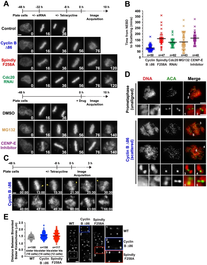
Figure 1
Chromosome scattering results from a prolonged metaphase arrest and is characterized by uncoordinated loss of chromatid cohesion.
( A ) Selected images from time-lapse imaging sequences of cells expressing histone H2b:mRFP. Time (in mins) is indicated on the lower right of each panel; time 0 indicates NEBD. The scheme for each s...
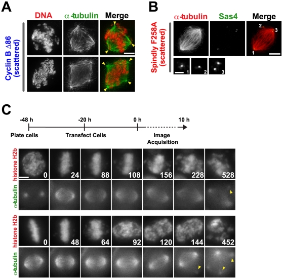
Figure 2
Chromosome scattering results in spindle defects that are secondary to loss of cohesion.
( A, B ) Immunofluorescence images of scattered cells stained with DNA and (A) u03b1-tubulin [arrowheads denote all spindle poles] and (B) centriole marker Sas4. Scale bar, 5 u00b5m; magnified image 1...
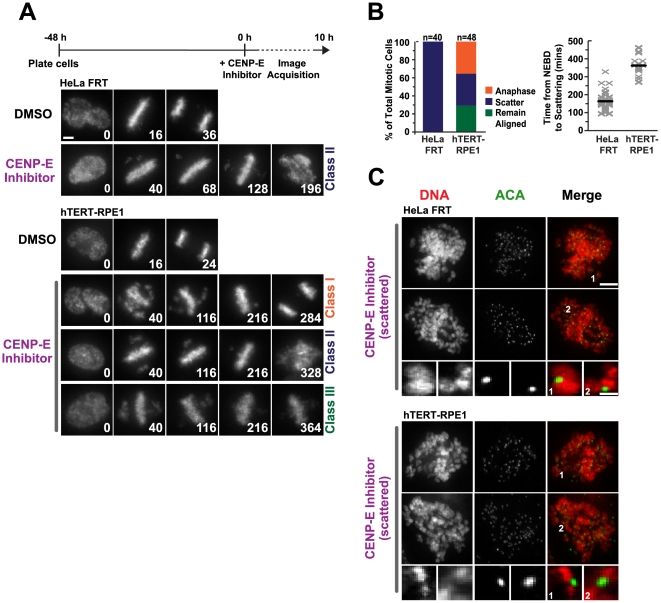
Figure 3
Chromosome scattering occurs in karotypically normal cells.
( A ) Selected images from time-lapse imaging sequences of cells expressing histone H2b:mRFP for HeLa FRT and hTERT-RPE1 cells treated with a CENP-E inhibitor. Time (in mins) is indicated on the lower...
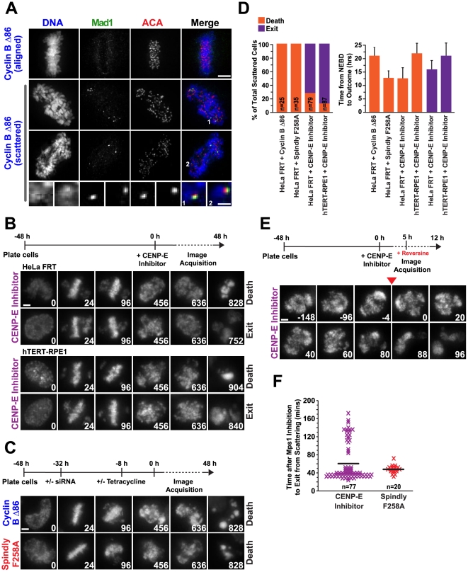
Figure 4
The spindle assembly checkpoint is reactivated upon loss of cohesion and is required to maintain the scattered state.
( A ) Immunofluorescence images of metaphase-aligned and scattered cells expressing Cyclin B u039486 stained for DNA, Mad1 and ACA. No Mad1 is seen on fully aligned chromosomes, whereas Mad1 relocaliz...
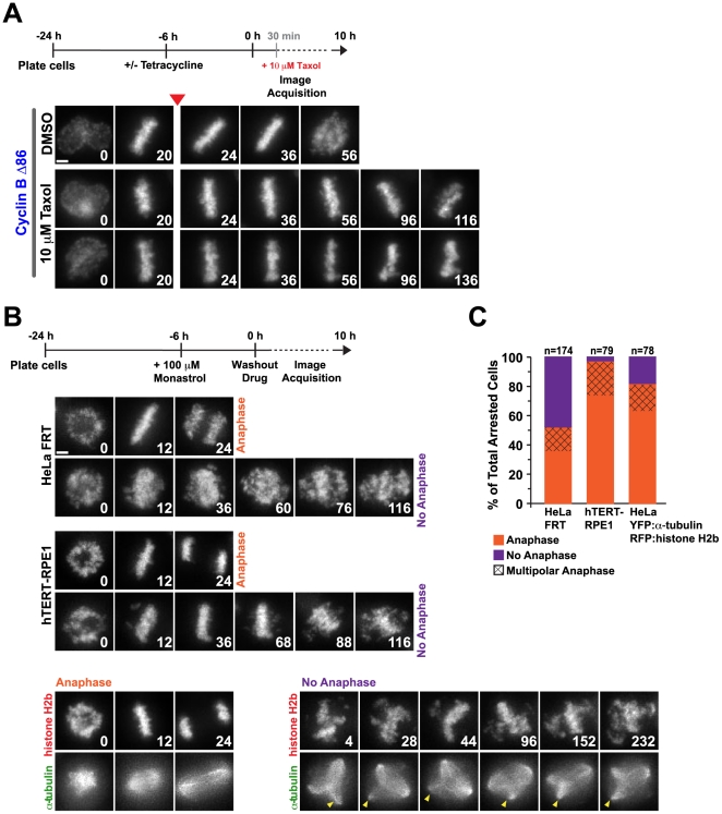
Figure 5
Biorientation with dynamic microtubule-dependent tension at kinetochores contributes to the onset of chromosome scattering.
( A ) Selected images from time-lapse imaging sequences of Cyclin B u039486 HeLa FRT cells expressing histone H2b:mRFP. The experimental scheme is depicted above the images. Taxol was added to an asyn...
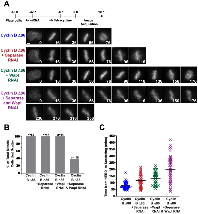
Figure 6
Chromosome scattering is prevented by inhibition of the two pathways that remove cohesion.
( A ) Selected images from time-lapse imaging sequences of Cyclin B u039486 HeLa FRT cells expressing histone H2b:mRFP and subjected to the indicated perturbations. The experimental scheme is depicted...
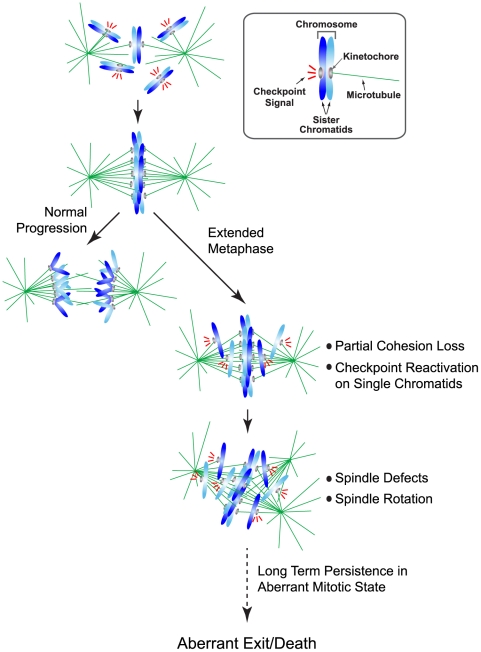
Figure 7
Model describing the chromosome scattering phenotype.
In an unperturbed mitosis, sister chromatids align along the metaphase plate and lose their cohesion in a coordinated manner such that all sisters are separated simultaneously. In contrast, during a p...
Figure images are served from the NIH/NLM PubMed Central Open Access Subset or Europe PMC; copyright remains with the publishers and authors.
💬 Discussion
0 commentsNo comments yet. Be the first to start a discussion!
Leave a Comment