Abstract
Histone variant H3.3 is deposited in chromatin at active sites, telomeres, and pericentric heterochromatin by distinct chaperones, but the mechanisms of regulation and coordination of chaperone-mediated H3.3 loading remain largely unknown. We show here that the chromatin-associated oncoprotein DEK regulates differential HIRA- and DAAX/ATRX-dependent distribution of H3.3 on chromosomes in somatic cells and embryonic stem cells. Live cell imaging studies show that nonnucleosomal H3.3 normally destined to PML nuclear bodies is re-routed to chromatin after depletion of DEK. This results in HIRA-dependent widespread chromatin deposition of H3.3 and H3.3 incorporation in the foci of heterochromatin in a process requiring the DAXX/ATRX complex. In embryonic stem cells, loss of DEK leads to displacement of PML bodies and ATRX from telomeres, redistribution of H3.3 from telomeres to chromosome arms and pericentric heterochromatin, induction of a fragile telomere phenotype, and telomere dysfunction. Our results indicate that DEK is required for proper loading of ATRX and H3.3 on telomeres and for telomeric chromatin architecture. We propose that DEK acts as a "gatekeeper" of chromatin, controlling chromatin integrity by restricting broad access to H3.3 by dedicated chaperones. Our results also suggest that telomere stability relies on mechanisms ensuring proper histone supply and routing.
🔬 Techniques
🧬 Organisms
✨ Fluorophores
🧪 Sample Preparation
🔬 Cell Lines
🏭 Microscope Brands
🧪 Reagent Suppliers
🔎 Objectives
💻 Software Details
🏛️ Research Organizations (ROR)
Affiliated research institutions:
📋 Methods
Cells
MSCs were cultured in DMEM/F12 medium containing GlutaMAX (Gibco) and 20% fetal calf serum ( Delbarre et al. 2013 ).
Mouse ESCs strain
ES129.1 were cultured in DMEM/15% fetal calf serum, essential amino acids, 10 3 U/mL of leukemia-inhibiting factor, and 0.1 mM β-mercaptoethanol. ESCs were induced to differentiate by removal of leukemia inhibitory factor and addition of retinoic acid ( Chang et al. 2013 ).
Transfection of plasmids and siRNAs
Transfection of mouse
ESCs with siRNAs was done using Lipofectamine 2000 (Invitrogen). Mouse Dek siRNAs (DEK ON-target Plus siRNA) were from Thermo Scientific.
Mouse Hira siRNAs
(Mss205135) were from Life Technologies. Transfection of MSCs was done by nucleofection ( Delbarre et al. 2013 ). For expression of proteins in siRNA-treated cells, a first siRNA transfection was followed 96 h later by a second transfection with both siRNA and the relevant plasmid. Plasmids encoding H3.3-EGFP, H3.3-mCherry, mCherry-PML, and mCherry-DAXX were as described ( Delbarre et al. 2013 ). The EGFP-PML plasmid was a gift from Dr. Harutaka Katano. The EGFP-DEK plasmid was a gift from Dr. Ferdinand Kappes ( Kappes et al. 2001 ). siRNAs to human genes were DEK : CCUUCUGGCAAACCAUUGCCGAAAU; HIRA : GAGCAGAACCUUGUGAAAGAGCUGA; DAXX : GAUCAUCGUGCUCUCAGACUCUGAU; ATRX : GAUAUUGCAGAGAAAUUCCUAAAGA; PML : GGAAGGUCAUCAAGAUGGAGU; Control siRNA: UUCUCCGAACGUGUCACGUTT.
Antibodies
Antibodies to TERF1 were as described ( Iwano et al. 2004 ), antibodies to PML (5E10) were a gift from Dr. Roel van Driel, human autoimmune serum CREST6 was from the Murdoch Children’s Research Institute (Melbourne), and antibodies to CBX3 were from Dr. Brigitte Buendia. The following antibodies were from one or several sources: DAXX (sc-7152, sc-7001), ATRX (sc-15408), DEK (sc-136222): Santa Cruz Biotechnology; DEK (16448-AP): BioSite; TERF2 (05-521), H3K9me3 (05-1250), HIRA (04-1488), H3.3 (09-838): Millipore; H3.3S31p (39367): Active Motif; H3K9me3 (pAb-056-050): Diagenode; CENPA (2186): Cell Signaling Technologies; PML (ab53773, ab72137, ab50637), H3 (ab1791), H4 (ab10158): Abcam; alpha-tubulin (T5168), FLAG (F1804): Sigma; gamma-H2AX (H2AXS139p) (05-636): Millipore; GFP (11814460001): Roche; Alexa Fluor 488 anti-goat (A-11055): Invitrogen; DyLight 549 anti-rabbit (711-505-152). Cy3-, Cy2-, AMCA-, Alexa Fluor 488 anti-mouse-, and HRP-conjugated: Jackson Laboratories. For immunofluorescence, primary antibodies were diluted 1:100 except TERF2 (1:200), DEK (1:50), and CENPA (1:400). Secondary antibodies were diluted 1:200 except DyLight 549 anti-rabbit (1:800) and AMCA-conjugated (1:100). For immunoblotting, antibodies were diluted 1:1000 except CBX3 (1:500), H3 (1:5000), and HRP-conjugated (1:7000). A peptide competition assay to assess specificity of the anti-H3.3S31p antibody was done by immunofluorescence analysis as described earlier ( Wong et al. 2009 ).
Show full methods section
Cells
MSCs were cultured in DMEM/F12 medium containing GlutaMAX (Gibco) and 20% fetal calf serum ( Delbarre et al. 2013 ).
Mouse ESCs strain
ES129.1 were cultured in DMEM/15% fetal calf serum, essential amino acids, 10 3 U/mL of leukemia-inhibiting factor, and 0.1 mM β-mercaptoethanol. ESCs were induced to differentiate by removal of leukemia inhibitory factor and addition of retinoic acid ( Chang et al. 2013 ).
Transfection of plasmids and siRNAs
Transfection of mouse
ESCs with siRNAs was done using Lipofectamine 2000 (Invitrogen). Mouse Dek siRNAs (DEK ON-target Plus siRNA) were from Thermo Scientific.
Mouse Hira siRNAs
(Mss205135) were from Life Technologies. Transfection of MSCs was done by nucleofection ( Delbarre et al. 2013 ). For expression of proteins in siRNA-treated cells, a first siRNA transfection was followed 96 h later by a second transfection with both siRNA and the relevant plasmid. Plasmids encoding H3.3-EGFP, H3.3-mCherry, mCherry-PML, and mCherry-DAXX were as described ( Delbarre et al. 2013 ). The EGFP-PML plasmid was a gift from Dr. Harutaka Katano. The EGFP-DEK plasmid was a gift from Dr. Ferdinand Kappes ( Kappes et al. 2001 ). siRNAs to human genes were DEK : CCUUCUGGCAAACCAUUGCCGAAAU; HIRA : GAGCAGAACCUUGUGAAAGAGCUGA; DAXX : GAUCAUCGUGCUCUCAGACUCUGAU; ATRX : GAUAUUGCAGAGAAAUUCCUAAAGA; PML : GGAAGGUCAUCAAGAUGGAGU; Control siRNA: UUCUCCGAACGUGUCACGUTT.
Antibodies
Antibodies to TERF1 were as described ( Iwano et al. 2004 ), antibodies to PML (5E10) were a gift from Dr. Roel van Driel, human autoimmune serum CREST6 was from the Murdoch Children’s Research Institute (Melbourne), and antibodies to CBX3 were from Dr. Brigitte Buendia. The following antibodies were from one or several sources: DAXX (sc-7152, sc-7001), ATRX (sc-15408), DEK (sc-136222): Santa Cruz Biotechnology; DEK (16448-AP): BioSite; TERF2 (05-521), H3K9me3 (05-1250), HIRA (04-1488), H3.3 (09-838): Millipore; H3.3S31p (39367): Active Motif; H3K9me3 (pAb-056-050): Diagenode; CENPA (2186): Cell Signaling Technologies; PML (ab53773, ab72137, ab50637), H3 (ab1791), H4 (ab10158): Abcam; alpha-tubulin (T5168), FLAG (F1804): Sigma; gamma-H2AX (H2AXS139p) (05-636): Millipore; GFP (11814460001): Roche; Alexa Fluor 488 anti-goat (A-11055): Invitrogen; DyLight 549 anti-rabbit (711-505-152). Cy3-, Cy2-, AMCA-, Alexa Fluor 488 anti-mouse-, and HRP-conjugated: Jackson Laboratories. For immunofluorescence, primary antibodies were diluted 1:100 except TERF2 (1:200), DEK (1:50), and CENPA (1:400). Secondary antibodies were diluted 1:200 except DyLight 549 anti-rabbit (1:800) and AMCA-conjugated (1:100). For immunoblotting, antibodies were diluted 1:1000 except CBX3 (1:500), H3 (1:5000), and HRP-conjugated (1:7000). A peptide competition assay to assess specificity of the anti-H3.3S31p antibody was done by immunofluorescence analysis as described earlier ( Wong et al. 2009 ).
Immunological procedures
For immunofluorescence, MSCs were fixed with ice-cold methanol for 6 min, or 3% paraformaldehyde for 15 min where indicated, permeabilized with 0.1% Triton X-100/2% bovine serum albumin/0.01% Tween 20, incubated with primary antibodies for 45 min, washed in PBS/0.01% Tween 20/2% bovine serum albumin, and incubated with secondary antibodies for 45 min. DNA was stained with 0.1 μg/mL DAPI and coverslips mounted in Mowiol 4-88 (Polysciences). Images were acquired on an Olympus IX71 upright microscope fitted with the DeltaVision system, and processed with ImageJ 1.42q ( Delbarre et al. 2013 ). ESCs were cultured for 1 h with colcemid, incubated in 75 mM KCl, spun onto slides, and incubated in KCM (120 mM KCl, 20 mM NaCl, 10 mM Tris-HCl, pH 7.2, 0.5 mM EDTA, 0.1% Triton X-100, protease inhibitors) followed by 5 min in KCM/0.5% Triton X-100 ( Chang et al. 2013 ). Slides were blocked in KCM/1% bovine serum albumin and incubated with primary and secondary antibodies for 1 h at 37°C. Slides were washed 3× 5 min in KCM, fixed in KCM/4% formaldehyde. DNA was stained with DAPI and slides mounted in Vectashield. Immunofluorescence analyses and image processing were done using a Zeiss Axioimager.M1 microscope/AxioCam MRm camera and the AxioVs40v4.6.1.0 software. PML or DEK was immunoprecipitated from MSC cell lysates prepared by probe sonication in 20 mM Tris-HCl (pH 7.5), 150 mM NaCl, 1 mM EDTA, 1 mM EGTA, 1% Triton X-100, and protease inhibitors. The lysate was sedimented at 10,000 g for 15 min at 4°C and the supernatant used for immunoprecipitation using anti-PML (10 µg) or anti-DEK (3 µg) antibodies after preclearing with Protein G Dynabeads. Immune complexes were washed five times in PBS/0.1% Triton X-100 and dissolved in SDS sample buffer prior to SDS-PAGE. Western blotting was done as described after resolving samples by 4%–20% SDS-PAGE ( Delbarre et al. 2013 ). Fluorescence recovery after photobleaching (FRAP) FRAP was done in a ∼2-μm diameter area using a SuperApochromat 60×/1.35 objective and an Olympus confocal microscope fitted with a SIM scanner as described ( Delbarre et al. 2013 ). Forty-eight hours after transfection of H3.3-EGFP, FRAP images were taken every 10 sec, with the first acquisition taken 10 sec before bleaching. Data analysis including measures and normalization was as described previously ( Delbarre et al. 2013 ). Fluorescence in situ hybridization (FISH) For telomere FISH ( Chang et al. 2013 ), ESCs treated with colcemid were suspended in 75 mM KCl, fixed in ice-cold methanol:acetic acid (3:1), dropped onto slides and air-dried. Slides were dehydrated in 70%, 95%, 100% ethanol series, denatured in 70% formamide/2× SSC at 70°C, and hybridized with a Cy3-conjugated telomere DNA probe (PNA Bio) in 50% formamide, 0.5% blocking reagent (Gibco), and 10 mM Tris-HCl, pH 7.2. Reverse transcription PCR Total RNA was isolated and DNase-treated before cDNA synthesis (High-Capacity cDNA Reverse Transcription Kit; Applied Biosystems). Real-time PCR was done using SYBR Green in a Roche Light Cycler Real-Time PCR System. Data were analyzed using the ΔC T method after normalization against C T values for Gapdh and Actb . Relative fold difference in RNA level was expressed as 2 −ΔΔCT . TERRA primers were 5′-GGTTTTTGAGGGTGAGGGTGAGGGTGAGGGTGAGGGT-3′ and 5′-TCCCGACTATCCCTATCCCTATCCCTATCCCTATCCCTA-3′. Actb primers were 5′-TCCCTGGAGAAGAGCTACGA-3′ and 5′-AGCACTGTGTTGGCGTACAG-3′, and Gapdh primers were 5′-AAGTATGATGACATCAAGAAGGTCCT-3′ and 5′-AGCCCAGGATGCCCTTTAGT-3′.
Cell and chromatin fractionation
After collection, cells were lysed for 20 min at 4°C in digestion buffer (250 mM sucrose, 50 mM Tris-HCl pH 7.5, 5 mM MgCl 2 , 1 mM CaCl 2 , 5 mM Na-butyrate, 1 mM DTT, 1 mM PMSF, and protease inhibitors) containing 0.5% Triton X-100. After sedimentation, the supernatant (S1 fraction) was collected. The pellet was washed in digestion buffer and resuspended in digestion buffer containing 0.167 units of MNase (Sigma-Aldrich) per µg DNA for 10 min at 37°C. Digestion was stopped by addition of EDTA to 5 mM, and the MNase-soluble nucleosomal fraction (S2) was recovered after sedimentation at 11000 g for 10 min at 4°C. DNA was recovered by methanol-chloroform-isoamyl alcohol extraction from aliquots of the S1, S2, and P2 (MNase-insoluble) fractions to assess digestion (data not shown). Proteins were purified from the same fractions, from the water-soluble phase after extraction with methanol-chloroform-isoamyl alcohol, and resolved by 4%–20% gradient SDS-PAGE.
Chromatin immunoprecipitation
Cells were harvested, crosslinked with 1% paraformaldehyde for 10 min, and quenched by adding glycine to 0.25 M. Cells were washed in PBS and lysed in 10 mM Tris pH 8, 10 mM NaCl, and 0.2% NP40. Nuclei were sedimented and resuspended in 50 mM Tris pH 8, 10 mM EDTA, and 1% SDS, and sonicated with a Bioruptor (Diagenode) to obtain chromatin fragments of ≤500 bp. Chromatin was diluted in 20 mM Tris pH 8, 2 mM EDTA, 150 mM NaCl, 1% Triton X-100, and 0.01% SDS, and precleared with Protein G magnetic beads at 4°C. Precleared chromatin was immunoprecipitated with antibody-bound beads at 4°C overnight. After washes, ChIP DNA was eluted, treated with RNase A and Proteinase K, and reverse crosslinked at 65°C overnight. DNA was extracted with phenol-chloroform and precipitated using tRNA and glycogen as carriers. Quantitative PCR was done using the following primers: 5′-GGTTTTTGAGGGTGAGGGTGAGGGTGAGGGTGAGGGT-3′ and 5′-TCCCGACTATCCCTATCCCTATCCCTATCCCTATCCCTA-3′.
Immunological procedures
For immunofluorescence, MSCs were fixed with ice-cold methanol for 6 min, or 3% paraformaldehyde for 15 min where indicated, permeabilized with 0.1% Triton X-100/2% bovine serum albumin/0.01% Tween 20, incubated with primary antibodies for 45 min, washed in PBS/0.01% Tween 20/2% bovine serum albumin, and incubated with secondary antibodies for 45 min. DNA was stained with 0.1 μg/mL DAPI and coverslips mounted in Mowiol 4-88 (Polysciences). Images were acquired on an Olympus IX71 upright microscope fitted with the DeltaVision system, and processed with ImageJ 1.42q ( Delbarre et al. 2013 ). ESCs were cultured for 1 h with colcemid, incubated in 75 mM KCl, spun onto slides, and incubated in KCM (120 mM KCl, 20 mM NaCl, 10 mM Tris-HCl, pH 7.2, 0.5 mM EDTA, 0.1% Triton X-100, protease inhibitors) followed by 5 min in KCM/0.5% Triton X-100 ( Chang et al. 2013 ). Slides were blocked in KCM/1% bovine serum albumin and incubated with primary and secondary antibodies for 1 h at 37°C. Slides were washed 3× 5 min in KCM, fixed in KCM/4% formaldehyde. DNA was stained with DAPI and slides mounted in Vectashield. Immunofluorescence analyses and image processing were done using a Zeiss Axioimager.M1 microscope/AxioCam MRm camera and the AxioVs40v4.6.1.0 software. PML or DEK was immunoprecipitated from MSC cell lysates prepared by probe sonication in 20 mM Tris-HCl (pH 7.5), 150 mM NaCl, 1 mM EDTA, 1 mM EGTA, 1% Triton X-100, and protease inhibitors. The lysate was sedimented at 10,000 g for 15 min at 4°C and the supernatant used for immunoprecipitation using anti-PML (10 µg) or anti-DEK (3 µg) antibodies after preclearing with Protein G Dynabeads. Immune complexes were washed five times in PBS/0.1% Triton X-100 and dissolved in SDS sample buffer prior to SDS-PAGE. Western blotting was done as described after resolving samples by 4%–20% SDS-PAGE ( Delbarre et al. 2013 ).
📊 Figures
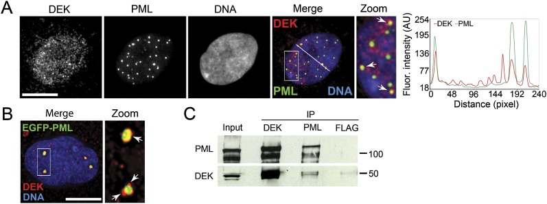
Figure 1.
DEK associates with PML NBs. ( A ) Immunofluorescence localization of DEK and PML in nuclei of human MSCs. Graph shows fluorescence intensity of PML and DEK along the line drawn on the merged image. (...
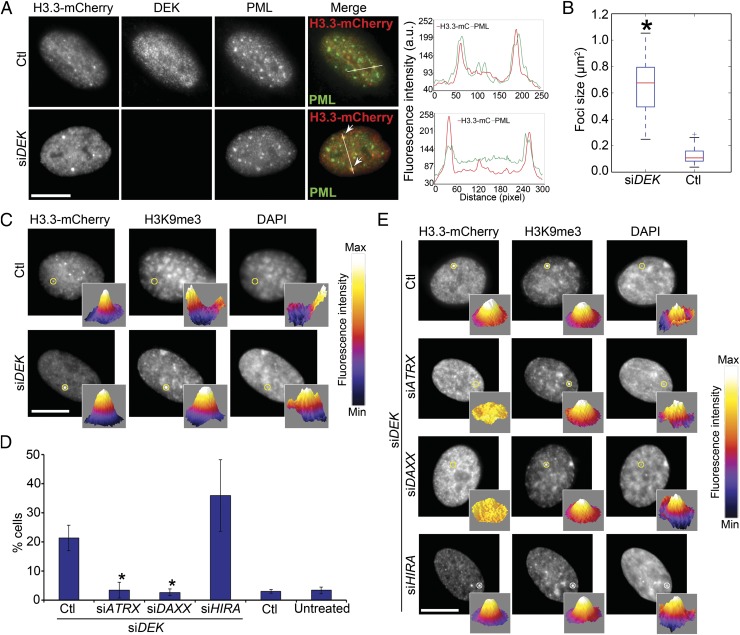
Figure 2.
Depletion of DEK inhibits recruitment of H3.3-EGFP to PML NBs and promotes its loading on chromatin. ( A ) H3.3 forms large foci colocalizing with PML after DEK depletion. Localization of H3.3-mCherry...
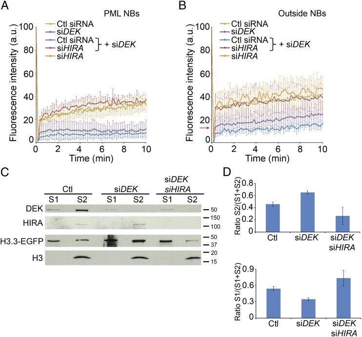
Figure 3.
Depletion of DEK inhibits recruitment of H3.3-EGFP to PML NBs and promotes its loading on chromatin. ( A ) Loss of DEK by siRNA inhibits targeting of H3.3-EGFP to PML NBs. FRAP analysis of H3.3-EGFP a...
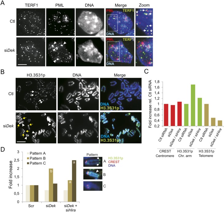
Figure 4.
Loss of DEK in ESCs results in displacement of PML from telomeres, reduced H3.3 loading at telomeres, and HIRA-dependent H3.3 deposition on chromosome arms. ( A ) Immunolocalization of TERF1 and PML i...
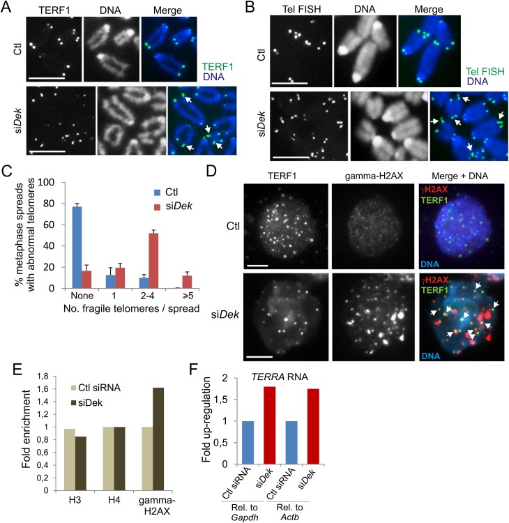
Figure 5.
DEK depletion in ESCs induces fragile telomeres. Immunofluorescence detection of TERF1 ( A ) and telomere FISH analysis of telomere structure ( B ), on metaphase chromosomes from control and DEK-deple...
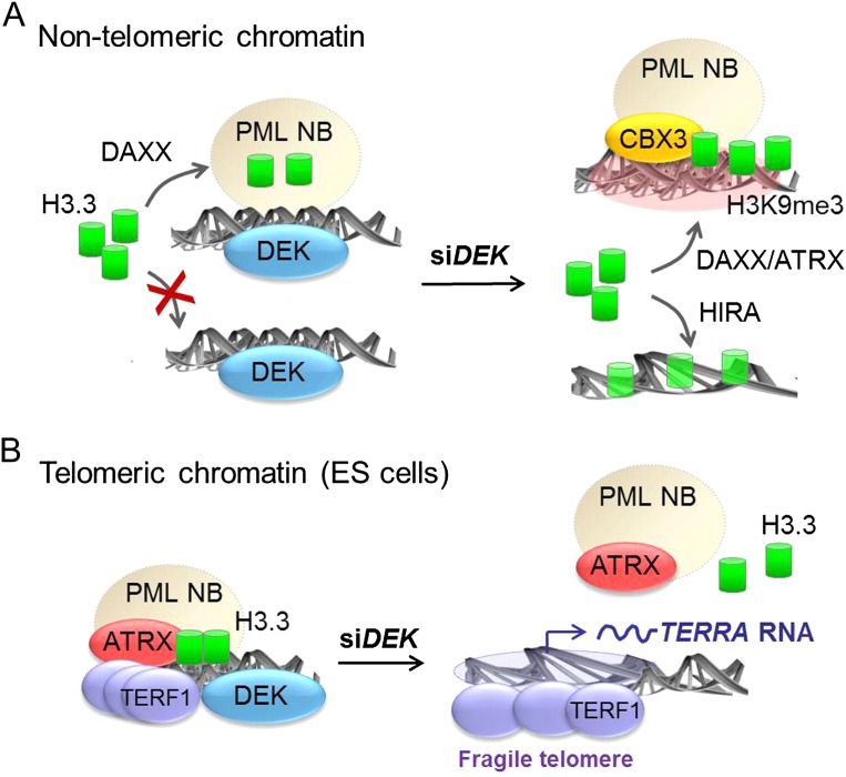
Figure 6.
Model of regulation of loading of H3.3 on chromatin by DEK. ( A ) Outside telomeres, DEK prevents chromatin loading of nonnucleosomal H3.3, which is recruited to PML NBs by DAXX ( Delbarre et al. 2013...
Figure images are served from the NIH/NLM PubMed Central Open Access Subset or Europe PMC; copyright remains with the publishers and authors.
💬 Discussion
0 commentsNo comments yet. Be the first to start a discussion!
Leave a Comment