Abstract
Considerable efforts are being undertaken to elucidate the processes of ribosome biogenesis. Although various preribosomal RNP complexes have been isolated and molecularly characterized, the order of ribosomal protein (r-protein) addition to the emerging ribosome subunits is largely unknown. Furthermore, the correlation between the ribosome assembly pathway and the structural organization of the dedicated ribosome factory, the nucleolus, is not well established. We have analyzed the nucleolar localization of several early binding r-proteins in human cells, applying various methods, including live-cell imaging and electron microscopy. We have located all examined r-proteins (S4, S6, S7, S9, S14, and L4) in the granular component (GC), which is the nucleolar region where later pre-ribosomal RNA (rRNA) processing steps take place. These results imply that early binding r-proteins do not assemble with nascent pre-rRNA transcripts in the dense fibrillar component (DFC), as is generally believed, and provide a link between r-protein assembly and the emergence of distinct granules at the DFC-GC interface.
🔬 Techniques
🧬 Organisms
✨ Fluorophores
🧪 Sample Preparation
🔬 Cell Lines
🏭 Microscope Brands
🧪 Reagent Suppliers
🔎 Objectives
💻 Software Details
🏛️ Research Organizations (ROR)
Affiliated research institutions:
📋 Methods
Materials and methods cDNA, plasmids, and transfection The following cDNAs were cloned by RT-PCR using total RNA isolated from Hep2 cells: S4 (gi:39812410), S6 (gi:20381195), S7 (gi:15431308), S9 (gi:550022), S14 (gi:14141191), L4 (gi:16579884), fibrillarin (gi:14763877), B23 (gi: 12803184), and Imp3 (gi:70908369). The cDNAs were cloned into the pCR2.1-Topo vector (Invitrogen), sequenced, and subcloned into the following vectors: pEGFP-N1, pEGFP-C1 or -C3, pDsRed-Monomer-N1, pCMV-Myc (all obtained from CLONTECH Laboratories, Inc.), pQ-C-His, and pVenus. pVenus was produced by site-directed mutagenesis of pEYFP (CLONTECH Laboratories, Inc.) as described by Nagai et al. (2002) . Transfections were performed using Effectene (QIAGEN) or Fugene (Roche) according to the manufacturers' instructions. Transiently transfected cells were analyzed 24–72 h after transfection. The clone of Hep2 cells stably expressing S4-GFP was isolated after a 3-wk selection period with 0.75 mg/ml G418.
Antibodies Hybridoma supernatant
S14-39 was raised against His-tagged S14. S14-His fusion protein was expressed from the vector pQ-C-His in Escherichia coli XLI-blue and purified using Talon metal affinity resin (BD Biosciences). Further antibodies used were mAb 72B9 against fibrillarin ( Reimer et al., 1987a ), autoimmune serum S18 against pol I ( Reimer et al., 1987b ), anti-GFP (Sigma-Aldrich), c-Myc mAb (BD Biosciences), and QM (C-17) against L10 (Santa Cruz Biotechnology, Inc.).
Indirect immunofluorescence and live-cell microscopy
Hep2 cells grown on coverslips were fixed with 2% formaldehyde in PBS and permeabilized with 0.2% Triton X-100 in PBS and incubated for 30 min with the primary antibodies and 15 min with the appropriate Texas red–conjugated secondary antibodies (Dianova). In some experiments, cells were treated with 0.04 μg/ml AMD for 2–3 h or with 50 μg/ml DRB for 5–6 h before fixation or live-cell imaging. For live-cell microscopy, cells were grown on glass-bottomed dishes (WPI). Images were taken with a confocal laser-scanning microscope (TCS-SP or TCS-SP2; Leica) equipped with 63×/1.4 NA oil-immersion objectives and a 37°C/5% CO 2 incubation chamber. High-resolution live-cell images ( Fig. 1, e and i–j ; and Fig. S1) were subject to noise reduction and background subtraction using ImageJ (NIH; http://rsb.info.nih.gov/ij/ ). Images were merged and assembled in Photoshop (Adobe).
Show full methods section
Materials and methods cDNA, plasmids, and transfection The following cDNAs were cloned by RT-PCR using total RNA isolated from Hep2 cells: S4 (gi:39812410), S6 (gi:20381195), S7 (gi:15431308), S9 (gi:550022), S14 (gi:14141191), L4 (gi:16579884), fibrillarin (gi:14763877), B23 (gi: 12803184), and Imp3 (gi:70908369). The cDNAs were cloned into the pCR2.1-Topo vector (Invitrogen), sequenced, and subcloned into the following vectors: pEGFP-N1, pEGFP-C1 or -C3, pDsRed-Monomer-N1, pCMV-Myc (all obtained from CLONTECH Laboratories, Inc.), pQ-C-His, and pVenus. pVenus was produced by site-directed mutagenesis of pEYFP (CLONTECH Laboratories, Inc.) as described by Nagai et al. (2002) . Transfections were performed using Effectene (QIAGEN) or Fugene (Roche) according to the manufacturers' instructions. Transiently transfected cells were analyzed 24–72 h after transfection. The clone of Hep2 cells stably expressing S4-GFP was isolated after a 3-wk selection period with 0.75 mg/ml G418.
Antibodies Hybridoma supernatant
S14-39 was raised against His-tagged S14. S14-His fusion protein was expressed from the vector pQ-C-His in Escherichia coli XLI-blue and purified using Talon metal affinity resin (BD Biosciences). Further antibodies used were mAb 72B9 against fibrillarin ( Reimer et al., 1987a ), autoimmune serum S18 against pol I ( Reimer et al., 1987b ), anti-GFP (Sigma-Aldrich), c-Myc mAb (BD Biosciences), and QM (C-17) against L10 (Santa Cruz Biotechnology, Inc.).
Indirect immunofluorescence and live-cell microscopy
Hep2 cells grown on coverslips were fixed with 2% formaldehyde in PBS and permeabilized with 0.2% Triton X-100 in PBS and incubated for 30 min with the primary antibodies and 15 min with the appropriate Texas red–conjugated secondary antibodies (Dianova). In some experiments, cells were treated with 0.04 μg/ml AMD for 2–3 h or with 50 μg/ml DRB for 5–6 h before fixation or live-cell imaging. For live-cell microscopy, cells were grown on glass-bottomed dishes (WPI). Images were taken with a confocal laser-scanning microscope (TCS-SP or TCS-SP2; Leica) equipped with 63×/1.4 NA oil-immersion objectives and a 37°C/5% CO 2 incubation chamber. High-resolution live-cell images ( Fig. 1, e and i–j ; and Fig. S1) were subject to noise reduction and background subtraction using ImageJ (NIH; http://rsb.info.nih.gov/ij/ ). Images were merged and assembled in Photoshop (Adobe).
Ribosome isolation and immunoblotting
Ribosomes from ∼10 7 Hep2 cells were isolated as previously described ( Elkon et al., 1986 ). Ribosomal subunits were separated by sucrose gradient (10–40%) centrifugation for 16 h at 23,000 rpm in an SW41 rotor (Beckman Coulter). Fractions containing the small and large ribosomal subunits were analyzed by SDS-PAGE, and immunoblots were performed as previously described ( Gareiss et al., 2005 ).
Immunogold electron microscopy
Hep2 cells expressing myc-tagged r-proteins and grown on coverslips were briefly washed in PBS, fixed for 10 min at RT in 2% formaldehyde in PBS (freshly prepared from paraformaldehyde), and washed again in PBS (3 × 5 min). Free aldehyde groups were quenched by incubation in 50 mM ammonium chloride in PBS for 15 min, followed by PBS wash. For postembedding labeling, the cells were dehydrated through an ascending ethanol series and infiltrated with LR White (Plano) according to the manufacturer's instructions. Finally, a Beem capsule filled with the unpolymerized resin was invertedly placed on the coverslip. After polymerization at 40°C for 72 h, the glass coverslip was removed and ultrathin sections were cut and placed on nickel grids. For immunolabeling, the sections were washed in PBS and then in PBS containing 0.1% Tween 20 and 1% BSA (PBS1), followed by incubation with anti-myc monoclonal antibody for 1 h at RT at a concentration of 2 μg/ml in PBS1. After several wash steps in PBS1 and PBS2 (PBS containing 0.1% Tween 20 and 0.1% BSA), Nanogold anti-mouse Fab′ conjugates were added for 1 h (Nanoprobes; 1:20 in PBS2). Then, the grids were rinsed with PBS2 and PBS, postfixed with 1.25% glutaraldehyde in PBS for 2 min, and rinsed in distilled water. Finally, the gold particles were silver enhanced using the R-Gent SE-EM kit (Aurion). The sections were rinsed in distilled water and contrasted with uranyl acetate and lead citrate. For preembedding labeling, the fixed cells were permeabilized for 10 min with 0.05% Triton X-100 in PBS and washed with PBS containing 0.2% BSA (PBS3). Incubation with the first and secondary antibodies was as detailed above except that PBS3 was used throughout. After several wash steps in PBS3 followed by PBS, cells were fixed in 2% glutaraldehyde in PBS (10 min at 4°C), washed in distilled water, and silver enhanced as described. After several wash steps in distilled water, cells were dehydrated and flat embedded in Epon 812 (Serva). Ultrathin sections were stained according to standard methods and examined in an electron microscope (EM10A; Carl Zeiss MicroImaging, Inc.) operating at 80 kV. Endogenous S14, fibrillarin, and RNA polymerase I were localized in Hep2 cells with mAb S14-39, mAb 72B9, and autoimmune serum S18, respectively, following the preembedding labeling protocol outlined above. Online supplemental material Fig. S1 shows the localization of IMP3 in the granular compartment of the nucleolus. Online supplemental material is available at http://www.jcb.org/cgi/content/full/jcb.200612048/DC1 .
Online supplemental material Fig. S1 shows the localization of IMP3 in the granular compartment of the nucleolus. Online supplemental material is available at http://www.jcb.org/cgi/content/full/jcb.200612048/DC1 .
Supplementary Material [Supplemental Material Index]
📊 Figures
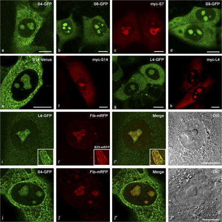
Figure 1.
Localization of early binding r-proteins in human Hep2 cells by confocal microscopy after transient expression as fusions with fluorescent proteins or with a myc epitope. (au2013h) The distribution of...
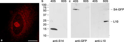
Figure 2.
Characterization of mAb S14-39 raised against recombinant S14. (a) The antibodies stain the nucleolus and the cytoplasm of Hep2 cells. (b) In immunoblots, the mAb recognizes a 15-kD polypeptide of the...
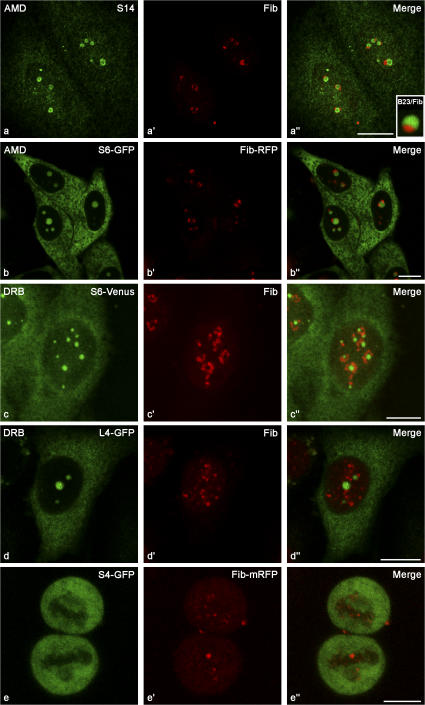
Figure 3.
Early binding r-proteins are restricted to the GC and absent from the DFC of nucleoli. After AMD treatment, the two major nucleolar components, GC and DFC, segregate into separate structures (au2033, ...
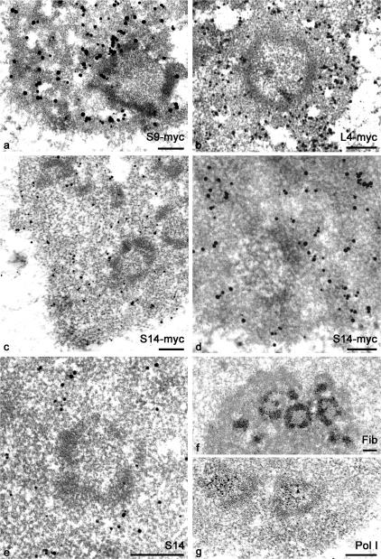
Figure 4.
Immunogold electron microscopy showing the intranucleolar distribution of early binding r-proteins in Hep2 cells. FCs with the surrounding electron-dense DFC layer are clearly identified in all figure...
Figure images are served from the NIH/NLM PubMed Central Open Access Subset or Europe PMC; copyright remains with the publishers and authors.
💬 Discussion
0 commentsNo comments yet. Be the first to start a discussion!
Leave a Comment