Abstract
BACKGROUND: Chondrosarcomas are malignant cartilage-forming tumours of bone. Because of their resistance to conventional chemotherapy and radiotherapy, currently no treatment strategies exist for unresectable and metastatic chondrosarcoma. Previously, PI3K/AKT/GSK3β and Src kinase pathways were shown to be activated in chondrosarcoma cell lines. Our aim was to investigate the role of these kinases in chemoresistance and migration in chondrosarcoma in relation to TP53 mutation status. METHODS: We used five conventional and three dedifferentiated chondrosarcoma cell lines and investigated the effect of PI3K/AKT/GSK3β pathway inhibition (enzastaurin) and Src pathway inhibition (dasatinib) in chemoresistance using WST assay and live cell imaging with AnnexinV staining. Immunohistochemistry on tissue microarrays (TMAs) containing 157 cartilaginous tumours was performed for Src family members. Migration assays were performed with the RTCA xCelligence System. RESULTS: Src inhibition was found to overcome chemoresistance, to induce apoptosis and to inhibit migration. Cell lines with TP53 mutations responded better to combination therapy than wild-type cell lines (P=0.002). Tissue microarray immunohistochemistry confirmed active Src (pSrc) signalling, with Fyn being most abundantly expressed (76.1%). CONCLUSION: These results strongly indicate Src family kinases, in particular Fyn, as a potential target for the treatment of inoperable and metastatic chondrosarcomas, and to sensitise for doxorubicin especially in the presence of TP53 mutations.
🔬 Techniques
✨ Fluorophores
🧪 Sample Preparation
🔬 Cell Lines
🏭 Microscope Brands
🧪 Reagent Suppliers
💻 Software Details
🏛️ Research Organizations (ROR)
Affiliated research institutions:
📋 Methods
We used five conventional and three dedifferentiated chondrosarcoma cell lines and investigated the effect of PI3K/AKT/GSK3 β pathway inhibition (enzastaurin) and Src pathway inhibition (dasatinib) in chemoresistance using WST assay and live cell imaging with AnnexinV staining.
Immunohistochemistry on tissue microarrays
(TMAs) containing 157 cartilaginous tumours was performed for Src family members. Migration assays were performed with the RTCA xCelligence System.
Materials and Methods Compounds Doxorubicin and cisplatin were obtained from the in-house hospital pharmacy in a 0.9% NaCl solution. Therapeutic concentrations of doxorubicin in patients are 5–50 μ M with an in vitro range of 1–10 μ M , for cisplatin these are 3–13 μ M with an in vitro range of 1–50 μ M ( Shrivastav et al , 1980 ). The PKC inhibitor enzastaurin ( Faul et al , 2003 ; Eli Lilly, IN, USA) and the Src inhibitor dasatinib ( Lombardo et al , 2004 ) (Bristol-Meyers Squibb, Princeton, NJ, USA) were dissolved in DMSO.
Cell culture
Chondrosarcoma cell lines ( Table 1 ), as well as MCF-7 and HeLa cell lines were cultured in RPMI 1640 (Gibco, Invitrogen Life Technologies, Scotland, UK) supplemented with 1% L -glutamax, 1% penicillin–streptomycin (100 U ml −1 ) and 10% heat-inactivated fetal calf serum (Gibco, Invitrogen Life Technologies, Scotland, UK). Cells were grown at 37 °C in a humidified incubator with 95% air and 5% CO 2 . Cells were cultured until they started multiplying stably. Chondrogenic phenotype was confirmed using RT–PCR for collagen I, IIB, III and X, aggrecan and SOX9 ( Cleton-Jansen et al , 2005 ). Identity of cell lines was confirmed using the Cell ID System after completion of experiments (Promega Benelux BV, Leiden, The Netherlands).
Show full methods section
We used five conventional and three dedifferentiated chondrosarcoma cell lines and investigated the effect of PI3K/AKT/GSK3 β pathway inhibition (enzastaurin) and Src pathway inhibition (dasatinib) in chemoresistance using WST assay and live cell imaging with AnnexinV staining.
Immunohistochemistry on tissue microarrays
(TMAs) containing 157 cartilaginous tumours was performed for Src family members. Migration assays were performed with the RTCA xCelligence System.
Materials and Methods Compounds Doxorubicin and cisplatin were obtained from the in-house hospital pharmacy in a 0.9% NaCl solution. Therapeutic concentrations of doxorubicin in patients are 5–50 μ M with an in vitro range of 1–10 μ M , for cisplatin these are 3–13 μ M with an in vitro range of 1–50 μ M ( Shrivastav et al , 1980 ). The PKC inhibitor enzastaurin ( Faul et al , 2003 ; Eli Lilly, IN, USA) and the Src inhibitor dasatinib ( Lombardo et al , 2004 ) (Bristol-Meyers Squibb, Princeton, NJ, USA) were dissolved in DMSO.
Cell culture
Chondrosarcoma cell lines ( Table 1 ), as well as MCF-7 and HeLa cell lines were cultured in RPMI 1640 (Gibco, Invitrogen Life Technologies, Scotland, UK) supplemented with 1% L -glutamax, 1% penicillin–streptomycin (100 U ml −1 ) and 10% heat-inactivated fetal calf serum (Gibco, Invitrogen Life Technologies, Scotland, UK). Cells were grown at 37 °C in a humidified incubator with 95% air and 5% CO 2 . Cells were cultured until they started multiplying stably. Chondrogenic phenotype was confirmed using RT–PCR for collagen I, IIB, III and X, aggrecan and SOX9 ( Cleton-Jansen et al , 2005 ). Identity of cell lines was confirmed using the Cell ID System after completion of experiments (Promega Benelux BV, Leiden, The Netherlands).
Cell viability assay
Chondrosarcoma cell lines were plated in 96-well plates for viability assessment (2 × 10 4 –2 × 10 5 cells per well depending on growth rate) and allowed to grow and adhere overnight after which the respective drugs were added in their corresponding concentrations. Combination assays were performed as described ( van Oosterwijk et al , 2012b ) with alternating treatments combining enzastaurin, dasatinib and/or doxorubicin. All experiments were performed in triplicate and at least three times. Graphs show data from one representative experiment. Error bars indicate the s.d.
Immunoblotting
Immunoblotting using AKT, pAKT, Fyn (Cell Signaling, Leiden, the Netherlands) and pSrc antibody (pSrc pY418, Invitrogen Life Technologies, Bleiswijk, the Netherlands) to investigate the Src and PI3K/AKT signalling pathway and p53 (Do7, Dako, Heverlee, Belgium), MDM2 (IF2, Zymed, Bleiswijk, the Netherlands) and p21 (Santa Cruz, Heidelberg, Germany) was performed as previously described ( Schrage et al , 2009 ), using 20 μ g of each sample.
Mutation analysis
To identify mutations in AKT1, direct sequencing was performed as described ( Pansuriya et al , 2011 ), using DNA derived from 57 tumours, 8 cell lines and 1 primary culture (L3310) using forward primer 3′-TAGAGTGTGCGTGGCCTCTCA-5′ and reverse primer 3′-CTGAATCCCGAGAGGCCAA-5′ to screen for hotspot mutations in the AKT1-E17K pleckstrin homology domain.
Apoptosis assay and immunofluorescence
Apoptosis assay and immunofluorescence for caspase 3 and cytochrome C were performed as described ( Puigvert et al , 2010 ; van Oosterwijk et al , 2012b ). In short, 20 000 chondrosarcoma cells were grown in black 96-well microclear plates (Greiner, Sigma-Aldrich, Zwijndrecht, the Netherlands) to perform a live cell apoptosis assay ( Puigvert et al , 2010 ), with AnnexinV-Alexa633 conjugate using the BD Pathway 855 (Becton Dickinson, Breda, the Netherlands). Time series were quantified using in-house-developed macros for Image-Pro Plus (Media Cybernetics, Bethesda, MD, USA). Drugs were added 0, 24, and 48 h before imaging and Annexin V-Alexa633 conjugate was added immediately before imaging. For all treatments, a pan-caspase inhibitor, z-VAD-fmk (Bachem-Holding AG, Weil am Rhein, Germany), was added 30 min before drug addition and imaging to establish apoptosis specificity of the assay. Before imaging, live nuclei were stained with HOECHST-33342 at 100 ng ml −1 . All experiments were performed in triplicate and at least three times. Error bars show s.d. from one representative experiment. Migration assays The RTCA xCelligence system (Roche Applied Sciences, Almere, the Netherlands), based on cell–electrode substract impedance detection technology, was used for migration assays. For migration assays, lower wells of the SIM plates (migration plates) were filled with growth medium (20% fetal calf serum in RPMI). Cell lines were plated at a density of 80 000 cells per well in the top wells in empty buffer (RPMI only) containing 0, 0.2, 0.4, 0.6, 0.8 or 1.0 μ M dasatinib. SIM plates were loaded into the RTCA station in the cell culture incubator immediately after plating and cell index was acquired every 5 min. Cell index as acquired by the software was set to 100% migration after flattening of the slope. Experiments were performed in triplicate. Tissue microarray (TMA) construction and clinicopathological data Tissue microarrays were constructed from formalin-fixed, paraffin-embedded tissue using standard procedures ( Kononen et al , 1998 ) using a 2.0-mm diameter punch-automated tissue arrayer (3DHistech Ltd, Budapest, Hungary). Each array contained three cores per tumour wherever possible including seven control tissues (skin, colon, tonsil, prostate, mamma carcinoma, spleen and liver). Using a tape-transfer system (Instrumedics, Hackensack, NJ, USA), 4- μ m sections were transferred to glass slides. All specimens in this study were handled according to the ethical guidelines described in ‘Code for Proper Secondary Use of Human Tissue in The Netherlands' of the Dutch Federation of Medical Scientific Societies. A total of 157 patients with cartilaginous tumours were selected from the archives of the Leiden University Medical Centre. Selected cases included 137 conventional chondrosarcomas (central chondrosarcoma, n =92; peripheral chondrosarcoma, n =45) and 20 benign cartilage tumours (osteochondroma, n =9; enchondroma, n =11). Only primary tumours were selected. Histology was reviewed by an experienced bone tumour pathologist (JVMGB). Clinicopathological data are shown in Table 2 . Total follow-up was available for 136 of 157 patients, with 14 patients showing metastasis at completion of this study. Histological grading of chondrosarcoma was performed according to Evans et al (1977 ). Rare chondrosarcoma subtypes were excluded.
Immunohistochemistry
Immunohistochemistry was performed on the TMAs. Slides were incubated with antibodies against Src, Lck, Fyn, Yes and phosphorylated Src (pSrc, recognises active Src family members phosphorylated at Y419). Details of antibodies and procedures are provided in Supplementary Table 1 . Immunohistochemical reactions were performed according to standard laboratory methods ( Bovée et al , 2000 ) and visualised using DAB+ Substrate Chromogen System (Dako, Heverlee, Belgium). Tissue microarray slides were scanned using a high-resolution Mirax Desk Instrument (Zeiss, Mirax 3DHistech) and scored independently by two observers (JVMGB and JGvO) and discrepancies were discussed. Staining intensity (0=absent, 1=weak, 2=moderate, 3=strong) and extent of the staining (0=0%, 1=1–24%, 2=25–49%, 3=50–74% and 4=75–100%) were assessed. Staining was considered high (score ⩾4) or low (score
📊 Figures
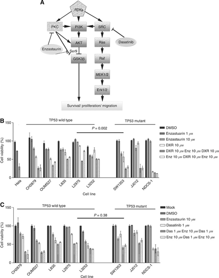
Figure 1
Chondrosarcoma cell lines are not sensitive to PKC inhibition. ( A ) Schematic representation of activation of PI3K and Src pathway by RTKs. Receptor tyrosine kinases can activate PKC, phosphatidylino...
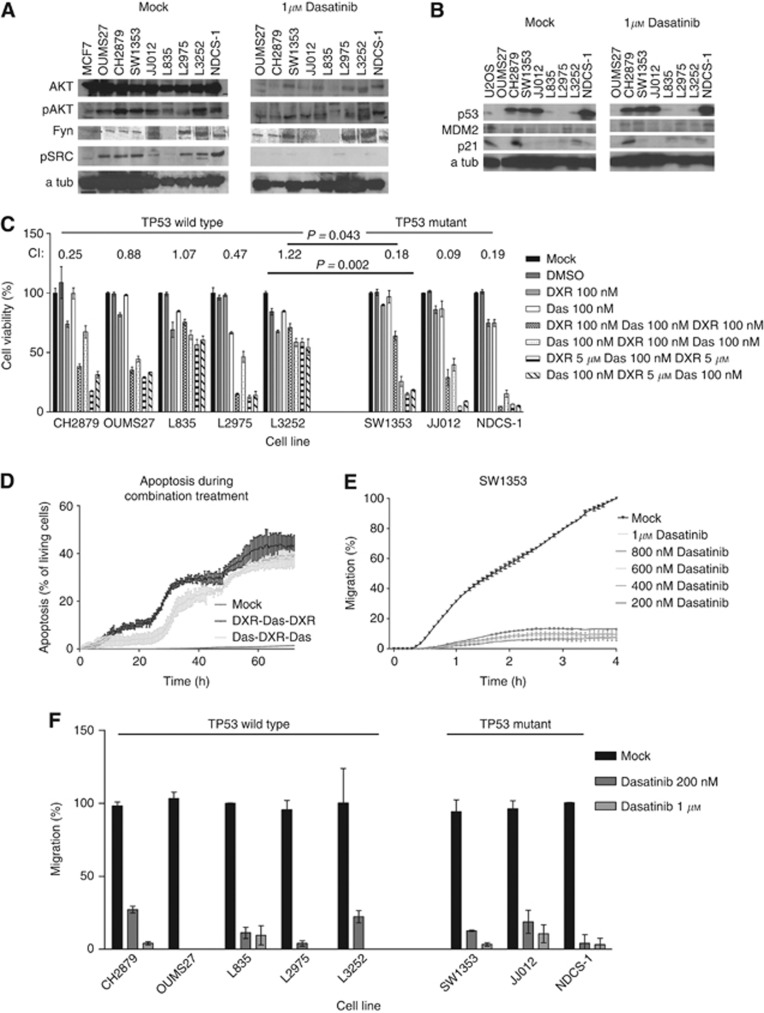
Figure 2
The Src pathway is involved in chondrosarcoma chemoresistance. ( A ) Immunoblotting showing AKT, pAKT, Fyn, pSrc and loading control u03b1 -tubulin (a tub) for untreated chondrosarcoma cell lines and ...
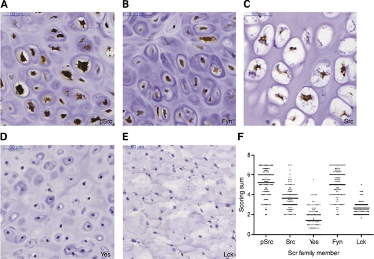
Figure 3
Immunohistochemistry demonstrating expression of Src family members in conventional chondrosarcoma tissue. ( A ) High pSrc expression in grade I chondrosarcoma. ( B ) High-intensity nuclear Fyn expres...
Figure images are served from the NIH/NLM PubMed Central Open Access Subset or Europe PMC; copyright remains with the publishers and authors.
💬 Discussion
0 commentsNo comments yet. Be the first to start a discussion!
Leave a Comment