Abstract
Abstract Background Although evidence suggests that the prevalence of Parkinson’s disease (PD) is lower in smokers than in non-smokers, the mechanisms of nicotine-induced neuroprotection remain unclear. Stimulation of the α7 nicotinic acetylcholine receptor (α7-nAChR) seems to be a crucial mechanism underlying the anti-inflammatory potential of cholinergic agonists in immune cells, including astrocytes, and inhibition of astrocyte activation has been proposed as a novel strategy for the treatment of neurodegenerative disorders such as PD. The objective of the present study was to determine whether nicotine-induced neuroprotection in the 1-methyl-4-phenyl-1,2,3,6-tetrahydropyridine (MPTP) mouse model occurs via α7-nAChR-mediated inhibition of astrocytes. Methods Both in vivo (MPTP) and in vitro (1-methyl-4-phenylpyridinium ion (MPP+) and lipopolysaccharide (LPS)) models of PD were used to investigate the role(s) of and possible mechanism(s) by which α7-nAChRs protect against dopaminergic neuron loss. Multiple experimental approaches, including behavioral tests, immunochemistry, and stereology experiments, astrocyte cell cultures, reverse transcriptase PCR, laser scanning confocal microscopy, tumor necrosis factor (TNF)-α assays, and western blotting, were used to elucidate the mechanisms of the α7-nAChR-mediated neuroprotection. Results Systemic administration of nicotine alleviated MPTP-induced behavioral symptoms, improved motor coordination, and protected against dopaminergic neuron loss and the activation of astrocytes and microglia in the substantia nigra. The protective effects of nicotine were abolished by administration of the α7-nAChR-selective antagonist methyllycaconitine (MLA). In primary cultured mouse astrocytes, pretreatment with nicotine suppressed MPP+-induced or LPS-induced astrocyte activation, as evidenced by both decreased production of TNF-α and inhibition of extracellular regulated kinase1/2 (Erk1/2) and p38 activation in astrocytes, and these effects were also reversed by MLA. Conclusion Taken together, our results suggest that α7-nAChR-mediated inhibition of astrocyte activation is an important mechanism underlying the protective effects of nicotine.
🔬 Techniques
✨ Fluorophores
🧪 Sample Preparation
🔬 Cell Lines
🏭 Microscope Brands
🧪 Reagent Suppliers
🏛️ Research Organizations (ROR)
Affiliated research institutions:
📋 Methods
Animals and treatments
All experiments were carried out in accordance with the National Institutes of Health Guide for the Care and Use of Laboratory Animals (publication number 85–23, revised 1985) and the Society for Neuroscience Guidelines for the Care and Use of Animals in Neuroscience Research , and were approved by the Institutional Animal Care and Use Committee of Nanjing Medical University [ 5 ]. Male C57BL/6 black mice (8 to 10-weeks old, weighing 24 to 28 g) were used. All animals were housed in groups of five per cage under standard laboratory conditions with free access to food and water, constant room temperature (22°C) and humidity (50 to 60 %), and a natural day/night cycle. Nicotine, methyllycaconitine citrate (MLA), and MPTP were dissolved in sterile saline (0.9 % NaCl). Mice were randomly divided into different groups as described below. All drugs were obtained from Sigma Chemical Co. (St. Louis, MO, USA). Control mice received saline only. Mice in the treatment group received four intraperitoneal injections of MPTP-HCl 20 mg/kg at 2-hour intervals on the same day. For nicotine treatment, mice were given intraperitoneal injections of nicotine (0.25 or 0.5 mg/kg) five times a day at 2-hour intervals over a 2-week period (1 week before, during, and 1 week after MPTP administration). The nicotine was injected 30 minutes before each MPTP injection. MLA 5.0 mg/kg, used for nicotine antagonism studies, was injected 30 minutes before nicotine administration twice a day for 2 weeks (1 week before, during, and 1 week after MPTP administration) [ 10 , 11 ]. Furthermore, cohorts of mice were treated with nicotine 0.5 mg/kg or MLA 5.0 mg/kg alone five times a day at 2-hour intervals for 2 weeks to observe whether those drugs alone influenced behavioral symptoms, dopaminergic neuron degeneration, and/or astrocyte activation.
Show full methods section
Animals and treatments
All experiments were carried out in accordance with the National Institutes of Health Guide for the Care and Use of Laboratory Animals (publication number 85–23, revised 1985) and the Society for Neuroscience Guidelines for the Care and Use of Animals in Neuroscience Research , and were approved by the Institutional Animal Care and Use Committee of Nanjing Medical University [ 5 ]. Male C57BL/6 black mice (8 to 10-weeks old, weighing 24 to 28 g) were used. All animals were housed in groups of five per cage under standard laboratory conditions with free access to food and water, constant room temperature (22°C) and humidity (50 to 60 %), and a natural day/night cycle. Nicotine, methyllycaconitine citrate (MLA), and MPTP were dissolved in sterile saline (0.9 % NaCl). Mice were randomly divided into different groups as described below. All drugs were obtained from Sigma Chemical Co. (St. Louis, MO, USA). Control mice received saline only. Mice in the treatment group received four intraperitoneal injections of MPTP-HCl 20 mg/kg at 2-hour intervals on the same day. For nicotine treatment, mice were given intraperitoneal injections of nicotine (0.25 or 0.5 mg/kg) five times a day at 2-hour intervals over a 2-week period (1 week before, during, and 1 week after MPTP administration). The nicotine was injected 30 minutes before each MPTP injection. MLA 5.0 mg/kg, used for nicotine antagonism studies, was injected 30 minutes before nicotine administration twice a day for 2 weeks (1 week before, during, and 1 week after MPTP administration) [ 10 , 11 ]. Furthermore, cohorts of mice were treated with nicotine 0.5 mg/kg or MLA 5.0 mg/kg alone five times a day at 2-hour intervals for 2 weeks to observe whether those drugs alone influenced behavioral symptoms, dopaminergic neuron degeneration, and/or astrocyte activation.
Behavioral tests
The effects of MPTP on movement were evaluated by a pole test [ 3 ]. Briefly, a cork ball (diameter 25 mm) was fixed to the top of a vertical, wooden rough-surfaced pole (diameter 10 mm, height 500 mm). The mouse was placed head upward on the cork ball, and the following activities were recorded: turning downward on the ball, climbing down the upper half of the pole, climbing down the lower half of the pole. The activities were scored as follows: 3 for recorded times of less than 3 seconds, 2 if less than 6 seconds, and 1 if less than 6 seconds. Results were expressed as the total score.
Immunochemistry and stereology
Animals were anaesthetized using chloral hydrate, and perfused with 0.9% NaCl, followed by cold 4% paraformaldehyde in 0.1 mol/l phosphate buffer (pH 7.4). The brains were dissected out and maintained in 4 % paraformaldehyde overnight. Brains were cryopreserved in 30% sucrose in PBS and stored at −70°C until required. Free-floating sections encompassing the entire midbrain were cut on a cryostat. Sections were processed for tyrosine hydroxylase (TH), GFAP, and Mac-1 (marker for microglia [ 4 ]) immunohistochemistry as described below. After incubation for 1 h in 10 % BSA with 0.3% Triton X-100 in 0.01 mol/l PBS, the tissue sections (30 mm) were incubated with primary antibodies overnight at 4°C. The primary antibodies used in this study were a mouse antibody against TH (1:3000, Sigma Chemical Co.), goat antibody against GFAP (1:1000; Millipore Corp., Billerica, MA, USA), and rat anti-mouse Mac-1 polyclonal antibody (1:100, CD11b, AbD; Serotec, Oxford, UK). Immunostaining was visualized by using 3,3′-diaminobenzidine, and sections were then counterstained with hematoxylin [ 5 , 6 , 12 ]. All cell counts were performed by researchers blinded to the experimental status of the animals. The total number of TH-immunoreactive (IR) neurons, GFAP-IR astrocytes, and Mac-1-IR microglia in the substantia nigra pars compacta (SNpc) were counted from six mice per group using an optical fractionator [ 13 ], which is an unbiased method of cell counting that is not affected by either the volume of reference or the size of the counted elements (Stereo Investigator software, Microbrightfield, Colchester, VT, USA). In this method, TH-IR neurons, GFAP-IR astrocytes, and Mac-1-IR microglia were counted in the SNpc of every fourth section (30 mm) throughout the entire extent of the SNpc. Each midbrain section was viewed at low power (×10 objective), and the SNpc was outlined in accordance with the established anatomical landmark. Then, at a random starting point, the number of TH-positive neurons, GFAP-positive astrocytes and Mac-1-IR microglia were counted at high power (×100, oil immersion). To avoid double counting of cells with unusual shapes, each type of cell (TH-IR neurons, GFAP-IR astrocytes and Mac-1-IR microglia) was counted only when its nucleus were optimally visualized, which occurred in only one focal plane. After all the appropriate cells were counted, the total numbers of TH-IR neurons, GFAP-IR astrocytes, and Mac-1-IR microglia in the SNpc were calculated using the formula described by West [ 13 ]. Sampling grid dimensions were 120 × 120 x 5 mm (x, y, and z axes, respectively).
Astrocyte cell cultures
Primary cultures of mouse astrocytes were prepared from the midbrain of C57BL/6 black newborn mice 1 to 2 days after birth as previously described [ 6 ]. In brief, the mid brain was dissected under sterile conditions and the meninges were carefully removed. Brain tissues were dissociated in 0.25% trypsin (Gibco) for 10 minutes at 37°C. The cell suspension was separated by centrifugation at 240 g for 5 minutes, and the cells were transferred to poly-D-lysine pre-coated cell culture flasks in DMEM containing 10% FCS, 100 U/ml penicillin and 100 μg/ml streptomycin. The cultures were maintained at 37°C in a humidified atmosphere of 5% CO 2 and 95% air. Before the experiments, analyses showed that over 95% of the cells stained positively for the astrocytic marker GFAP (1:800; Abcam, Cambridge, MA, USA). All experiments were performed after approximately 12 to 15 days in culture.
Staining with α-bungarotoxin and confocal microscopy
Primary cultured astrocytes (as described above) were passaged in six-well tissue-culture plates at 5 × 10 5 cells per well, and then cultured for 24 hours. Thereafter, cultures were incubated with Alexa Fluor 488-conjugated α-bungarotoxin (2.5 μg/ml ; B13422 , Invitrogen Corp., OR, USA) at 4°C for 15 minutes. Immediately after incubation, these cells were washed with PBS three times, and then fixed for 15 minutes in 4% paraformaldehyde in PBS at room temperature. After fixation, cells were washed once with PBS and then mounted for viewing under a laser scanning confocal microscope (Meta 710 Laser Scanning Microscope, Carl Zeiss Inc., Thornwood, NY, USA). Reverse transcription-polymerase chain reaction and real-time PCR For RNA extraction and reverse transcription (RT)-PCR, total RNA was isolated (RNAiso TM Plus; TaKaRa Biotechnology, Dalian, China) from the SN brain region of mice after drug treatment. First-strand cDNA was synthesized from total RNA using a first-strand cDNA synthesis kit (TaKaRa Biotechnology) in accordance with the manufacturer’s instructions. PCR was performed on the equivalent cDNAs from each sample. Amplification was performed with the primer sets shown in Table 1 . The thermal cycling conditions for both sets of primers were 94°C for 5 minutes, followed by 30 cycles of 94°C for 30 seconds, 57°C for 30 seconds, and 72°C for 45 seconds, with a final extension step at 72°C for 10 minutes. The PCR products were then separated in a 3% agarose gel containing ethidium bromide, and analyzed using a gel imaging system (model 3500, Tanon Science and Technology Co., Shanghai, China). Each real-time PCR reaction was carried out in triplicate in a total volume of 20 μl with SYBR Green (Premix Ex Taq TM ; TaKaRa Biotechnology, Dalian, China) under the following conditions: stage 1, 95°C for 30 seconds (1 cycle); stage 2, 95°C for 5 seconds and 60°C for 20 seconds (40 cycles).
Determination of the cycle threshold
(Ct) value in a PCR amplification curve was performed using a real-time PCR system (LightCycler; Roche Diagnostics, Basel, Switzerland). Table 1 Primers used for amplification Name Genbank accession number Direction Sequence, 5′→3′ Size of product bp GADPH NM_008084.2 Forward TGTGTCCGTCGTGGATCTGA 150 Reverse TTGCTGTTGAAGTCGCAGGAG α7 subunit NM 007390.3 Forward AACCATGCGCCGTAGGACA 172 Reverse CTCAGCCACAAGCAGCATGAA GADPH, glyceraldehyde-3-phosphate dehydrogenase. Tumor necrosis factor-α assay Nicotine, LPS, MPP + and MLA were dissolved in buffered Hank’s buffered salt solution at neutral pH (7.0). All drugs were obtained from Sigma Chemical Co.. Cells were plated onto 12-well plates (1.5 ml, 1 × 10 6 /well), and allowed to adhere for 24 hours at 37°C before being subjected to various treatments. When performing treatments, we used FCS-free DMEM, and the amount of TNF-α in the culture medium was determined 24 hours after treatment using a mouse TNF-α ELISA kit (Beijing 4A Biotech Co. Ltd., Beijing, China) [ 12 ].
Western blotting
Cells were collected and homogenized in 200 μl lysis buffer. After incubation for 20 minutes on ice, cell lysates were separated by centrifugation, and the protein concentration in the extracts was determined by the Bradford assay. Proteins in the cell extracts were denatured with SDS sample buffer and separated by 10% SDS-PAGE. Proteins were transferred to nitrocellulose membranes using a wet transfer unit (Miniprotein-III; Bio-Rad Laboratories, Inc., Hercules, CA, USA). The membranes were incubated with 5% BSA dissolved in Tris-buffered saline with Tween 20 (TBS-T, 10 mmol/l Tris–HCl, 150 mmol/l NaCl, and 0.1% Tween 20, pH 7.5)) at room temperature for 1 hour, washed three times, and incubated with different antibodies (Erk1/2, phosphor-Erk1/2, p38 and phospho-p38, 1:1000; Cell Signaling Technology Inc., Beverly, MA USA; α7-nAChRs, 1:300, Santa Cruz Biotechnology Inc., Santa Cruz, CA USA) overnight at 41°C. The membranes were washed three times with TBS-T buffer, and incubated with secondary antibody for 1 hour, followed by four washes in TBS-T. Signal detection was performed with an enhanced chemiluminescence kit [ 6 , 12 ]. The results were scanned using a gel imaging system (GelMax Imager; Ultra-Violet Products Ltd., Upland, CA, USA) and measured using analyzing software (Gel-Pro Analyzer software; Media Cybernetics, Inc., Bethesda, MD, USA).
Statistical analyses
All values are expressed as mean ± standard error of the mean (SEM). Differences between means were analyzed using one-way or two-way ANOVA with time and treatment as the independent factors. When ANOVA showed significant differences, pairwise comparisons between means were further analyzed using the Newman-Keuls post hoc test. In all analyses, significance was set at P = 0.05 [ 3 , 5 ].
📊 Figures
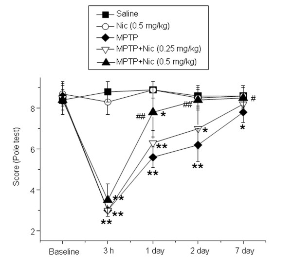
Figure 1
The effects of nicotine on 1-methyl-4-phenyl-1,2,3,6-tetrahydropyridine (MPTP)-induced mouse motor deficits. Pole-test scores for each animal were calculated after acute MPTP treatment at 3 hours, 1u2...
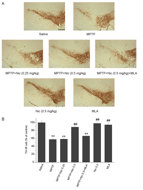
Figure 2
The effects of nicotine on 1-methyl-4-phenyl-1,2,3,6-tetrahydropyridine (MPTP)-induced dopaminergic neuronal loss in the mouse substantia nigra pars compacta (SNpc). (A) Tyrosine hydroxylase-immunorea...
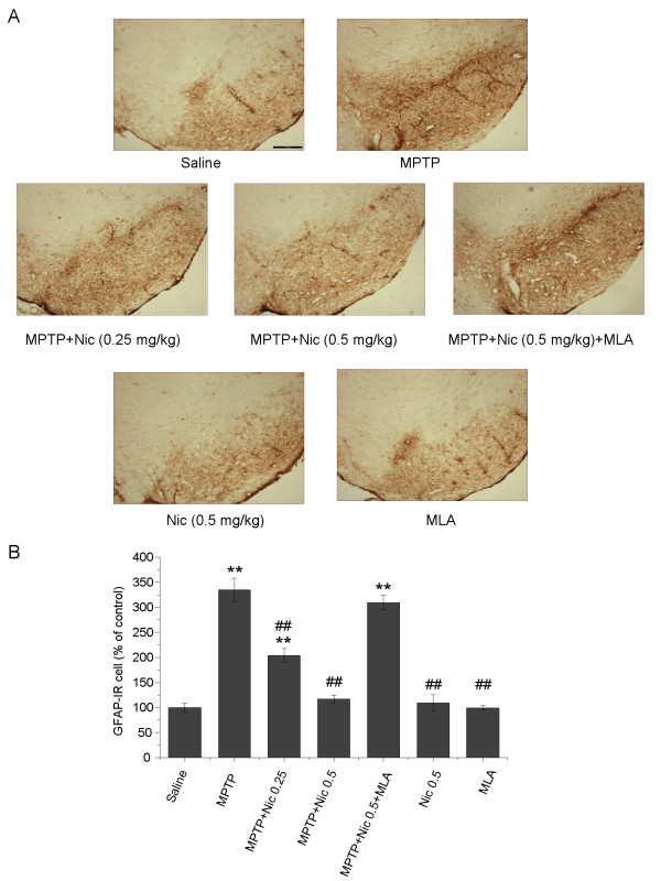
Figure 3
The effects of nicotine on 1-methyl-4-phenyl-1,2,3,6-tetrahydropyridine (MPTP)-induced astrocyte activation in the mouse substantia nigra pars compacta (SNpc). (A) Glial fibrillary acidic protein immu...
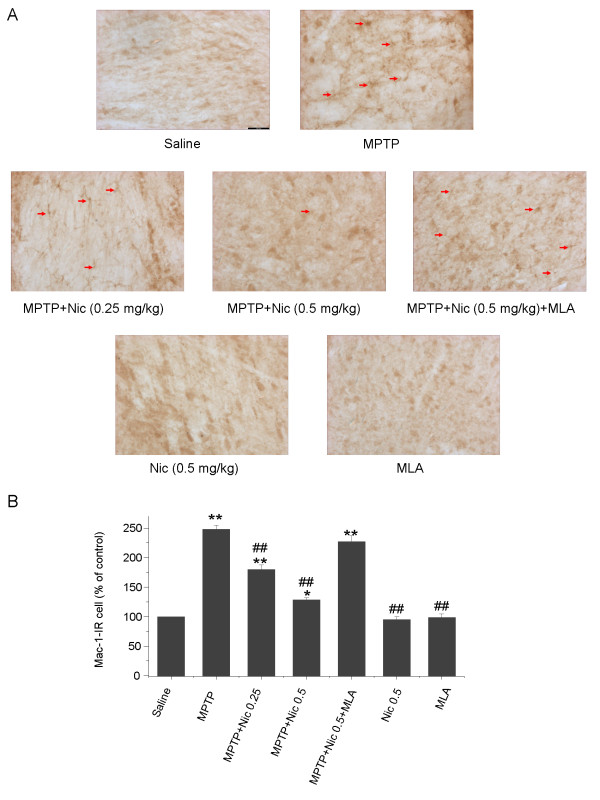
Figure 4
The effects of nicotine on 1-methyl-4-phenyl-1,2,3,6-tetrahydropyridine (MPTP)-induced microglia activation in the mouse substantia nigra pars compacta (SNpc). (A) Mac-1-IR microglia in mouse SNpc. Sc...
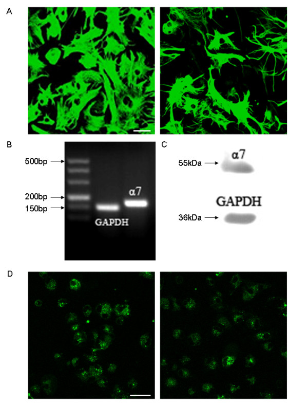
Figure 5
Expression of the u03b17-nicotinic acetylcholine receptor (u03b17-nAChR) subunit expression in primary astrocyte cell cultures. (A) Glial fibrillary acidic protein (GFAP) immunofluorescence staining o...
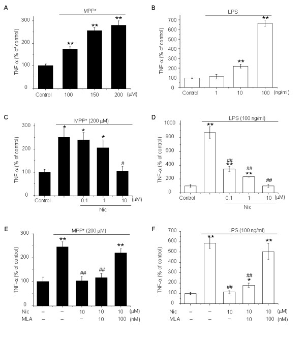
Figure 6
The effects of nicotine on enhanced production from astrocytes of tumor necrosis factor (TNF)-u03b1 induced by 1-methyl-4-phenylpyridinium ion (MPP + ) and lipopolysaccharide (LPS). (A) MPP + and (B) ...
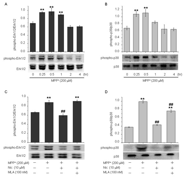
Figure 7
The effects of nicotine on activation of extracellular regulated kinase (Erk)1/2 and p38 mitogen-activated protein kinase (MAPK) induced by 1-methyl-4-phenylpyridinium ion (MPP + ) in astrocytes. MPP ...
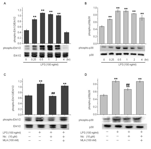
Figure 8
The effects of nicotine on lipopolysaccharide (LPS)-induced Erk1/2 and p38 mitogen-activated protein kinase (MAPK) activation in astrocytes. LPS also increased Erk1/2 and p38 MAPK phosphorylation, ref...
Figure images are served from the NIH/NLM PubMed Central Open Access Subset or Europe PMC; copyright remains with the publishers and authors.
💬 Discussion
0 commentsNo comments yet. Be the first to start a discussion!
Leave a Comment