Abstract
BACKGROUND: Single-particle cryo-electron microscopy (cryo-EM) has become a mainstream tool for the structural determination of biological macromolecular complexes. However, high-resolution cryo-EM reconstruction often requires hundreds of thousands of single-particle images. Particle extraction from experimental micrographs thus can be laborious and presents a major practical bottleneck in cryo-EM structural determination. Existing computational methods for particle picking often use low-resolution templates for particle matching, making them susceptible to reference-dependent bias. It is critical to develop a highly efficient template-free method for the automatic recognition of particle images from cryo-EM micrographs. RESULTS: We developed a deep learning-based algorithmic framework, DeepEM, for single-particle recognition from noisy cryo-EM micrographs, enabling automated particle picking, selection and verification in an integrated fashion. The kernel of DeepEM is built upon a convolutional neural network (CNN) composed of eight layers, which can be recursively trained to be highly "knowledgeable". Our approach exhibits an improved performance and accuracy when tested on the standard KLH dataset. Application of DeepEM to several challenging experimental cryo-EM datasets demonstrated its ability to avoid the selection of un-wanted particles and non-particles even when true particles contain fewer features. CONCLUSIONS: The DeepEM methodology, derived from a deep CNN, allows automated particle extraction from raw cryo-EM micrographs in the absence of a template. It demonstrates an improved performance, objectivity and accuracy. Application of this novel method is expected to free the labor involved in single-particle verification, significantly improving the efficiency of cryo-EM data processing.
🔬 Techniques
🔭 Microscopes
💻 Software
✨ Fluorophores
🏭 Microscope Brands
📷 Detectors
💻 Software Details
💾 Data Repositories
🏛️ Research Organizations (ROR)
Affiliated research institutions:
📊 Figures
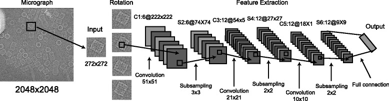
Fig. 1
The architecture of the convolutional neural network used in DeepEM. The convolutional layer and the subsampling layer are abbreviated as C and S, respectively. C1:6@222u00d7222 means that it is a con...
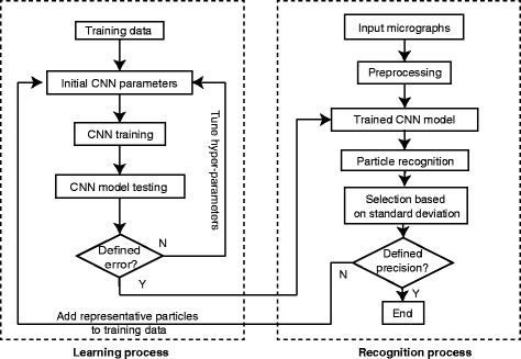
Fig. 2
The workflow diagram of the DeepEM algorithm. The dashed box on the left represents the learning process; the dashed box on the right represents the recognition process
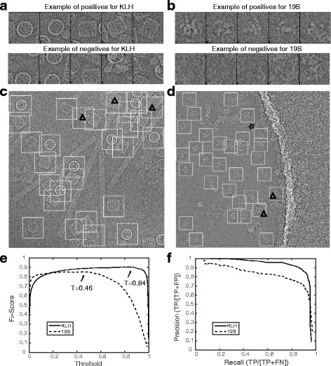
Fig. 3
The DeepEM results for the KLH and 19S regulatory particle datasets. a and b Examples of positive and negative particle images selected for the CNN training in conjunction with the KLH and 19S dataset...
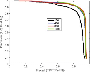
Fig. 4
Impact of the training image number on the precision-recall curve. The black , blue , red and green curves were obtained with the training datasets including 100, 400, 800 and 1200 positive or negativ...
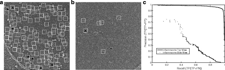
Fig. 5
Two challenging examples of automated particle recognition. a A typical micrograph showing high-density top views of the inflammasome complex. Considerable ice contaminants and overlapping particles a...
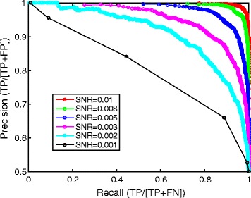
Fig. 6
Effect of the signal-to-noise ratio (SNR) on the precision-recall curves. Three synthetic datasets were generated through computational simulation of micrographs containing single-particle images with...
Figure images are served from the NIH/NLM PubMed Central Open Access Subset or Europe PMC; copyright remains with the publishers and authors.
💬 Discussion
0 commentsNo comments yet. Be the first to start a discussion!
Leave a Comment