Abstract
Expansion microscopy (ExM) physically magnifies biological specimens to enable nanoscale-resolution imaging using conventional microscopes. Current ExM methods permeate specimens with free-radical-chain-growth-polymerized polyacrylate hydrogels, whose network structure limits the local isotropy of expansion as well as the preservation of morphology and shape at the nanoscale. Here we report that ExM is possible using hydrogels that have a more homogeneous network structure, assembled via non-radical terminal linking of tetrahedral monomers. As with earlier forms of ExM, such 'tetra-gel'-embedded specimens can be iteratively expanded for greater physical magnification. Iterative tetra-gel expansion of herpes simplex virus type 1 (HSV-1) virions by ~10× in linear dimension results in a median spatial error of 9.2 nm for localizing the viral envelope layer, rather than 14.3 nm from earlier versions of ExM. Moreover, tetra-gel-based expansion better preserves the virion spherical shape. Thus, tetra-gels may support ExM with reduced spatial errors and improved local isotropy, pointing the way towards single-biomolecule accuracy ExM.
🔬 Techniques
🔭 Microscopes
🧬 Organisms
💻 Software
✨ Fluorophores
🧪 Sample Preparation
🔬 Cell Lines
🏭 Microscope Brands
🧪 Reagent Suppliers
💻 Software Details
💾 Data Repositories
🏛️ Research Organizations (ROR)
Affiliated research institutions:
📋 Methods
Synthesis of tetra-gel
(TG) monomers Monomer 1 was synthesized using a procedure modified from a previously described synthesis ( Supplementary Fig. 1 ) 30 . First, tetra-arm poly(t-butyl acrylate) with bromo terminal groups ( 4 ) was synthesized by atom transfer radical polymerization (ATRP). Next, tetra-arm poly(t-butyl acrylate) with azide terminal groups ( 5 ) was synthesized by replacing bromines of 4 with azides ( Supplementary Fig. 2 ). Finally, monomer 1 was synthesized by hydrolysis and neutralization of 5 to a final pH of ~7. Monomer 2’ , 2’’ , and 2’’’ were synthesized by N-hydroxysuccinimide (NHS) ester-based conjugation of the alkynes (DBCO-NHS, BCN-NHS, or DBCO-SS-NHS) to the terminal primary amines of tetra-arm polyethylene glycols (PEGs). A detailed procedure for the synthesis can be found in the Supplementary Methods .
Cell culture
HEK293FT cells (Thermo Fisher) were cultured in chambered coverglasses (CultureWell, Thermo Fisher) to a confluency of 60–80% 1 , fixed, and immunostained 1 , 4 , 8 . Briefly, the cells were treated with 3% (w/v) formaldehyde and 0.1% (w/v) glutaraldehyde in phosphate buffered saline (PBS, 1x unless otherwise noted) for 10 min at room temperature before the subsequent quenching, blocking, immunostaining, and expansion procedure. HeLa cells (ATCC CCL-2) were plated on coverglasses coated with Matrigel (BD Sciences) to a confluency of 50–90% and fixed 1 , 4 , 8 , 14 . The cells were treated with PBS + 3% (w/v) formaldehyde + 0.1% (w/v) glutaraldehyde for 10 min at room temperature before the subsequent quenching, blocking, and expansion procedure. All the cells were not subjected to additional authentication and were not tested for mycoplasma contamination. A detailed procedure for the immunostaining can be found in the Supplementary Methods . Thy1-YFP mouse brain slice All procedures involving Thy1-YFP-H transgenic mice (Jackson Laboratory) were carried out in accordance with the US National Institutes of Health Guide for the Care and Use of Laboratory Animals and approved by the MIT Committee on Animal Care. All the animals were housed in group in standardized cages (temperature: 20–22 °C, humidity: 30–70%) with a 12-hour light/12-hour dark cycle with unrestricted access to food and water. 50–100 μm coronal brain slices of Thy1-YFP-H mice of 2–4 months old, both male and female, were prepared and immunostained for expansion 4 , 8 , 13 . A detailed procedure for the immunostaining can be found in the Supplementary Methods . General procedure for gelation, digestion, and expansion Fixed (and immunostained) cells and tissues were incubated in ~0.1–0.2 mg/mL NHS-azide in PBS overnight at room temperature and washed with PBS twice. To form the gelling solution, the two monomer solutions were mixed at a close to 1:1 molar ratio, and an additional amount of water was added to adjust the final concentration of monomer 1 to ~3.3% (w/v). For example, 10 µL each of monomer 1 and monomer 2’ (both ~200 mg/mL) and 40 µl of water were mixed to yield the gelling solution. Gelation was carried out for 1–2 hours at 37 °C (blank gels) or overnight at 4 °C (cell and tissue samples) in a gelation chamber 4 , 8 . The gelled cell and tissue samples were incubated in digestion buffer with proteinase K (8 units/mL) (New England BioLabs; 1:100 dilution) overnight at room temperature 4 , 8 and expanded in an excess amount of water three times, each time for 20 min. Expansion of HeLa cells (pre-expansion immunostaining and iterative expansion) Fixed HeLa cells were stained with primary antibodies, oligo-conjugated secondary antibodies, and azide-modified tertiary oligos as previously described 1 , 14 . The cells were gelled with a cleavable TG gelling solution prepared by mixing monomer 1 , monomer 2’’’ , and water. The gelled samples were incubated in digestion buffer with Proteinase K at 8 units/mL overnight at room temperature with gentle shaking before de-hybridization of the oligos from the gel-anchored oligos. The expanded samples were re-embedded in N,N′-bis(acryloyl)cystamine (BAC)-crosslinked non-expanding gel, hybridized with 1 st linker oligos, re-embedded in N,N′-diallyl L-tartardiamide (DATD)-crosslinked expanding gel, and incubated in BAC-cleaving buffer. For fluorescence readout, the samples were incubated with fluorophore-conjugated locked nucleic acid (LNA) oligos and expanded in water. A detailed procedure for the expansion can be found in the Supplementary Methods . Expansion of HSV-1 virions (direct-labelling and iterative expansion) Purified HSV-1 virion stock 47 was diluted before being drop-casted onto a plasma cleaned #0 circular 12-mm coverslip. After 15 min of incubation at room temperature, the virions were fixed in 4% PFA in PBS for 10 min. Azide-modified oligos were directly conjugated to the virion envelope proteins via SoluLink bioconjugation chemistry as previously described 1 . The virions were gelled, digested, expanded, and hybridized with fluorophore-conjugated LNA oligos using a similar procedure to that of the HeLa cell expansion. For 3-round iterative expansion, BAC-cleaved samples were re-embedded in DATD-crosslinked non-expanding gel, hybridized with 2 nd linker oligos, re-embedded in bis-crosslinked expanding gel, and incubated in DATD-cleaving buffer before the LNA oligo-conjugation, expansion, and imaging. A detailed procedure for the expansion and for the sodium polyacrylate/polyacrylamide gel (PAAG) control can be found in the Supplementary Methods . Expansion of HIV and VSV virions (direct labelling and iterative expansion) Purified HIV and VSV virions were immobilized, fixed, conjugated with oligos, gelled, and expanded following the 2-round iterative expansion protocol of HSV-1 virions.
Show full methods section
Synthesis of tetra-gel
(TG) monomers Monomer 1 was synthesized using a procedure modified from a previously described synthesis ( Supplementary Fig. 1 ) 30 . First, tetra-arm poly(t-butyl acrylate) with bromo terminal groups ( 4 ) was synthesized by atom transfer radical polymerization (ATRP). Next, tetra-arm poly(t-butyl acrylate) with azide terminal groups ( 5 ) was synthesized by replacing bromines of 4 with azides ( Supplementary Fig. 2 ). Finally, monomer 1 was synthesized by hydrolysis and neutralization of 5 to a final pH of ~7. Monomer 2’ , 2’’ , and 2’’’ were synthesized by N-hydroxysuccinimide (NHS) ester-based conjugation of the alkynes (DBCO-NHS, BCN-NHS, or DBCO-SS-NHS) to the terminal primary amines of tetra-arm polyethylene glycols (PEGs). A detailed procedure for the synthesis can be found in the Supplementary Methods .
Cell culture
HEK293FT cells (Thermo Fisher) were cultured in chambered coverglasses (CultureWell, Thermo Fisher) to a confluency of 60–80% 1 , fixed, and immunostained 1 , 4 , 8 . Briefly, the cells were treated with 3% (w/v) formaldehyde and 0.1% (w/v) glutaraldehyde in phosphate buffered saline (PBS, 1x unless otherwise noted) for 10 min at room temperature before the subsequent quenching, blocking, immunostaining, and expansion procedure. HeLa cells (ATCC CCL-2) were plated on coverglasses coated with Matrigel (BD Sciences) to a confluency of 50–90% and fixed 1 , 4 , 8 , 14 . The cells were treated with PBS + 3% (w/v) formaldehyde + 0.1% (w/v) glutaraldehyde for 10 min at room temperature before the subsequent quenching, blocking, and expansion procedure. All the cells were not subjected to additional authentication and were not tested for mycoplasma contamination. A detailed procedure for the immunostaining can be found in the Supplementary Methods . Thy1-YFP mouse brain slice All procedures involving Thy1-YFP-H transgenic mice (Jackson Laboratory) were carried out in accordance with the US National Institutes of Health Guide for the Care and Use of Laboratory Animals and approved by the MIT Committee on Animal Care. All the animals were housed in group in standardized cages (temperature: 20–22 °C, humidity: 30–70%) with a 12-hour light/12-hour dark cycle with unrestricted access to food and water. 50–100 μm coronal brain slices of Thy1-YFP-H mice of 2–4 months old, both male and female, were prepared and immunostained for expansion 4 , 8 , 13 . A detailed procedure for the immunostaining can be found in the Supplementary Methods . General procedure for gelation, digestion, and expansion Fixed (and immunostained) cells and tissues were incubated in ~0.1–0.2 mg/mL NHS-azide in PBS overnight at room temperature and washed with PBS twice. To form the gelling solution, the two monomer solutions were mixed at a close to 1:1 molar ratio, and an additional amount of water was added to adjust the final concentration of monomer 1 to ~3.3% (w/v). For example, 10 µL each of monomer 1 and monomer 2’ (both ~200 mg/mL) and 40 µl of water were mixed to yield the gelling solution. Gelation was carried out for 1–2 hours at 37 °C (blank gels) or overnight at 4 °C (cell and tissue samples) in a gelation chamber 4 , 8 . The gelled cell and tissue samples were incubated in digestion buffer with proteinase K (8 units/mL) (New England BioLabs; 1:100 dilution) overnight at room temperature 4 , 8 and expanded in an excess amount of water three times, each time for 20 min. Expansion of HeLa cells (pre-expansion immunostaining and iterative expansion) Fixed HeLa cells were stained with primary antibodies, oligo-conjugated secondary antibodies, and azide-modified tertiary oligos as previously described 1 , 14 . The cells were gelled with a cleavable TG gelling solution prepared by mixing monomer 1 , monomer 2’’’ , and water. The gelled samples were incubated in digestion buffer with Proteinase K at 8 units/mL overnight at room temperature with gentle shaking before de-hybridization of the oligos from the gel-anchored oligos. The expanded samples were re-embedded in N,N′-bis(acryloyl)cystamine (BAC)-crosslinked non-expanding gel, hybridized with 1 st linker oligos, re-embedded in N,N′-diallyl L-tartardiamide (DATD)-crosslinked expanding gel, and incubated in BAC-cleaving buffer. For fluorescence readout, the samples were incubated with fluorophore-conjugated locked nucleic acid (LNA) oligos and expanded in water. A detailed procedure for the expansion can be found in the Supplementary Methods . Expansion of HSV-1 virions (direct-labelling and iterative expansion) Purified HSV-1 virion stock 47 was diluted before being drop-casted onto a plasma cleaned #0 circular 12-mm coverslip. After 15 min of incubation at room temperature, the virions were fixed in 4% PFA in PBS for 10 min. Azide-modified oligos were directly conjugated to the virion envelope proteins via SoluLink bioconjugation chemistry as previously described 1 . The virions were gelled, digested, expanded, and hybridized with fluorophore-conjugated LNA oligos using a similar procedure to that of the HeLa cell expansion. For 3-round iterative expansion, BAC-cleaved samples were re-embedded in DATD-crosslinked non-expanding gel, hybridized with 2 nd linker oligos, re-embedded in bis-crosslinked expanding gel, and incubated in DATD-cleaving buffer before the LNA oligo-conjugation, expansion, and imaging. A detailed procedure for the expansion and for the sodium polyacrylate/polyacrylamide gel (PAAG) control can be found in the Supplementary Methods . Expansion of HIV and VSV virions (direct labelling and iterative expansion) Purified HIV and VSV virions were immobilized, fixed, conjugated with oligos, gelled, and expanded following the 2-round iterative expansion protocol of HSV-1 virions.
Imaging and visualization
All the expanded samples were imaged with a diffraction-limited spinning disk confocal microscope (CSU-W1, Yokogawa on Eclipse Ti-E microscope body, Nikon) with a CFI Apo LambdaS LWD 40x, 1.15 NA water-immersion objective (Nikon), controlled by NIS-Elements AR v4.60.00 (Nikon). The two-colour HeLa cell images and all the virion images were deconvolved with theoretical point-spread-functions (PSFs) (Huygens Essential for MacX11 built on Feb 7, 2013, SVI) before visualization and image analysis. Unless otherwise noted, all the 3D renderings were generated using Imaris x64 8.3 (Oxford Instruments). Part of the image datasets were analysed and visualized using ImageJ (Fiji) 2.10/1.53c. Part of the plots were generated using OriginPro8.1 (Origin).
Image analysis
For virion envelope protein analysis, first, single-particle averaged virion images were generated using a semi-automated image analysis pipeline implemented on MATLAB R2018a-R2020b (“Virus Particle Analysis”) 48 . Each virion was manually aligned, automatically cropped, calibrated with the expansion factor, and arithmetically averaged to generate the single-particle averaged images. Next, using Virus Particle Analysis, spatial arrangements of the virion envelope proteins were quantified. Radii in 8 directions (45 degrees apart) were measured for each virion as the distance from the particle centroid to the Gaussian-fitted centre of the envelope profile. After inspection to remove unfitted profiles, standard deviations of all the accepted radii within the same particle ( σ ) were reported as population statistics for virions with >= 3 accepted radii. For HSV-1 virion shape analysis, virions with >= 6 accepted radii (for eccentricity, virions with >= 6 accepted radii and with the accepted radii constituting at least 2 mutually perpendicular diameters) were used to assure an accurate approximation of the particle shapes. Eccentricity ( e ) was defined as the ratio of the minor axis to the major axis. Sphericity ( s ) was defined as the ratio of the radius of the inscribing circle to the radius of the circumscribing circle. The normalized circular standard deviation ( σ n ) was defined as the standard deviation of normalized radii ( R n ) within each particle. For microtubule analysis, the peak-to-peak distance between microtubule sidewalls was measured using a semi-automated algorithm implemented on MATLAB R2018a-R2020b (“Microtubule Peak-to-Peak Distance Analysis”) 48 . On maximum intensity z-projection images of beta-tubulin-stained HeLa cells, two points were manually selected along the centreline of a microtubule segment, and a 200 nm (in biological length) segment was cropped out from the selected segment. The line intensity profile along the 200 nm segment was fitted with two Gaussian functions to detect the two peaks in the fluorescence intensity, between which the distance was measured as the peak-to-peak distance of the microtubule sidewalls. A detailed description of image analysis can be found in the Supplementary Methods . Modelling and simulation of single-particle averaged HSV-1 virion images A model of spherical virions was developed on MATLAB R2020a-R2020b based on experimental parameters derived from individual HSV-1 virions, in order to simulate the single-particle averaged HSV-1 virion images and their quantitative metrics (“HSV-1 Averaged Particle Image Simulation”) 48 . A detailed description of the modelling can be found in the Supplementary Methods .
General procedure for gelation, digestion, and expansion Fixed (and immunostained) cells and tissues were incubated in ~0.1–0.2 mg/mL NHS-azide in PBS overnight at room temperature and washed with PBS twice. To form the gelling solution, the two monomer solutions were mixed at a close to 1:1 molar ratio, and an additional amount of water was added to adjust the final concentration of monomer 1 to ~3.3% (w/v). For example, 10 µL each of monomer 1 and monomer 2’ (both ~200 mg/mL) and 40 µl of water were mixed to yield the gelling solution. Gelation was carried out for 1–2 hours at 37 °C (blank gels) or overnight at 4 °C (cell and tissue samples) in a gelation chamber 4 , 8 . The gelled cell and tissue samples were incubated in digestion buffer with proteinase K (8 units/mL) (New England BioLabs; 1:100 dilution) overnight at room temperature 4 , 8 and expanded in an excess amount of water three times, each time for 20 min.
Supplementary Material 1 1673281_Supp_Vid1
📊 Figures
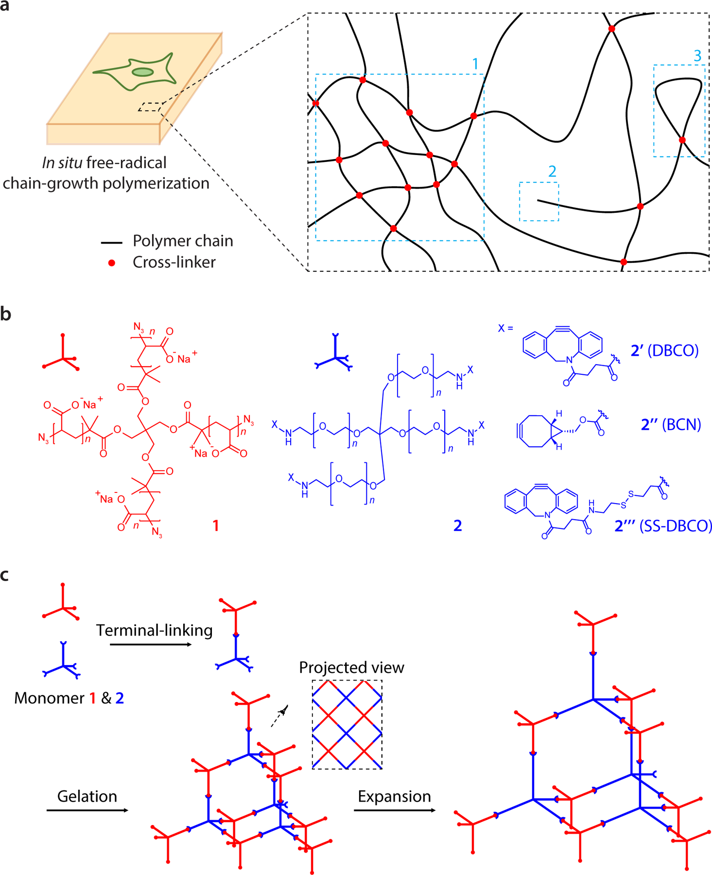
Figure 1.
Design and synthesis of tetra-gel (TG) for expansion microscopy.
a, Cell/tissue-hydrogel composites formed by in situ free-radical chain-growth polymerization are known to have structural inhomogeneities in the range of tens of nanometres due to (1) local fluctuati...
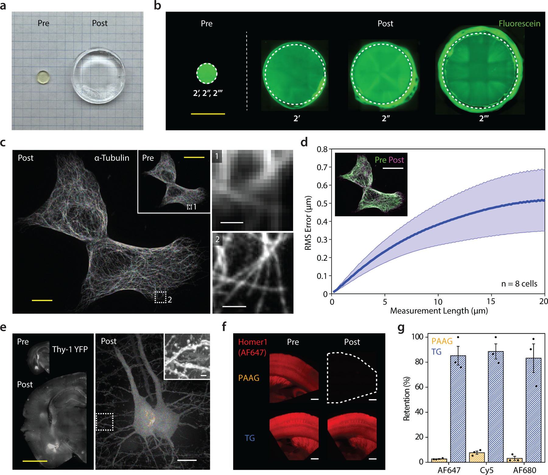
Figure 2.
TG-mediated expansion of cells and tissues.
a, Image of TG (using monomer 2u2019u2019u2019 ) as synthesized (left, pre-expansion) and after swelling in deionized water (right, post-expansion). The two gels were cast in circular molds with ident...
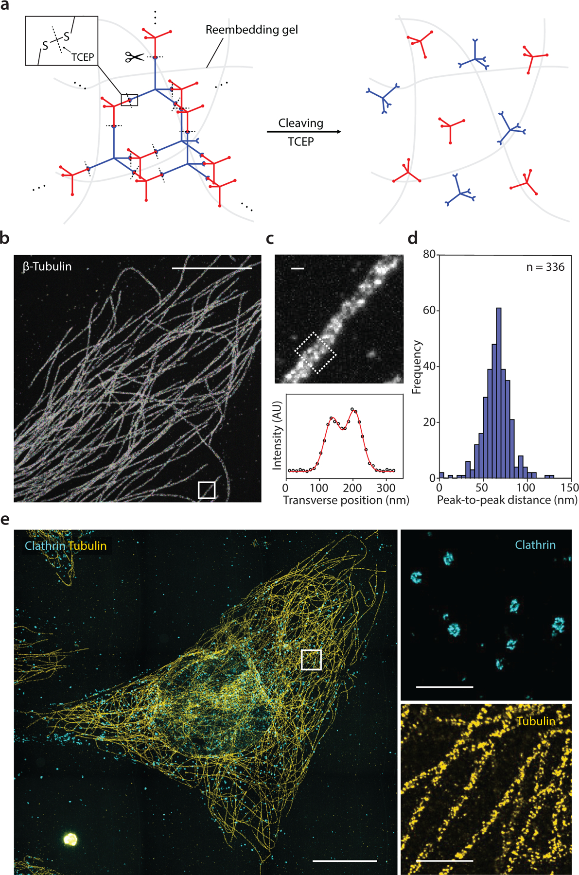
Figure 3.
TG-based iterative expansion.
a, Monomeric cleaving of TG (monomer 2u2019u2019u2019 ) after re-embedding in a second hydrogel. A reducing agent, tris(2-carboxyethyl)phosphine) (TCEP), was applied to cleave the disulfide bonds in t...
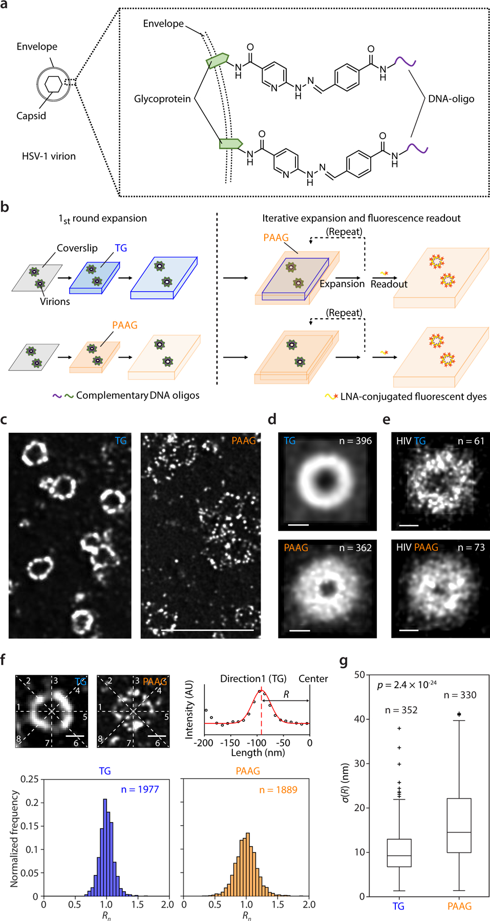
Figure 4.
Spatial errors introduced by TG-based vs. classical PAAG-based iterative expansion microscopy.
a, Short DNA oligos (22 bp) were covalently conjugated to the envelope proteins of herpes simplex virus type 1 (HSV-1) virions via hydrazone formation, which allows labelling transfer across multiple ...
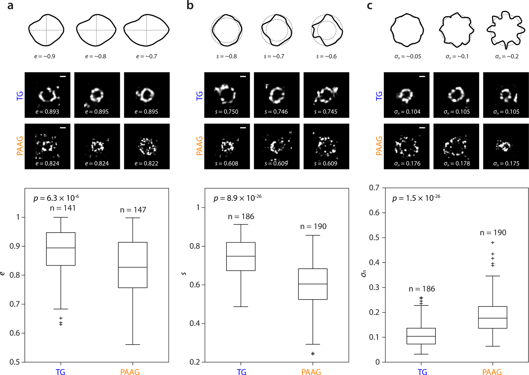
Figure 5.
Shape analysis of TG- vs PAAG-expanded HSV-1 virions.
a. Top, schematic examples of shapes with ~0.9, 0.8, and 0.7 eccentricity ( e ). Middle, HSV-1 virions expanded by TG- (top row) and PAAG-based (bottom row) 2-round iterative expansion, with the z-mid...
Figure images are served from the NIH/NLM PubMed Central Open Access Subset or Europe PMC; copyright remains with the publishers and authors.
💬 Discussion
0 commentsNo comments yet. Be the first to start a discussion!
Leave a Comment